

Electroporation controlled with real time imaging
a real-time imaging and electrode technology, applied in the field of electrode electrode control with real-time imaging, can solve the problem of not being closely monitored and controlled
- Summary
- Abstract
- Description
- Claims
- Application Information
AI Technical Summary
Benefits of technology
Problems solved by technology
Method used
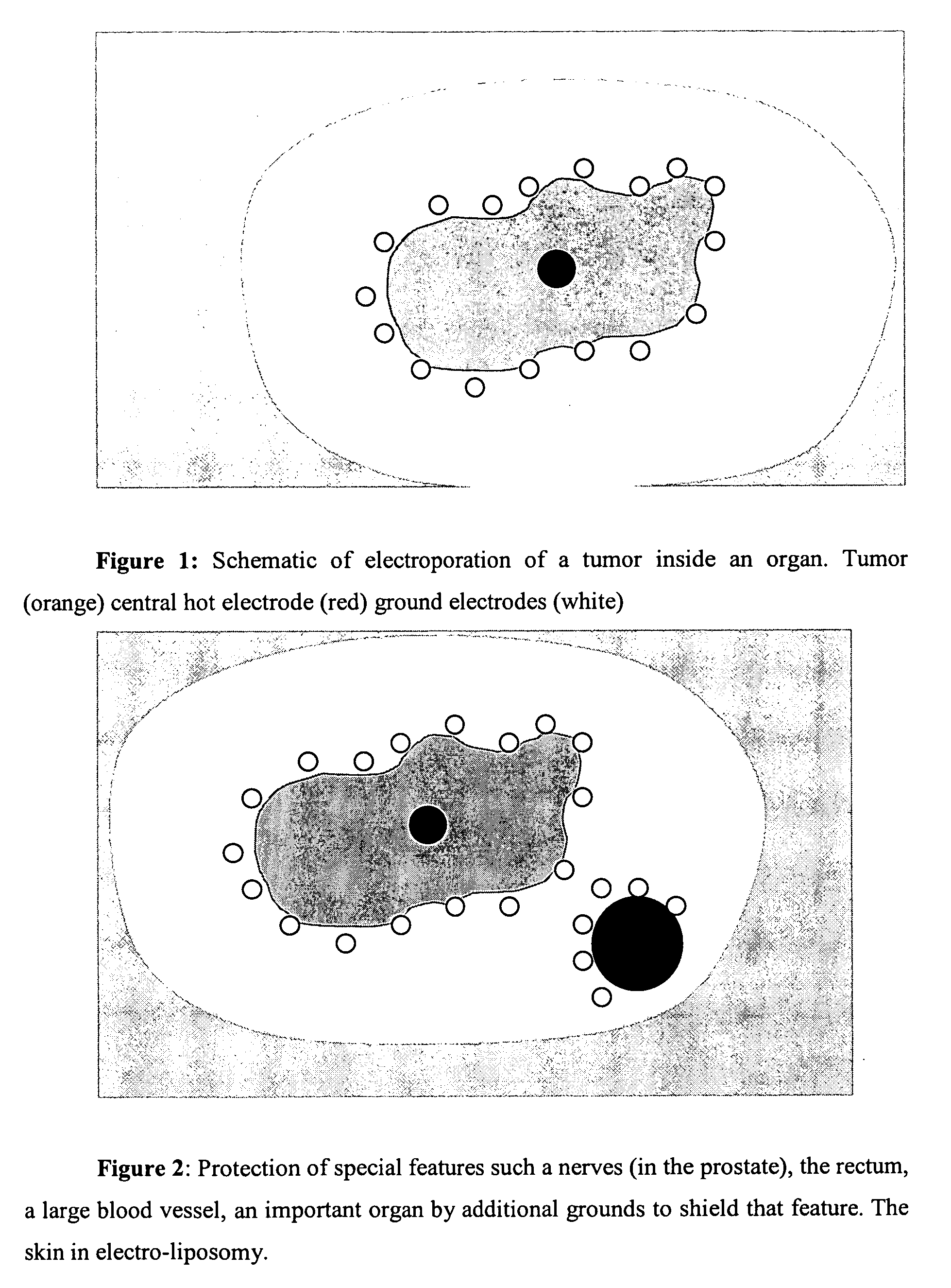
Image

Examples
example 1
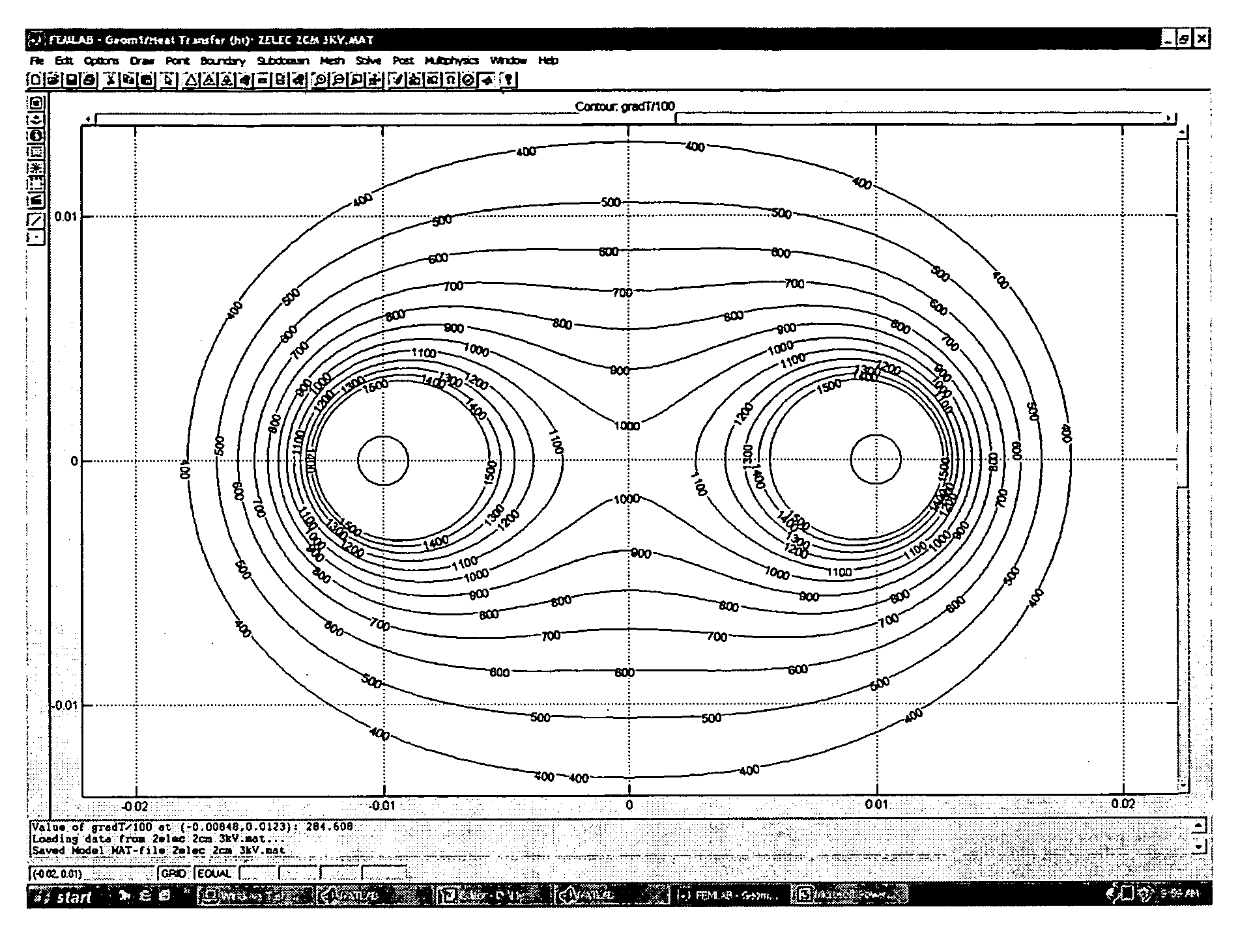
[0061] An experimental study with analytical components was performed on a pig liver. The study was conducted in accordance with Good Laboratory Practice regulations as set forth by the 21 Code of Federal Regulations (CFR) Part 58. Full QA oversight, GLP documentation, and a GLP report has provided for this study which fulfills the requirements for submission to federal and / or other agencies requesting non-clinical GLP documentation. The study was performed at Covance Research Products, Berkeley Calif.
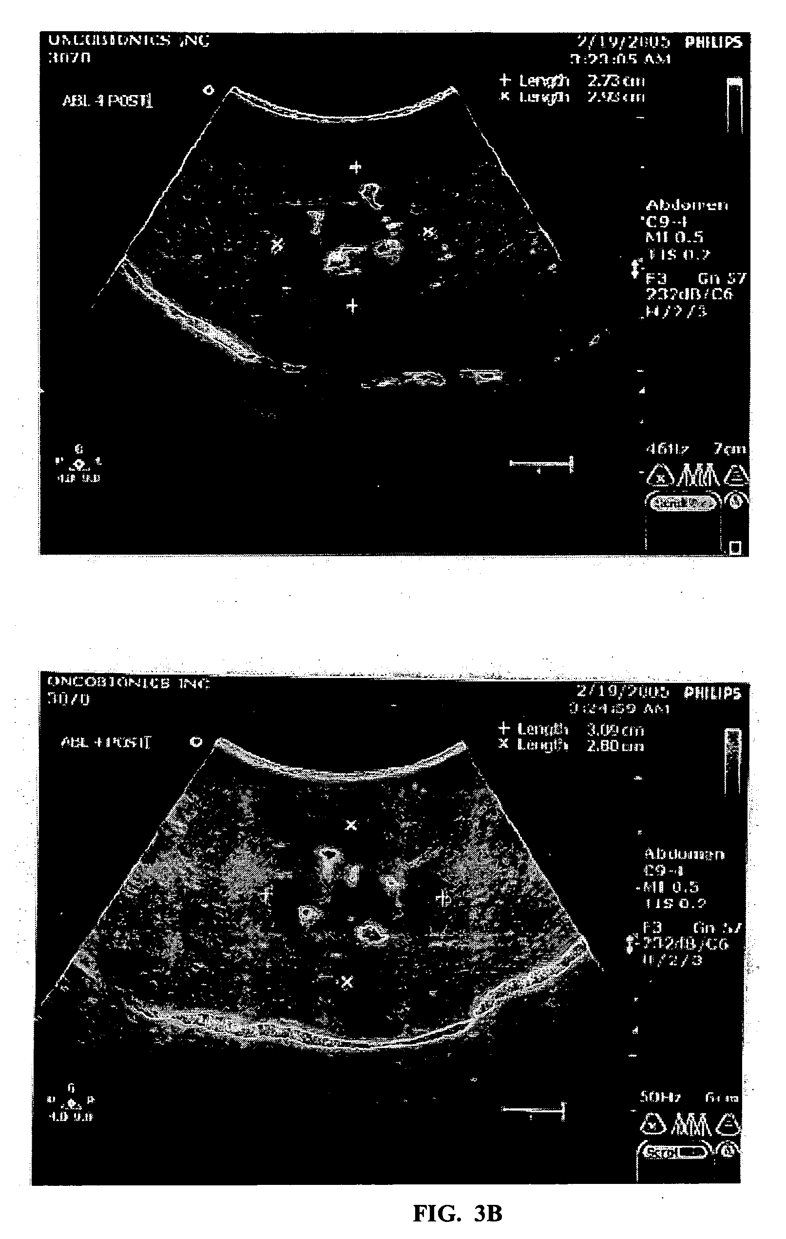
[0062] Five 100 lb pigs were used in this study. In a typical procedure the pig was anesthetized using general anesthesia. The was liver exposed by an open laparotomy incision. Between two and nine electrode needles were introduced in the liver at desired location under ultrasound monitoring. Approximately 20 different experiments with a variety of needle configuration placements and electroporation potentials were used with the goal of correlating electrical potentials, medical imagi...
PUM
 Abstract
Description
Claims
Application Information