
System and method of computer-aided detection

a computer-aided detection and detection system technology, applied in the field of computer-aided detection, can solve the problems of not always providing the correct diagnosis, the inability of automated diagnosis the inability of computer applications to identify most or all abnormalities, etc., to achieve the effect of facilitating further image processing

Active Publication Date: 2006-12-07
THE MEDIPATTERN CORP +1
View PDF37 Cites 313 Cited by
  • Summary
  • Abstract
  • Description
  • Claims
  • Application Information

AI Technical Summary

Benefits of technology

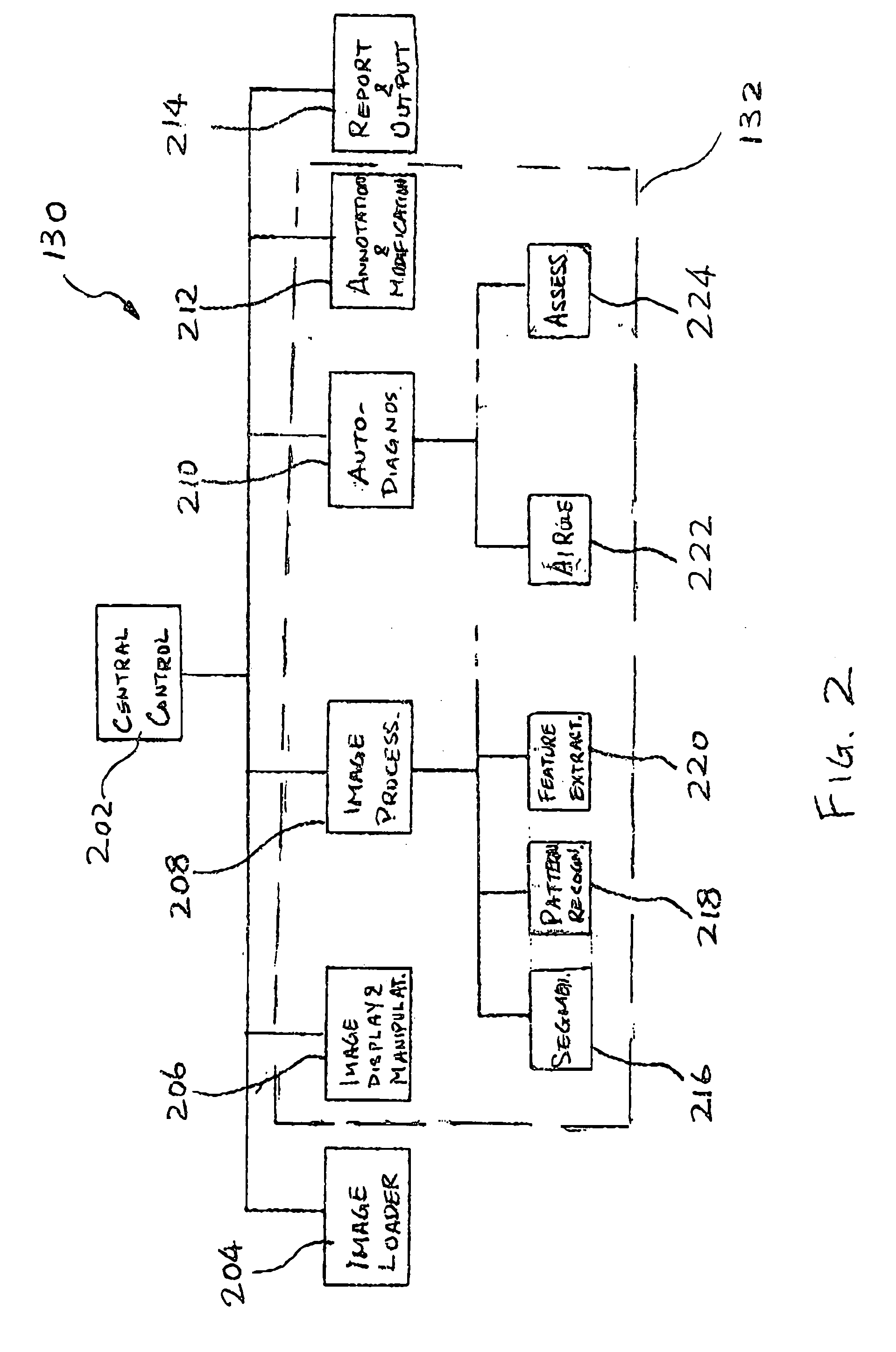
"The invention is a computer-aided system and method for detecting abnormalities in medical images and providing a diagnosis based on the identified features. The system includes an image processor that extracts relevant features from the medical image, a decision engine that generates an initial diagnosis, and an annotation and modification tool for the user to review and modify the features. The system can also analyze multiple medical images from different modalities and adjust acquisition conditions to improve image quality. The technical effects of the invention include improved accuracy and efficiency in detecting abnormalities and providing a diagnosis, assisting in the interpretation of medical images, and aiding in the diagnosis of rare or difficult-to-distinguish abnormalities."

Problems solved by technology

However, while computerized pattern recognition has seen tremendous advances in the past decade or so, sometimes, a computer application may still have difficulty in identifying most or all abnormalities.
As a radiologist may not place too high a confidence in results of automated detection, biopsy may be ordered, which sometimes turn out to be unnecessary.
Further, even if successful detection of all relevant characteristics in a medical image were possible, automated diagnosis may not always provide a correct diagnosis due to, for example, inadequacy or lack of sophistication of models underlying a diagnosis engine.
The foregoing creates challenges and constraints for all CAD systems for extracting, i.e., identifying characteristics and medical features in medical images and suggesting diagnosis based on characteristics automatically detected in the medical image.

Method used


Image

  • System and method of computer-aided detection
  • System and method of computer-aided detection
  • System and method of computer-aided detection

Examples


Embodiment Construction

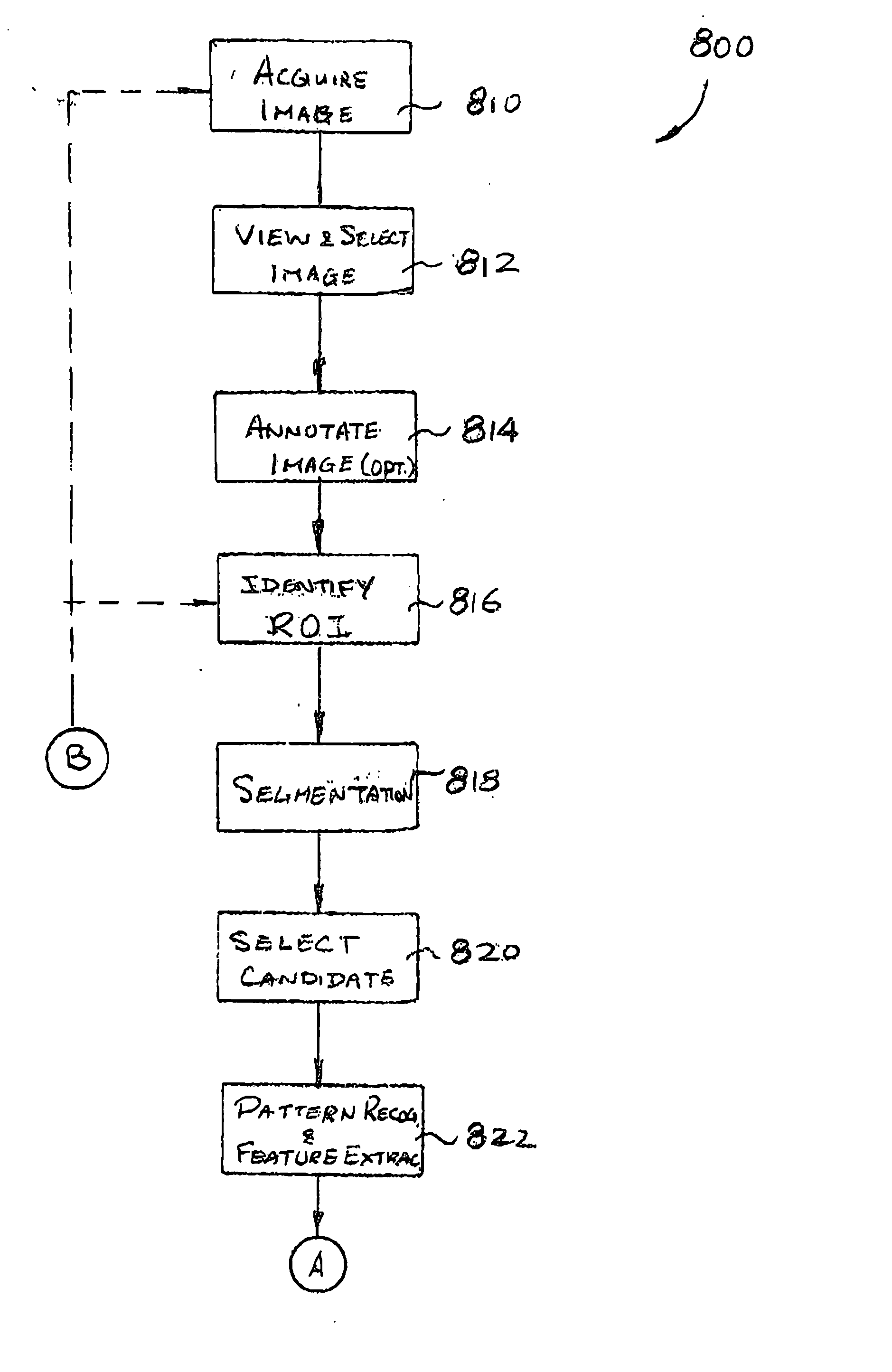
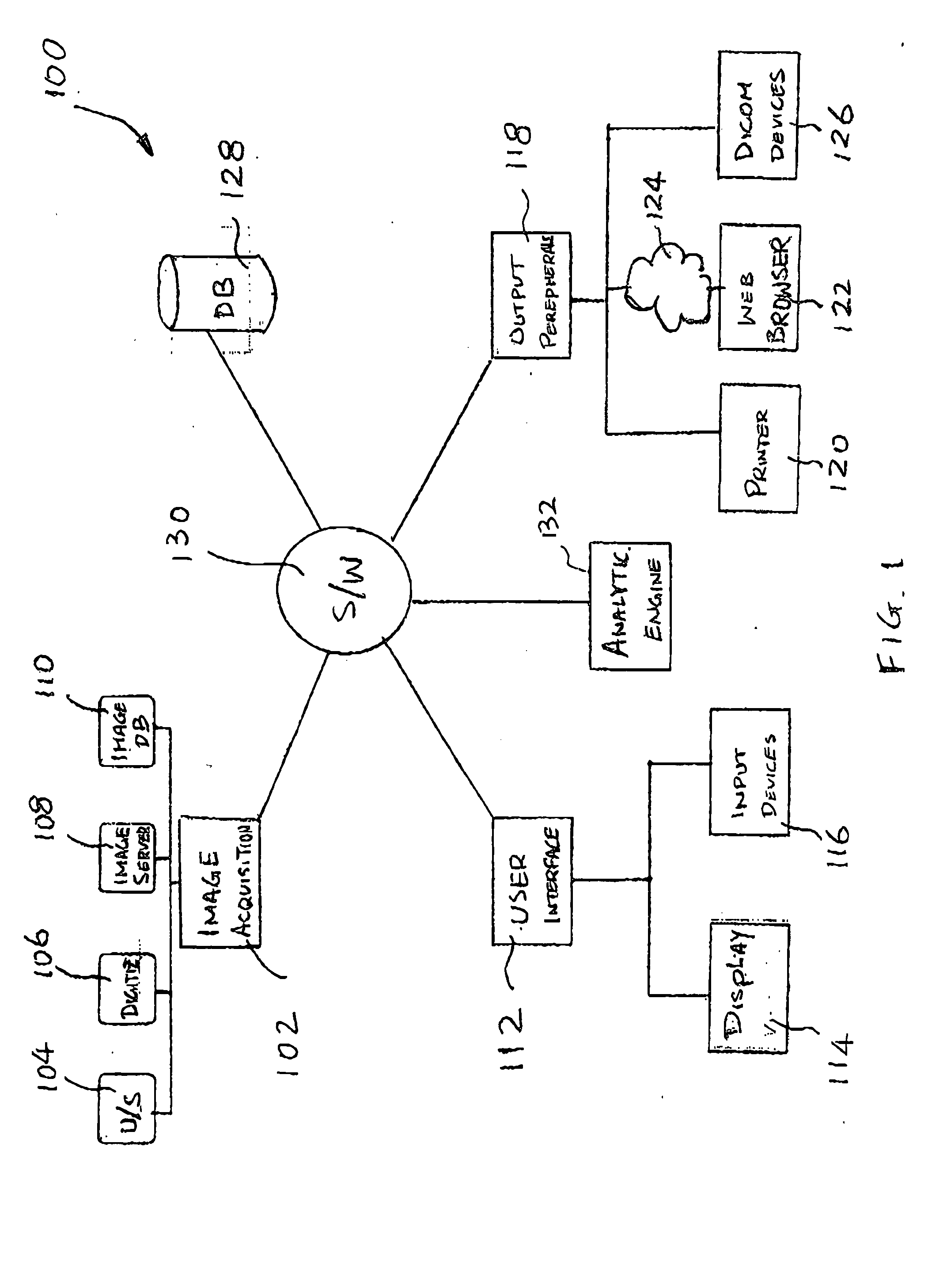
[0035] The description which follows and the embodiments described therein are provided by way of illustration of an example, or examples, of particular embodiments of the principles of the present invention. These examples are provided for the purposes of explanation, and not limitation, of those principles and of the invention. In the description which follows, like parts are marked throughout the specification and the drawings with the same respective reference numerals.

[0036]FIG. 1 shows a CAD system 100 that is controlled by a software system for automatically analyzing medical images, detecting, identifying and classifying physical, textural and morphological characteristics or other features of masses within medical images, providing computer-aided detection and assessment of suspected lesions for user selection, and allowing interactive feedback from a user to dynamically modify a list of detected features and the diagnosis computed therefrom. The user may be a technician, ...


PUM


Abstract

The invention provides a system and method for computer-aided detection (“CAD”). The invention relates to computer-aided automatic detection of abnormalities in and analysis of medical images. Medical images are analyzed, to extract and identify a set of features in the image relevant to a diagnosis. The system computes an initial diagnosis based on the set of identified features and a diagnosis model, which are provided to a user for review and modification. A computed diagnosis is dynamically re-computed upon user modification of the set of identified features. Upon a user selecting a diagnosis based on system recommendation, a diagnosis report is generated reflecting features present in the medical image as validated by the user and the user selected diagnosis.

Description

CROSS-REFERENCE TO RELATED APPLICATIONS [0001] This application claims priority from U.S. Provisional Application No. 60 / 686,397 filed on Jun. 2, 2005 and U.S. Provisional Application No. 60 / 738,999 filed on Nov. 23, 2005 which are hereby incorporated by reference.FIELD OF INVENTION [0002] The invention relates generally to the field of computer-aided detection (“CAD”) and analysis of abnormalities. In particular, the invention relates to automatic detection of abnormalities in and analysis of medical images and automated assessment thereof. BACKGROUND OF INVENTION [0003] With the emphasis on early detection of cancer, more and more people are taking part in early screening programs, such as mammography screening and in some parts of the world ultrasound screening for breast cancer. Some recent studies suggest that diagnostic breast ultrasonography may successfully help distinguish many benign from malignant solid lesions or nodules. For example, in “Solid breast nodules: use of son...

Claims


Application Information

Patent Timeline
Patent Type & Authority Applications(United States)
IPC IPC(8): G06K9/00G16H30/40G16H50/20G16Z99/00
CPCA61B6/00A61B8/00G06F19/321A61B6/5247G06T7/0012G06T2207/30068G06F19/345G16H50/20G16H30/40G16Z99/00G06T7/00
Inventor COLLINS, JEFFREYSAGHATELYAN, KARENLACHMANN, FREDERIC
Owner THE MEDIPATTERN CORP
Learn More