Drape Assembly For Surgical Microscope Assembly

a surgical microscope and assembly technology, applied in the field of surgical microscope drape assembly, can solve the problems of impracticality in sterilizing these microscopes, large and cumbersome assembly of microscopes,

Inactive Publication Date: 2007-03-22
INSIGHT INSTR
View PDF6 Cites 21 Cited by
  • Summary
  • Abstract
  • Description
  • Claims
  • Application Information

AI Technical Summary

Benefits of technology

[0008] A method of maintaining a sterile field around a surgical microscope is also provided. In one example the method includes providing a drape for covering the microscope, with the drape including a collar having a first opening through the drape for receiving an adapter. The collar includes a flap with a surface having an adhesive formed thereon. The adapter is attached to a lens housing of the microscope and the collar is passed along an exterior portion of the adapter so that part of the adapter extends through the collar opening. The flap of the collar is attached to the adapter or the optical component with the adhesive.

Problems solved by technology

These microscopes are typically large and cumbersome assemblies that are maintained in operating room environments although it is impractical to sterilize these microscopes.

Method used


Image

  • Drape Assembly For Surgical Microscope Assembly
  • Drape Assembly For Surgical Microscope Assembly
  • Drape Assembly For Surgical Microscope Assembly

Examples


Embodiment Construction

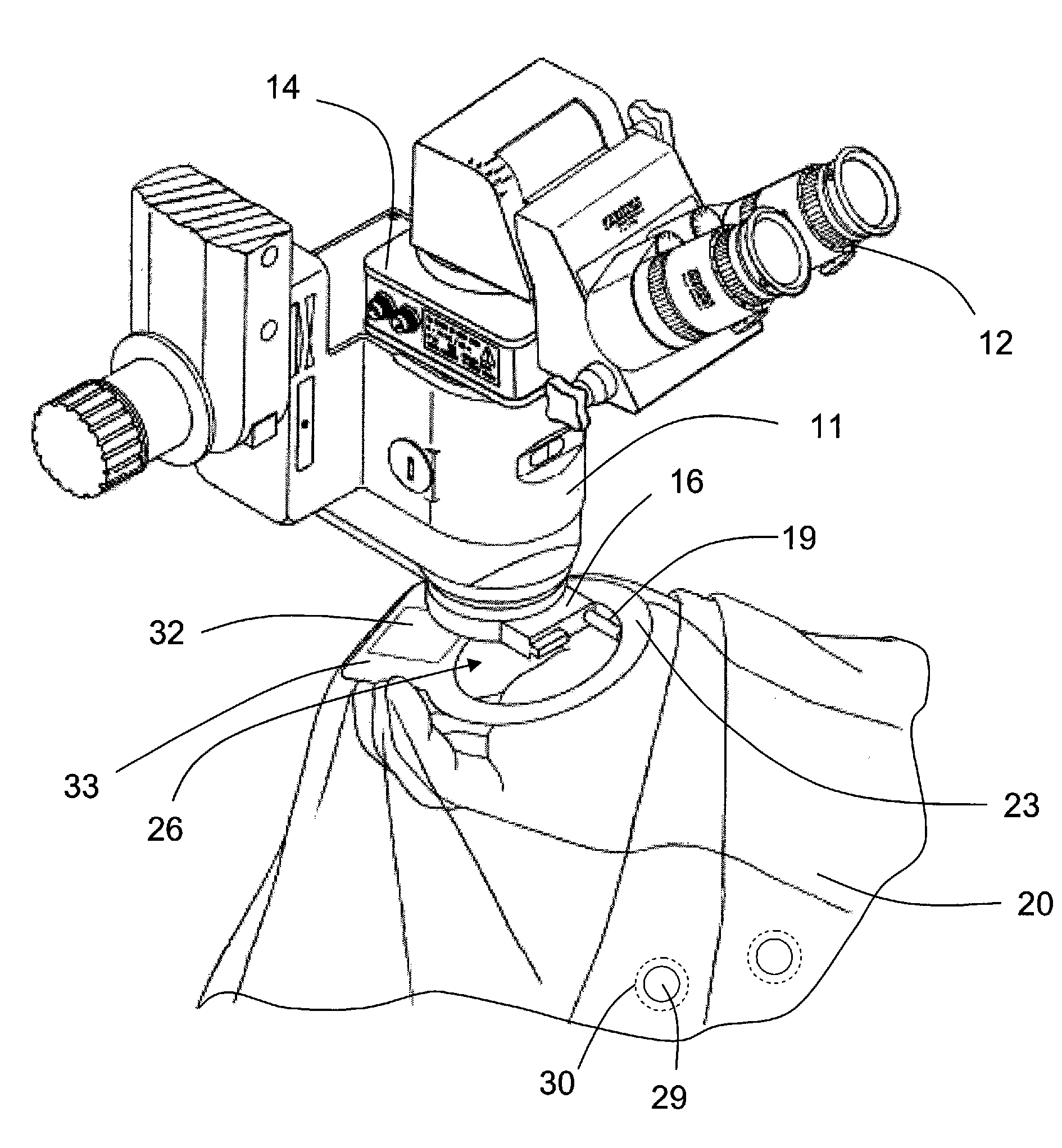
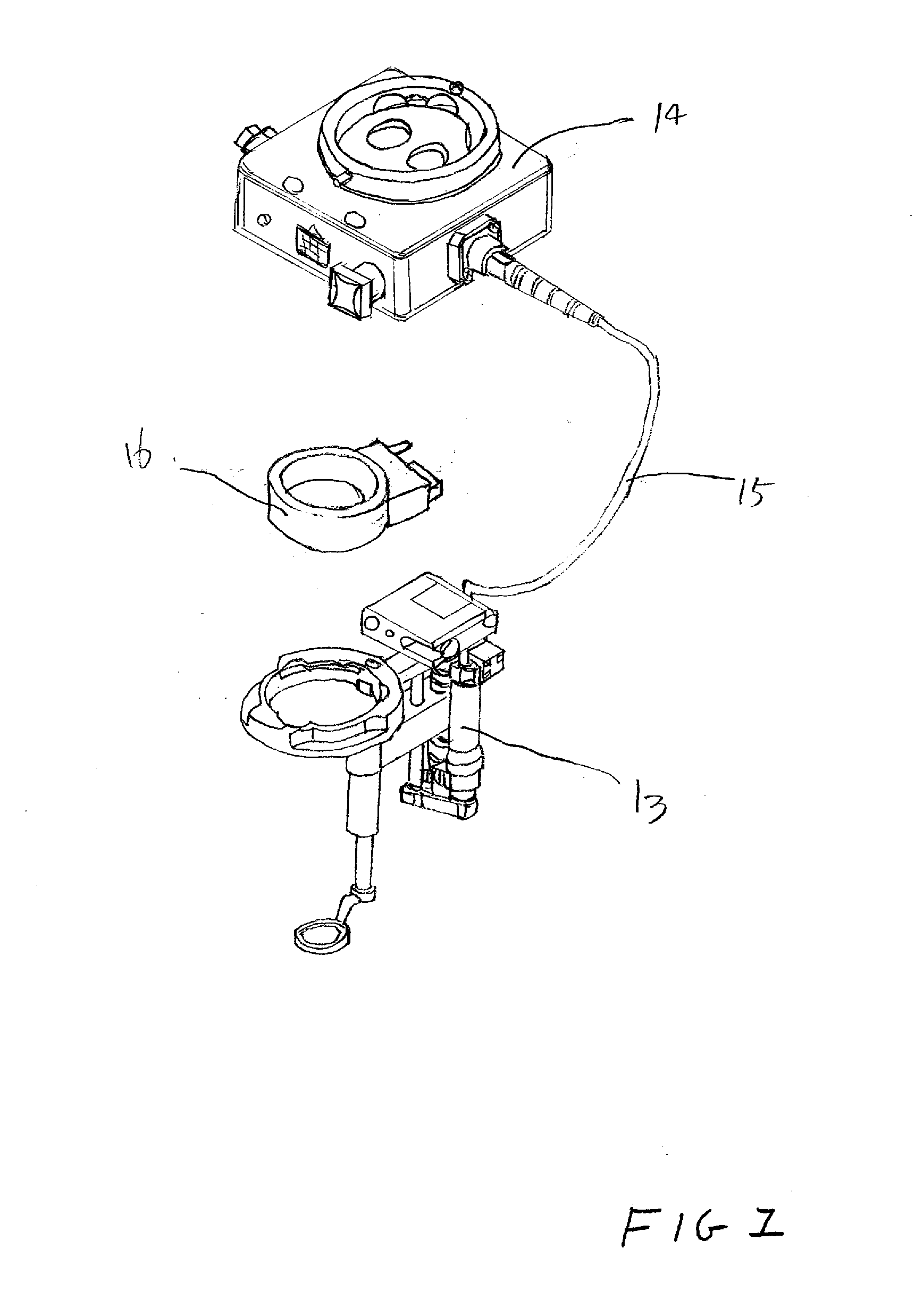
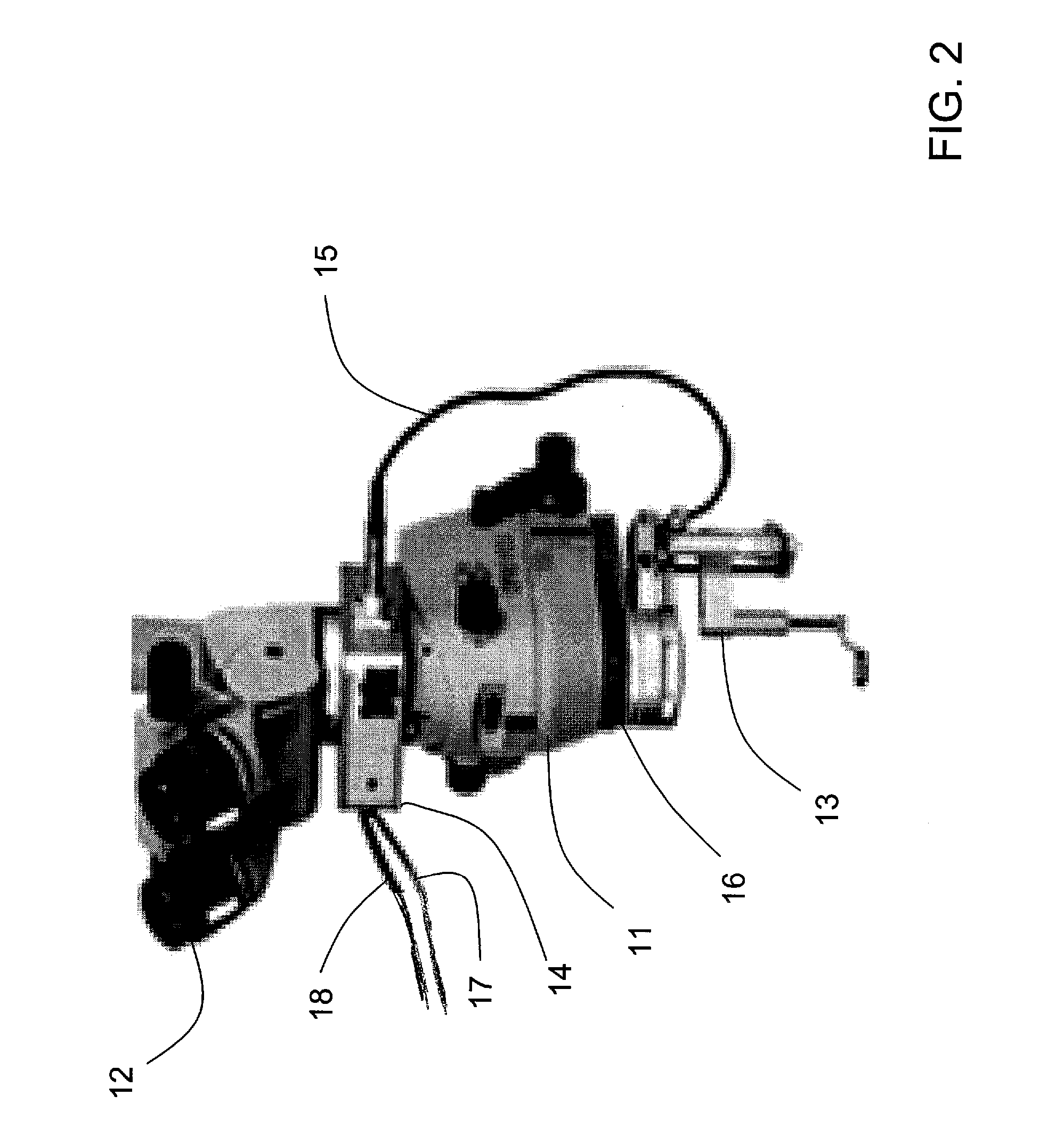
[0017] In the field of ophthalmic surgery, it is common to attach optical devices to conventional microscopes in order to further increase magnification, invert images, and facilitate stereoscopic viewing by a surgeon. One such device is a Binocular Indirect Ophthalmo-Microscope Stereoscopic Diagonal Inverter (BIOM / SDI) system. FIG. 1 illustrates a BIOM / SDI system 10 comprising a Binocular Indirect Ophthalmo-Microscope (BIOM) 13, a stereoscopic diagonal inverter (SDI) 14, a connection cable 15, and a conventional BIOM adapter 16. Normally, all of the foregoing elements, except the SDI, are sterilized before being brought into the sterilized operating room environment. FIG. 2 is a photograph of a surgical microscope 1 incorporating a BIOM / SDI system 10. The microscope 1 comprises a lens housing 11, a pair of ocular ports 12, the BIOM 13, the SDI 14, the optical cable 15 connecting the BIOM 13 and the SDI 14, the BIOM adapter 16, a power cable 17, and a switch cable 18.

[0018] Accordi...


PUM


Abstract

A drape assembly is provided for maintaining a sterile field around a surgical microscope. A drape has a first opening for receiving a protruding adapter connected to a lower end of the microscope and a collar is mounted in the first opening. An adhesive portion of the collar secures the drape to the adapter. The adapter becomes part of the optical system and may be referred to as an optical component.

Description

SPECIFIC DATA RELATED TO THE INVENTION [0001] This application claims the benefit of U.S. Provisional application No. 60 / 719,022 filed Sep. 21, 2005.BACKGROUND OF THE INVENTION [0002] 1. Field of the Invention [0003] The present invention relates to a surgical microscope drape assembly and, more particularly, to a drape which maintains a sterile field during the performance of a surgical procedure. [0004] 2. Background Art [0005] Stereoscopic microscopes are used extensively in surgical environments and, in particular, during eye surgery. These microscopes are typically large and cumbersome assemblies that are maintained in operating room environments although it is impractical to sterilize these microscopes. Accordingly, various types of surgical drapes have been developed to cover non-sterilized areas of the microscope while still allowing surgeons and staff to adjust the instrumentation and use such an optical assembly without interference from the drape. SUMMARY OF THE INVENTION...

Claims


Application Information

Patent Timeline
Patent Type & Authority Applications(United States)
IPC IPC(8): A61B19/08
CPCA61B19/081A61B46/10
Inventor LULOH, K. PETERANNEN, MICHAEL
Owner INSIGHT INSTR
Why Eureka
Try Eureka