Augmented reality surgical microscope and microscopy method

a surgical microscope and microscopy technology, applied in the field of augmented reality surgical microscope and microscopy method, can solve the problems of inability to capture one, cumbersome switching process, and distracting surgeons from surgery

Pending Publication Date: 2019-09-19
LEICA INSTR SINGAPORE PTE
View PDF4 Cites 20 Cited by
  • Summary
  • Abstract
  • Description
  • Claims
  • Application Information

AI Technical Summary

Benefits of technology

[0025]In the following, the invention is described in detail with reference to the opening drawing, using an exemplary embodiment. The specific combination of features shown in this embodiment is for explanatory purposes only and may be altered according to the above description of the various additional features. For example, a feature may be omitted if its technical effect is not needed in a particular application and vice versa: a feature may be added if, for a particular application, its technical effect is needed.

Problems solved by technology

However, they are only capable of capturing one mode at a time.
This switching process is cumbersome and distracts the surgeon from surgery.
Further, the surgeon essentially needs to combine all the information by memory, which is inevitably inaccurate, prone to mistakes, tiring and requires extensive training and experience.

Method used


Image

  • Augmented reality surgical microscope and microscopy method
  • Augmented reality surgical microscope and microscopy method
  • Augmented reality surgical microscope and microscopy method

Examples


Embodiment Construction

[0032]In the following, the invention is described with reference to FIGS. 1, 2 and 3.

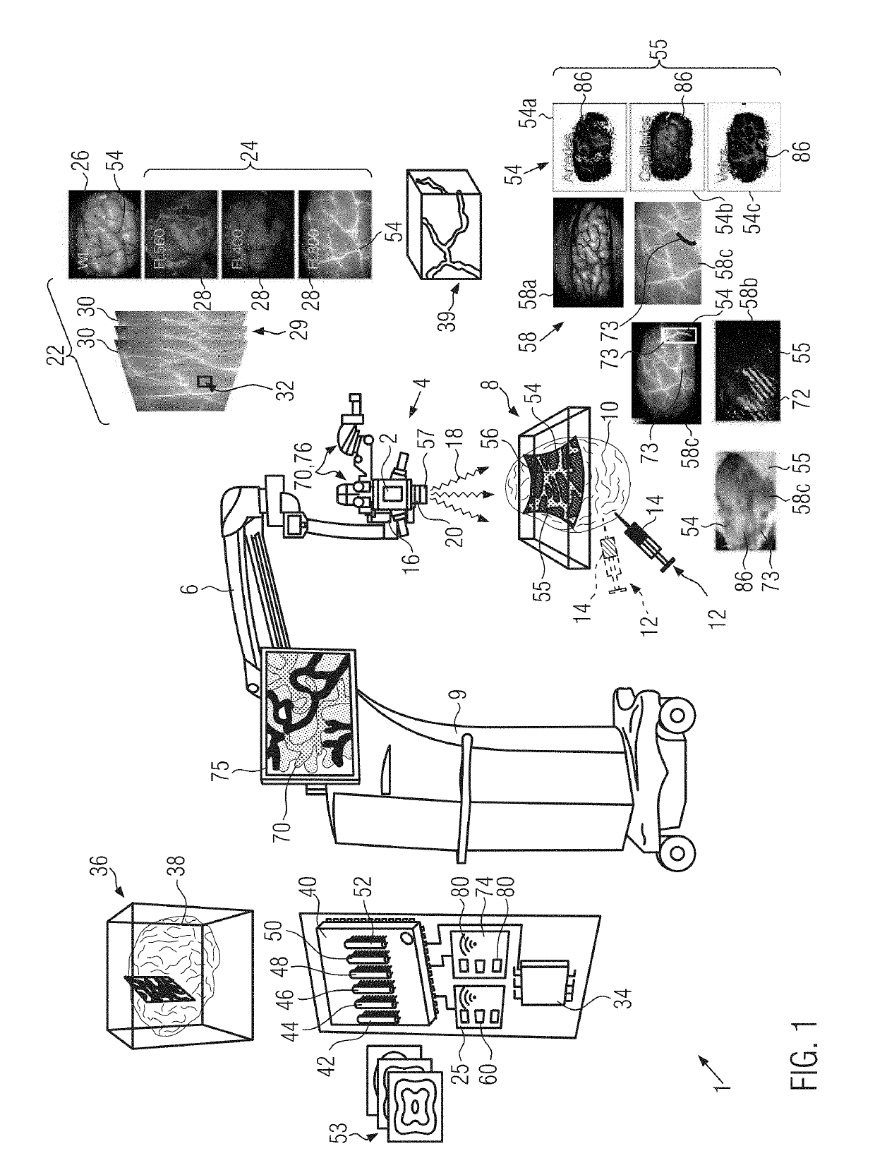
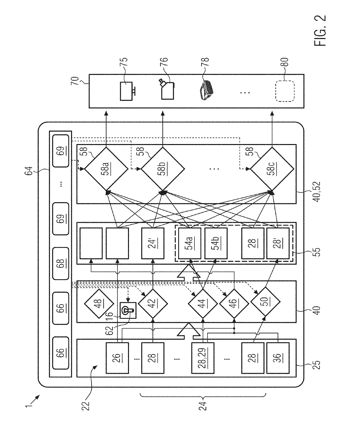
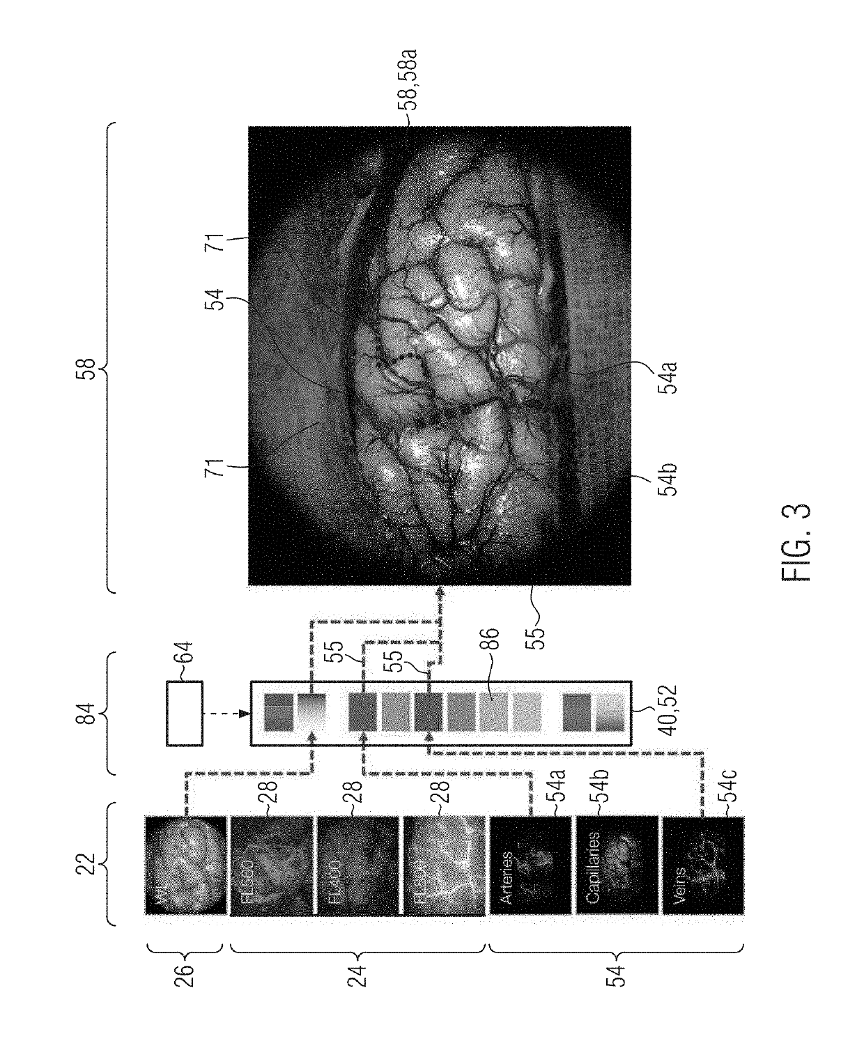
[0033]FIG. 1 shows an augmented reality surgical microscope 1. The microscope 1 is provided with at least one camera 2, which preferably comprises at least one multispectral or hyperspectral camera. The camera 2 is located in an optics holder 4 which is mounted on a movable cantilevered and / or telescopic arm 6 to be positioned freely above an object 8 to be observed during surgery. The arm 6 is supported on a frame 9 of the microscope 1, which may be stationary or which may be moved on wheels.

[0034]Typically, the object 8 comprises live tissue 10 which may have been provided with at least one bolus 12 of at least one fluorophore 14. The surgical microscope 1 may also comprise an illumination system 16, which may also be arranged within the optics holder 4. Illumination light 18 from the illumination system 16 may be guided through a microscope lens 20 onto the live tissue 10. The camera may also us...


PUM


Abstract

The invention relates to an augmented reality surgical microscope (1) having a camera (2), preferably a multispectral or hyperspectral camera. The augmented reality surgical microscope (1) retrieves input image data (22) of an object (8) comprising in particular live tissue (10). The input image data (22) comprise visible-light image data (26) as well as fluorescent-light image data (24) of at least one fluorophore (14). The augmented reality surgical microscope (1) permits the automatic identification and marking in pseudo-color (86) of various types of vascular plexus structures (54a 54b, 54c) and/or of blood flow direction (56) depending on the fluorescent-light image data (24). A pre-operative three-dimensional atlas (36) and/or ultrasound image data (39) may be elastically matched to the visible-light image data (26). The augmented reality surgical microscope (1) allows different combinations of the various image data to be output simultaneously to different displays (75). The invention also relates to a microscopy method and a non-transitory computer-readable storing a program causing a computer to execute the microscopy method.

Description

CROSS REFERENCE TO RELATED APPLICATIONS[0001]This application claims priority of European patent application number 18162165.7 filed Mar. 16, 2018, the entire disclosure of which is incorporated by reference herein.FIELD OF THE INVENTION[0002]The invention relates to an augmented reality surgical microscope and microscopy method.BACKGROUND OF THE INVENTION[0003]Current microscopes are able to capture images in different imaging modes, such as white light, fluorescent light of one of more fluorophores and polarized light. However, they are only capable of capturing one mode at a time. During surgery, the surgeon often needs to switch between different imaging modes in order to access all the necessary information. This switching process is cumbersome and distracts the surgeon from surgery. Further, the surgeon essentially needs to combine all the information by memory, which is inevitably inaccurate, prone to mistakes, tiring and requires extensive training and experience.SUMMARY OF ...

Claims


Application Information

Patent Timeline
Patent Type & Authority Applications(United States)
IPC IPC(8): A61B5/00G06T11/00G06K9/00G06T11/60G06F3/14A61B90/20A61B90/00
CPCG06K9/00134A61B2090/378A61B5/0071A61B90/36G06T2210/41A61B2090/365G06K2209/05G06T11/001G06F3/1423A61B2090/372A61B90/20G06T11/60A61B90/361G06T1/0007G02B21/0012G02B21/16A61B90/37A61B2090/364G06V20/693G06V2201/03
Inventor THEMELIS, GEORGE
Owner LEICA INSTR SINGAPORE PTE
Why Eureka
Try Eureka