Inhibition of placenta growth factor (PLGF) mediated metastasis and/or angiogenesis

a growth factor and placenta technology, applied in the field of inhibiting angiogenesis, can solve the problems of not always achieving durable responses or even cures, and the role of vegf and other members of the vegf family of growth factors in tumor recurrence or metastatic growth, so as to prevent tumors from metastasizing and inhibit or eliminate tumor metastasis

Inactive Publication Date: 2007-04-19
CENT FOR MOLECULAR BIOLOGY & MEDICINE +1
View PDF12 Cites 136 Cited by
  • Summary
  • Abstract
  • Description
  • Claims
  • Application Information

AI Technical Summary

Benefits of technology

[0011] The administration of PlGF ligands may be effective to inhibit or eliminate tumor metastasis, angiogenesis in solid tumors, tumor cell survival and / or tumor cell mobility. In preferred embodiments, administration of one or more PlGF ligands may prevent tumors from metastasizing or may result in regression or growth inhibition of existing tumors. The skilled artisan will realize that one or more PlGF ligands may be administered alone or alternatively in conjunction with other known therapeutic treatments for cancer and / or angiogenesis, such as chemotherapy, radiation therapy, immunotherapy, anti-VEGF agents and / or other known anti-angiogenesis agents and the like. In some embodiments, the PlGF ligands may be administered with a bispecific antibody, with one binding site for the PlGF ligand and a second binding site for a tumor antigen or other target. In other embodiments, PlGF ligands may be covalently attached to or provided as a fusion protein with an antibody, antibody fragment, monoclonal antibody, Fc fragment, Fc-binding protein or antibody binding protein.

Problems solved by technology

However, the role of VEGF and other members of the VEGF family of growth factors in tumor recurrence or metastatic growth has not been clearly established.
Yet, marked inhibition of VEGF does not always result in durable responses or even cure.

Method used


Image

  • Inhibition of placenta growth factor (PLGF) mediated metastasis and/or angiogenesis
  • Inhibition of placenta growth factor (PLGF) mediated metastasis and/or angiogenesis
  • Inhibition of placenta growth factor (PLGF) mediated metastasis and/or angiogenesis

Examples


example 1

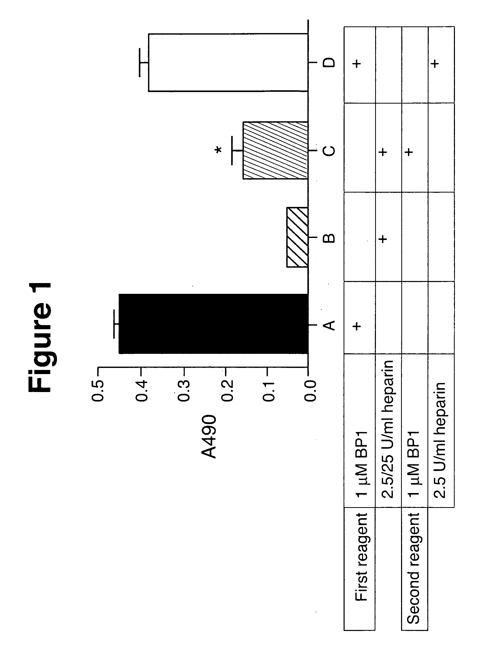
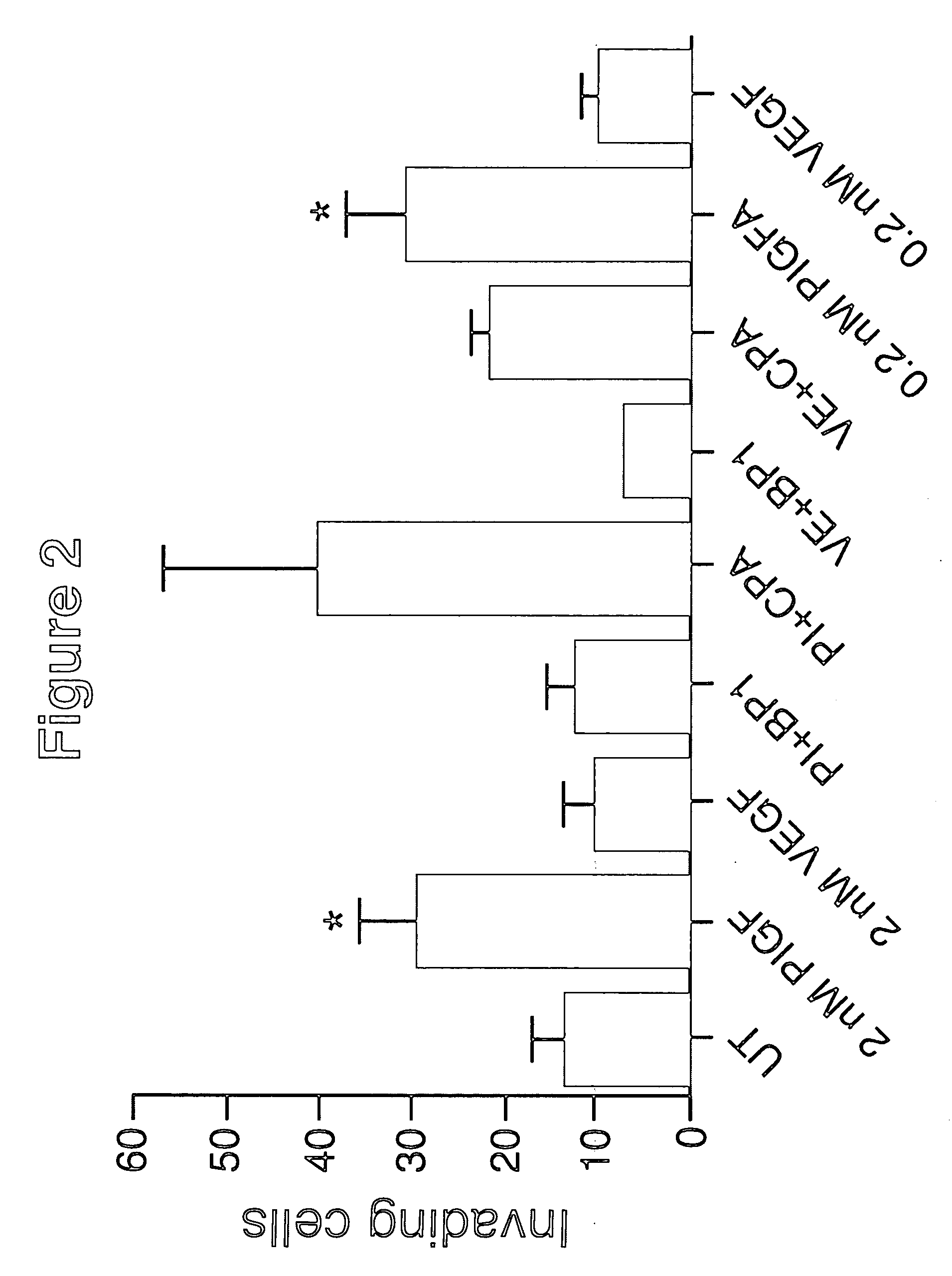
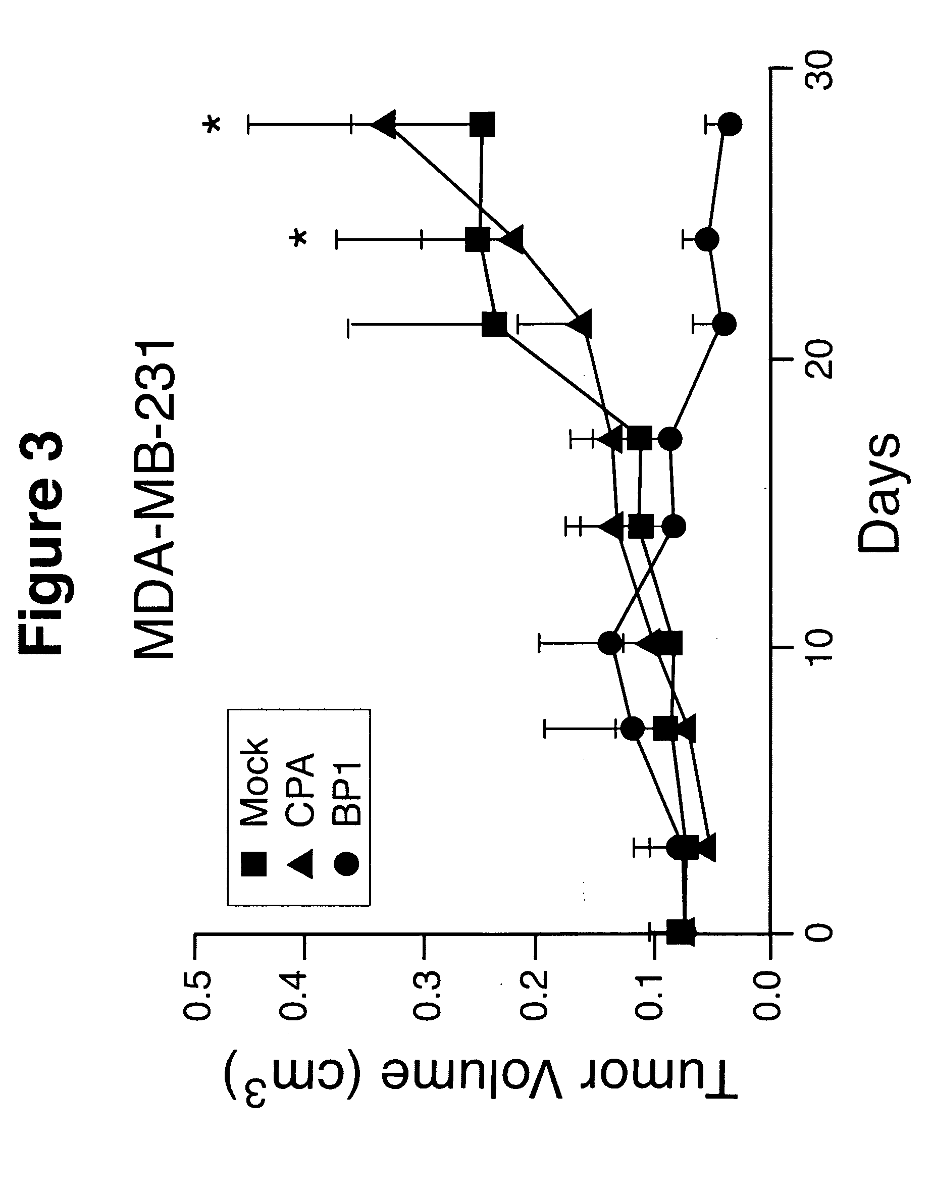
Effects of PlGF on Tumor Cell Growth, Mobility, Angiogenesis and Metastasis

Methods and Materials

[0228] Immunohistochemistry, Histopathology and Flow Cytometry

[0229] Flow cytometry was performed by standard methods using 1-5 μg / ml primary antibody, and 1:500 dilution of FITC-labeled secondary antibody (Biosource International, Camarillo, Calif.). Data were collected on a BD FACSCalibur flow cytometer (BD Biosciences) using Cell Quest software.

[0230] Paraffin-embedded primary breast cancer tissue arrays from US Biomax Inc. (Rockville, Md.) and TARP, NCI (Bethesda, Md.) were stained using standard immunohistochemistry procedures (Taylor et al., 2002a) for PlGF and VEGF expression. Tissue arrays were also probed for Flt-1 or NRP-1 (ABXIS, Seoul, Korea). Slides were deparaffinized, blocked with appropriate normal serum, and incubated with antibodies purchased from Santa Cruz Biotechnology, Inc. (Santa Cruz, Calif.). After incubation with the primary antibody, biotinylated secondary ...

example 2

Inhibition of PlGF Mediated Angiogenesis in a Mouse Corneal Assay

[0292] The ability of PlGF ligands to block PlGF-mediated angiogenesis in corneal tissues in vivo is investigated. Corneal micropockets are created using a modified von Graefe cataract knife in both eyes of male 5-6-wk-old C57B16 / J mice. Micropellets comprised of a sustained release formulation of polyglucuronic acid / polylactic acid containing 100 ng of PlGF or PlGF with 1 μM BP-1, BP-2, BP-3 or BP-4 are implanted into each corneal pocket. Eyes are examined by a slit-lamp biomicroscope on day 5 after pellet implantation. Vessel length and neovascularization are measured.

[0293] PlGF induces a strong angiogenic response with formation of a high density of microvessels. Addition of BP-1, BP-3 or BP-4 inhibits PlGF mediated angiogenesis in corneal tissues. Treatment of a subject of diabetic retinopathy or macular degeneration results in an inhibition of blood vessel formation and ameliorates the condition.

example 3

BP1 Fusion Proteins and Conjugates

[0294] For purposes of oral administration or in other embodiments, BP1 and its analogs may be recombinantly or chemically linked to a carrier protein. For linking peptides composed of the 20 common L-amino acids found in naturally occurring proteins, recombinant DNA methods are preferred. For linking peptide mimetics or peptides that contain D-amino acids, modified amino acids, or unnatural amino acids, only chemical conjugation methods are currently feasible. As disclosed herein, a general method of preparing conjugates of BP1 linked to the carbohydrates of the Fc of human IgG1 (hFc) is provided. Methods of constructing three exemplary fusion proteins comprised of BP1 and hFc are disclosed below.

[0295] A vector for expressing hFc in myeloma cells is prepared using standard recombinant DNA techniques. The expression vector shCD20-Fc-pdHL2 (FIG. 6) is used as the DNA template to amplify the coding sequences for Fc (hinge, CH2, and CH3 domains) by ...


PUM

PropertyMeasurementUnit
volumeaaaaaaaaaa
temperaturesaaaaaaaaaa
concentrationsaaaaaaaaaa

Abstract

The present invention concerns methods and compositions for inhibiting angiogenesis and / or tumor growth, survival and / or metastasis. In particular embodiments, the methods and compositions may concern ligands against placenta growth factor (PlGF), such as BP-1, BP-2, BP-3 or BP-4. Some methods may comprise administering one or more PlGF ligands, alone or in combination with one or more other agents, such as chemotherapeutic agents, other anti-angiogenic agents, immunotherapeutic agents or radioimmunotherapeutic agents to a subject. The PlGF ligands are effective to inhibit angiogenesis, tumor cell motility, tumor metastasis, tumor growth and / or tumor survival. In certain embodiments, PlGF ligands may be administered to subjects to ameliorate other angiogenesis related conditions, such as macular degeneration. In some embodiments, PlGF expression levels may be determined by any known method to select those patients most likely to respond to PlGF targeted therapies.

Description

RELATED APPLICATIONS [0001] The present application claims the benefit under 35 U.S.C. §119(e) of provisional U.S. Patent Application Ser. No. 60 / 728,292, filed on Oct. 19, 2005, the entire text of which is incorporated herein by reference.FEDERALLY FUNDED RESEARCH [0002] The studies disclosed herein were supported in part by grant DAMD 17-03-1-0758 from the U.S. Department of Defense (A.P.T.). The U.S. government may have certain rights to practice the subject invention.BACKGROUND [0003] 1. Field of the Invention [0004] The present invention concerns methods and compositions for inhibiting angiogenesis, particularly pathologic angiogenesis and / or growth and metastasis of tumors. In particular embodiments, the compositions and methods concern inhibitors targeted to placenta growth factor-2 (PlGF) and / or its receptor, Flt-1, including but not limited to peptides, antibodies, antibody fragments, humanized antibodies, chimeric antibodies, antibody analogs, aptamers, organic compounds a...

Claims


Application Information

Patent Timeline
Patent Type & Authority Applications(United States)
IPC IPC(8): A61K39/395A61K38/18
CPCA61K38/00C07K14/515C07K16/22A61P1/04A61P7/10A61P9/00A61P9/10A61P11/00A61P11/06A61P17/02A61P17/06A61P19/02A61P27/02A61P27/06A61P29/00A61P35/00A61P35/02A61P35/04A61P37/02A61P37/06A61P43/00A61K39/395C07K16/46
Inventor TAYLOR, ALICE P.GOLDENBERG, DAVID M.CHANG, CHIEN HSING
Owner CENT FOR MOLECULAR BIOLOGY & MEDICINE
Why Eureka
Try Eureka