Eureka AIR delivers breakthrough ideas for toughest innovation challenges, trusted by R&D personnel around the world.

Patient positioning device for a computer tomograph

Inactive Publication Date: 2007-05-03
SIEMENS AG
View PDF6 Cites 4 Cited by
  • Summary
  • Abstract
  • Description
  • Claims
  • Application Information

AI Technical Summary

Benefits of technology

[0007] A patient positioning device for a computer tomograph (CT) device and a CT device having such a patient positioning device are described. The patient positioning device ensures a stable position of the patient body in the examination opening of the gantry, is simultaneously flexibly adjustable in position, and has good accessibility to medical staff.

Problems solved by technology

At the same time, however, a complicated construction for supporting the patient body in the examination opening of the gantry cannot be used, because it would create interfering artifacts in the image.
However, the positioning device offers no possibility of height adjustment, and has a bulky construction below the patient, which makes accessibility more difficult.
Changes in the patient position, as the patient positioning device is moved to approach the gantry, which could cause movements on the part of the patient and hence interfering artifacts in the CT image data, are thus averted.

Method used


Image

  • Patient positioning device for a computer tomograph
  • Patient positioning device for a computer tomograph
  • Patient positioning device for a computer tomograph

Examples


Embodiment Construction

[0024] Exemplary embodiments may be better understood with reference to the drawings, but these examples are not intended to be of a limiting nature. Like numbered elements in the same or different drawings perform equivalent functions.

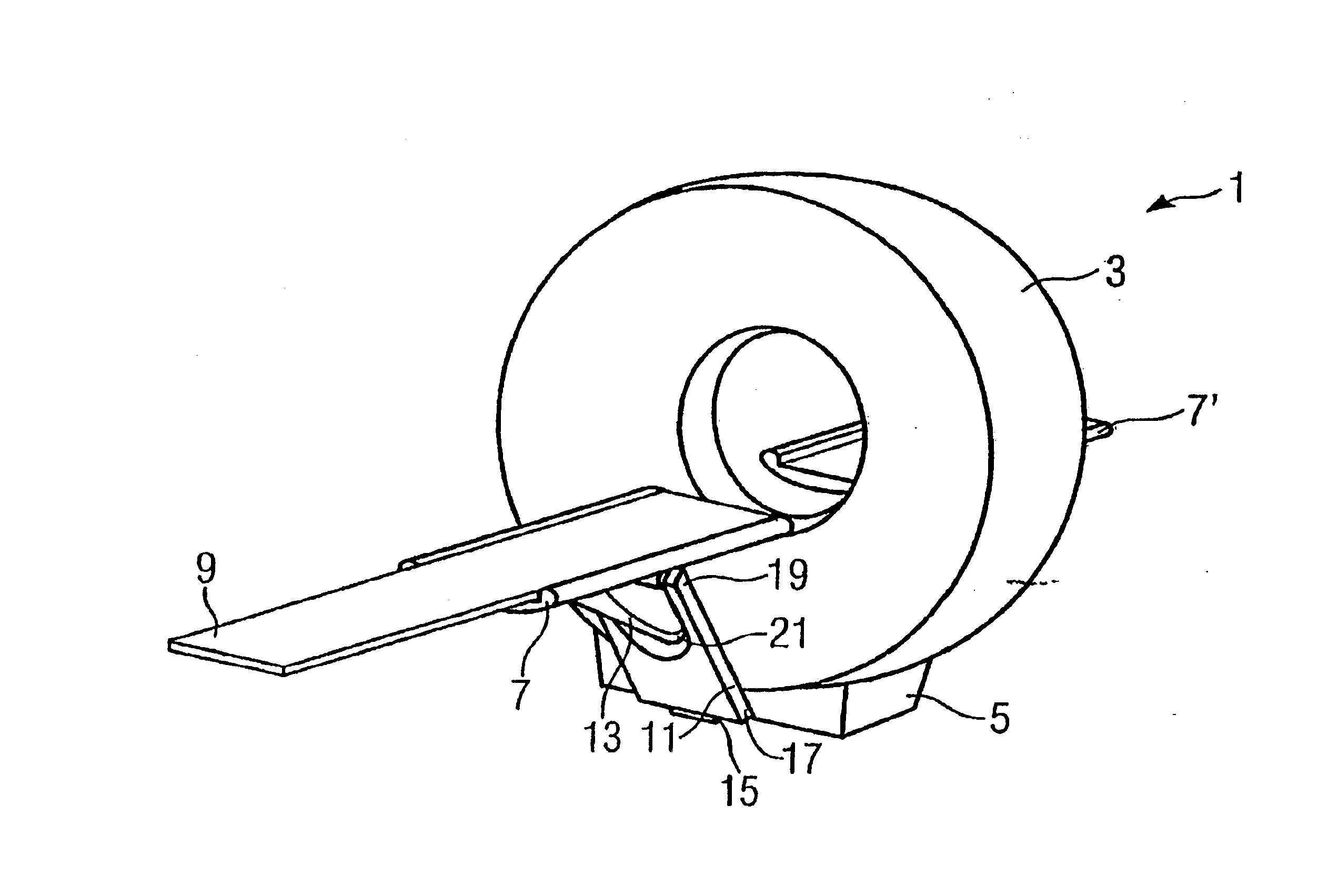
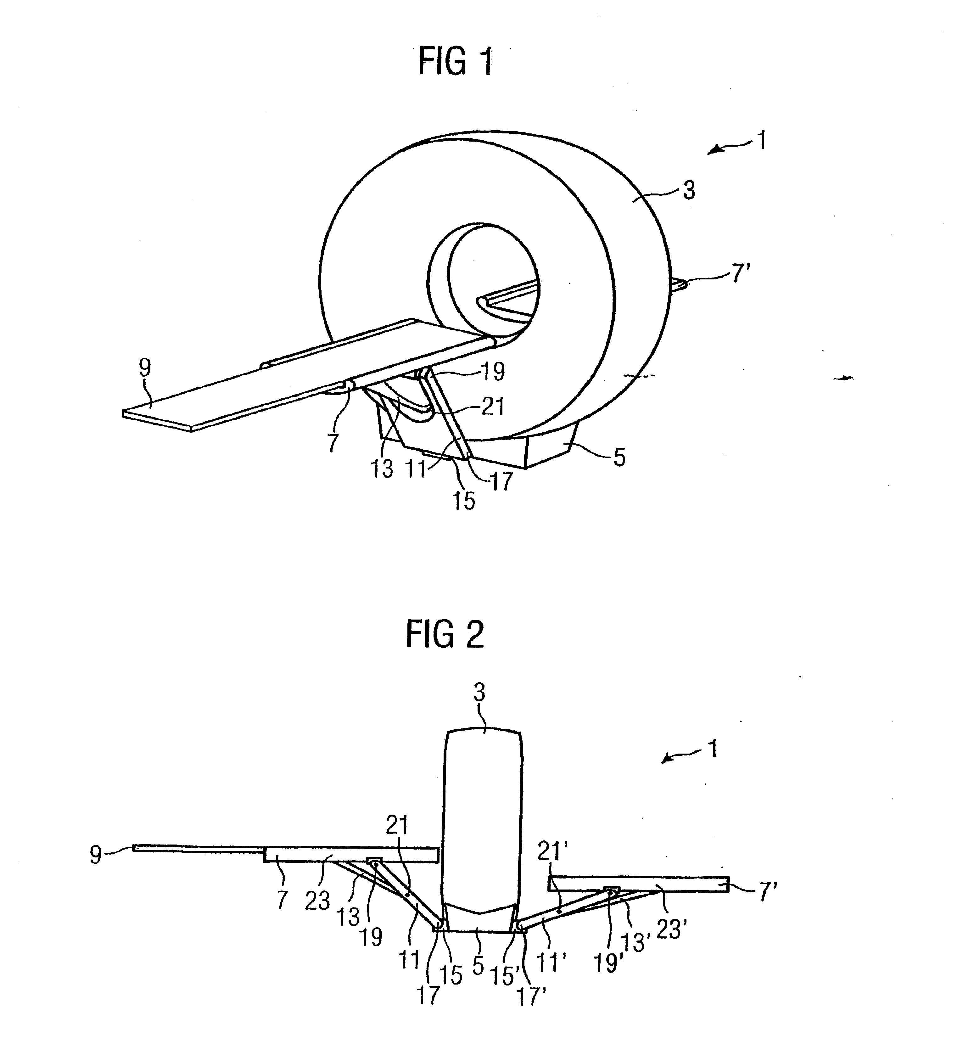
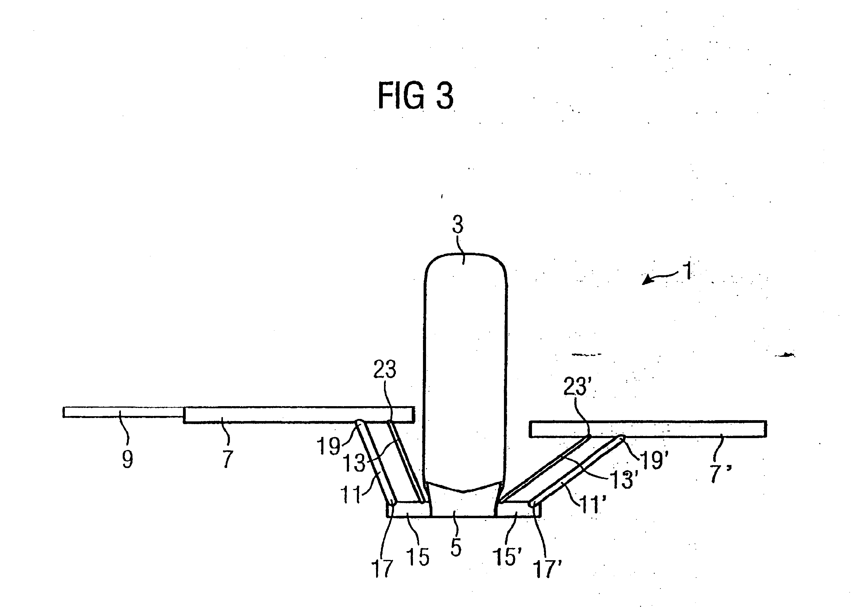
[0025] A CT device 1 with a patient positioning device is shown in a perspective view in FIG. 1. The CT device 1 has a gantry 3 with an examination opening. The gantry 3 rests on a base 5. A patient to be examined is placed on a patient bed 9, which is introduced into the bed guide 7. The bed guide 7 guides the patient bed 9 so that the patient bed is displaceable longitudinally, such that the patient bed 9 may be introduced into the examination opening or aperture in the gantry 3. Gantry 3 may have a patient positioning device on each side of the examination opening, and the patient bed 9, on being introduced into the aperture in gantry 3, is transferred the bed guide 7′ of the opposite patient positioning device disposed at the opposite side of the...


PUM


Abstract

A patient positioning device for a computer tomographic device having a gantry supported on a base is disclosed. The patient positioning device comprises a bed guide for a patient bed having an arm mounted on or to the base by means of a rotating joint which rotates about a horizontal axis. The bed guide is mounted to the arm by means of a rotating joint which rotates about a horizontal axis, and the height of the bed guide may be adjusted.

Description

TECHNICAL FIELD [0001] This application relates to a patient positioning device for a computer tomograph device and, to a computer tomograph device having a patient positioning device of this kind. BACKGROUND [0002] Computer tomograph devices (CTs), serve to make three-dimensional images or images of slices of a body. The image data are computed by a computer using two-dimensional X-ray projections of the body. For recording the two-dimensional raw image data, an X-ray beam source and an image detector are disposed diametrically opposite one another and rotate about the body to be examined. The rotation takes place inside a gantry which has an examination opening, or aperture, into which the body can be introduced. [0003] The position of the body in the gantry and the stability of the body position affect the CT image quality. A patient positioning device should therefore assure sufficiently stable support of the patient body and should not have any significant sagging, for instance...

Claims


Application Information

Patent Timeline
IPC IPC(8): A47B13/00A61B6/04
CPCA61B6/04A61B6/0457A61B6/0487
Inventor BARTELS, FRANKHELMREICH, GERHARDLEIDENBERGER, STEFAN
Owner SIEMENS AG
Why Eureka
Eureka Blog
Learn More