Method of dental microscopic procedure

a dental microscopic and x-ray technology, applied in the field of dental microscopic procedures, can solve the problems of limiting mobility, little to improve the quality, efficiency, safety and comfort of surgical procedures, and the surgery process, and achieve the effect of recording and manipulating the images obtained

Inactive Publication Date: 2007-05-03
YOO BEN +3
View PDF5 Cites 35 Cited by
  • Summary
  • Abstract
  • Description
  • Claims
  • Application Information

AI Technical Summary

Benefits of technology

[0017] An object of the invention is to provide a method of dental microscopic procedure, which enables dentists to perform dental procedures including surgery by seeing the live images on a screen and not looking into a small eyepiece.
[0018] Another object of the invention is to provide a method of dental microscopic procedure, which makes recording and manipulating the images obtained from the microscope and X-ray convenient.
[0030] The process of Panning Window indicates steps of displaying live video images on the window screen, locating an area of interest from a streaming live video screen, locating an area of interest from streaming live video image, manipulating the located window, and displaying the manipulated window screen. The Panning Window function allows a dentist to both magnify and scan an area without having to move, re-focus, and re-adjust the camera or patient. The Panning Window enables the dentist to easily scan a specific tooth, surrounding teeth, or gum line by using a mouse to drag the locator box, which indicates the selected area on the screen with a red outlined box, accordingly. The live and manipulated window screen is shown within the same display. By clicking on the Panning Window icon, the Panning Window will appear on the left side of the Capture Window. When the Panning Window appears, the Zoon In icon must be clicked. Once the image has been zoomed in, the dentist will be able to use the Panning Window to explore the patient's affected area by dragging the locator box within the Panning Window. The dentist can also use the mouse to move within the Panning Window to scan and magnify the activated area by the locator box.

Problems solved by technology

Current variations of the microscope disrupt the surgery process and do little to improve the quality, efficiency, safety, and comfort of surgical procedures.
Today, many dentists use microscopes, limiting mobility, and causing neck, shoulder, and eye strain.
Other telescopic microscopes require the dentist to adjust and constantly focus the scope to get an adequate image, interfering with the process of surgery and lending unnecessary difficulties during the operation.
Should the dentist or the patient move, or if the scope gets displaced, relocate the image, then the image becomes blurry and requires one to refocus.
It can be moved in virtually any direction and / or angle, unlike traditional scopes that are much more limited in directional / angle maneuverability.
Currently, available microscopes with attached video camera can only display a limited depth of field; thus making the practice of dentistry with the traditional microscope much more difficult.
No other equipment sold today allows this function.
Traditional equipment limits the dentist.
With a loupe and headlight he / she is tethered to the cord by the headlamp, resulting in poor ergonomics.
No other dental microscopic device on the market today can make that claim.

Method used


Image

  • Method of dental microscopic procedure
  • Method of dental microscopic procedure
  • Method of dental microscopic procedure

Examples


Embodiment Construction

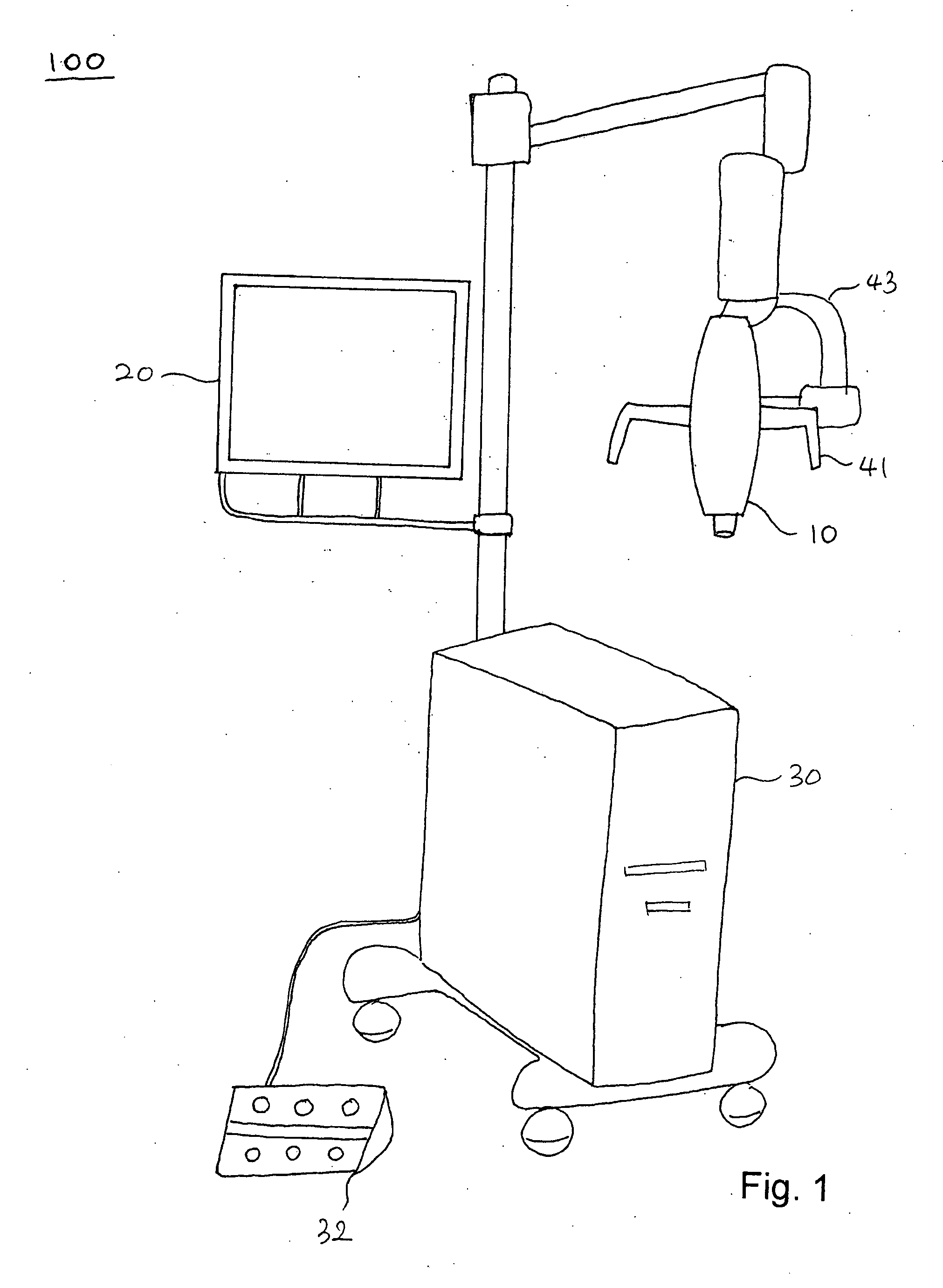
[0060]FIG. 1 shows a perspective view of a digital surgical scope 100 according to the present invention.

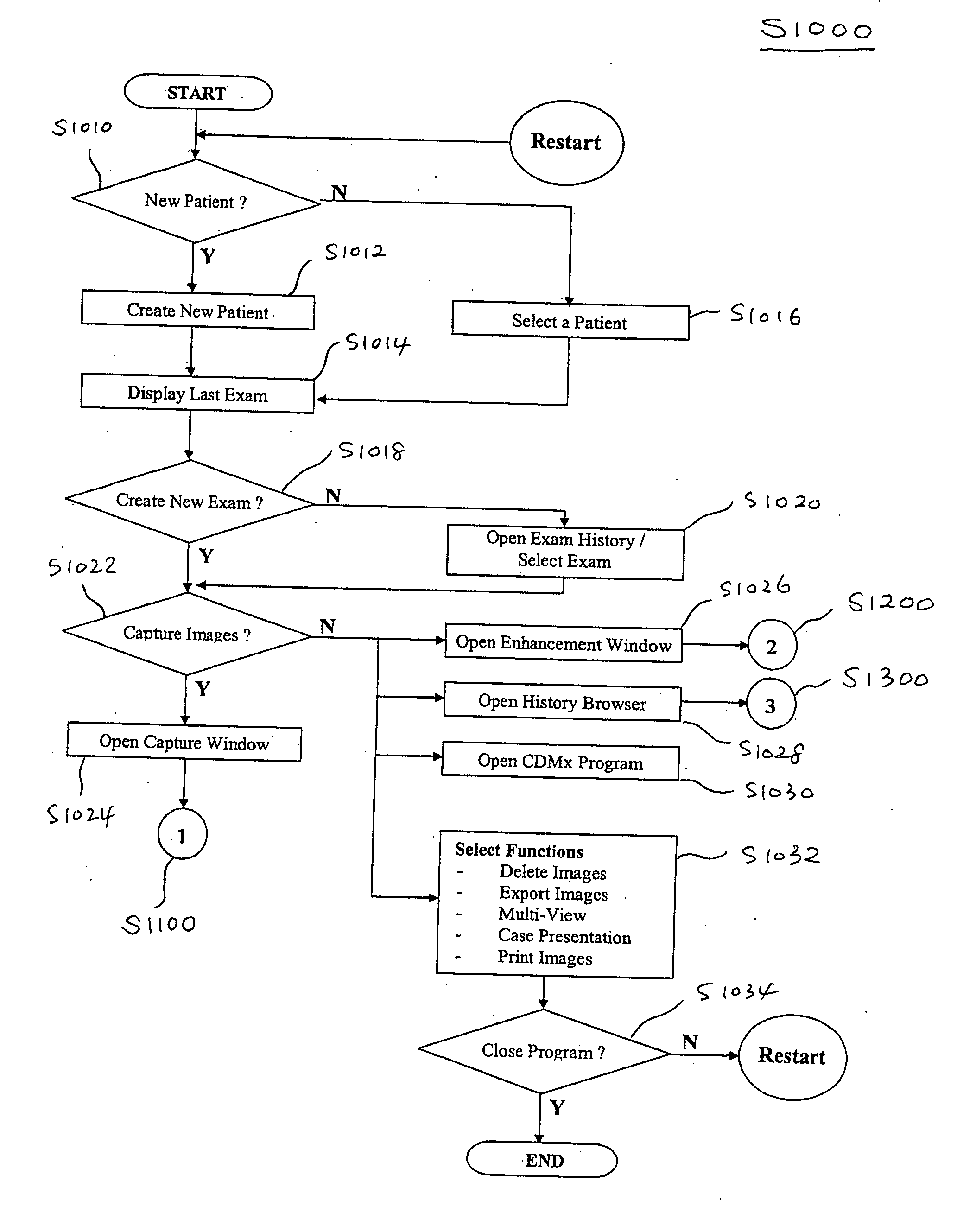
[0061]FIG. 2 shows a flow chart showing a method S1000 of dental microscopic procedure according to the present invention.

[0062] The method S1000 of dental microscopic procedure, using a digital surgical scope 100, includes steps for selecting (S1022) a function of the digital surgical scope 100 to perform, obtaining (S1100) the corresponding images from the image data taking device 12 or the image data processing / storing device 30 (refer to FIGS. 1, 7, and 8), processing (S1200, S1300) the images by the image data processing / storing device 30, and displaying (S1014) the images on the displays 20 as shown in FIG. 1 and FIG. 2.

[0063] The dental surgical procedure is done by seeing the images displayed on the display 20.

[0064] The digital surgical scope 100 includes a digital surgical scope 10, an image data taking device 12, an image data processing / storing device 30, and one ...


PUM


Abstract

A method of dental microscopic procedure, using a digital surgical scope, includes steps for selecting a function of the digital surgical scope to perform, obtaining the corresponding images from the image data taking device or the image data processing / storing device, processing the images by the image data processing / storing device, and displaying the images on the displays. The dental surgical procedure is done by seeing the images displayed on the screen. The digital surgical scope includes a microscope lens assembly for magnifying an optical image of an object and an in-line illumination device for illuminating the object along the same optical axis and from the same direction of the microscope lens assembly. The image data processing / storing device is for manipulating the image taken by the microscope lens assembly and the image data taking device.

Description

BACKGROUND OF THE INVENTION [0001] The present invention relates to a method of dental microscopic procedure, which enables dentists to perform dental procedures including surgery by seeing the images on a screen, and not looking into a small eyepiece. [0002] Furthermore, this invention relates to a method of dental microscopic procedure, which makes recording and manipulating images obtained from the digital surgical microscope, an image data taking device more convenient. [0003] Current variations of the microscope disrupt the surgery process and do little to improve the quality, efficiency, safety, and comfort of surgical procedures. Today, many dentists use microscopes, limiting mobility, and causing neck, shoulder, and eye strain. Other telescopic microscopes require the dentist to adjust and constantly focus the scope to get an adequate image, interfering with the process of surgery and lending unnecessary difficulties during the operation. Should the dentist or the patient mo...

Claims


Application Information

Patent Timeline
Patent Type & Authority Applications(United States)
IPC IPC(8): G06K9/00G16H20/40G16H30/20G16H30/40G16H40/63
CPCA61C1/082A61C9/004G02B21/0012G06F19/321G06Q50/24A61C9/0053G16H20/40G16H30/40G16H30/20G16H40/63A61B17/24G06F18/00
Inventor YOO, BENHAN, YOUNGSHIM, SUNGWOO, WON JOO
Owner YOO BEN
Why Eureka
Try Eureka