

Capsule type medical device
a medical device and capsule technology, applied in the field of capsules, can solve the problems of difficult passage through the organs, high cost, etc., and achieve the effect of reducing the risk of being caught when passing through the narrow bending part such as the small bowel
- Summary
- Abstract
- Description
- Claims
- Application Information
AI Technical Summary
Benefits of technology
Problems solved by technology
Method used
Image

Examples
first embodiment
The First Embodiment
[0069] Referring to the drawing, the first embodiment according to the capsule type medical device of the present invention will be described.
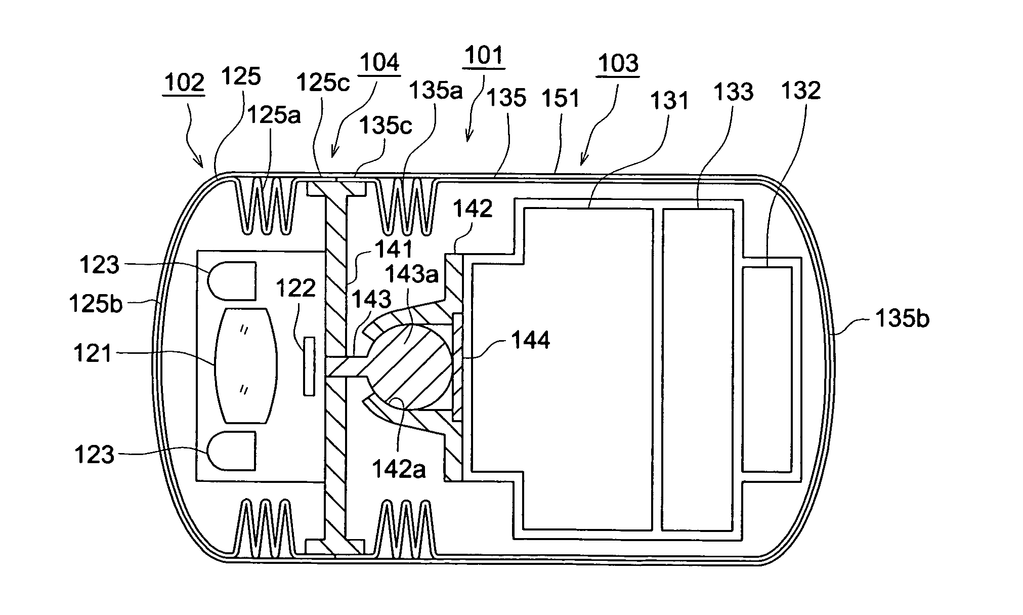
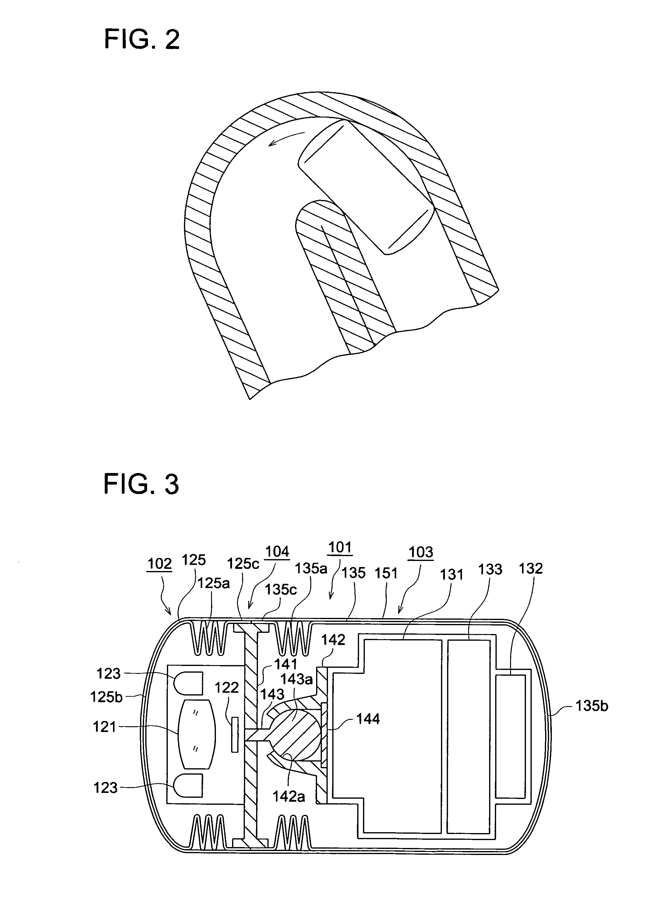
[0070] Initially, based on FIG. 3, the structure of the capsule type medical device will be described, however, FIG. 3 is a sectional view of the capsule type medical device before use.
[0071] The present capsule type medical device 101 is structured by the front part 102, rear part 103, and the connection part 104 connecting the front part 102 and the rear part 103.
[0072] In the front part 102, the image pick-up lens 121 structured in the wide angle, and for image picking-up in the organs of the person, image pick-up element 122 composed of CCD or CMOS, and for image-forming the image, picked-up by the image pick-up lens 121 and for photoelectric converting, and a plurality of illumination members 123 composed of LED, and for illuminating the organ to be image picked-up at the time of image picking-up, are housed, and th...
second embodiment
The Second Embodiment
[0095] Referring to the drawings, the second embodiment according the capsule type medical device of the present invention will be described below.
[0096] Initially, based on FIG. 7, the structure of the capsule type medical device will be described, and FIG. 7 is a sectional view of the capsule type medical device when the subject of the near distance is image picked-up in the front.
[0097] The present capsule type medical device 201 is structured by the front part 202, rear part 203, and the connection part 204 connecting the front part 202 and the rear part 203.
[0098] In the front part 202, the second lens 221, the image pick-up element 222 for image forming and photoelectric converting the picked-up image by the image pick-up optical system which will be described later, and a plurality of illumination members 223, composed of LED, for illuminating the organ to be picked-up at the time of the image pick-up, are housed, and they are held by the connection me...
third embodiment
The Third Embodiment
[0124] Referring to the drawings, the third embodiment according the capsule type medical device of the present invention will be described below.
[0125] When the image of the organs of the tested subject is observed by the inspection doctor by an external device, there is a case where an anxious disease is discovered at the periphery of the picked-up image. In this case, as described above, the lens may be changed to the wide lens and image picked-up so that the wide range is picked-up, however, there is a problem that the image becomes small and it becomes difficult to be observed.
[0126] In this case, when the inspection doctor operates the external device and the tilt signal is transmitted to the capsule type medical device 201, the control section in the image processing section 231 controls the micro-mini motor by the transmission and reception section 232, and each drive member 236 is moved. Then, the drive member 236 positioned in the direction in which t...
PUM
 Abstract
Description
Claims
Application Information