Magnetic resonance imaging apparatus

a magnetic resonance imaging and apparatus technology, applied in the field of magnetic resonance imaging apparatus, can solve the problems of reducing image quality, reducing image quality, and generating images with body move artifacts, so as to suppress the development of body move artifacts and improve image quality

Active Publication Date: 2007-05-31
GE MEDICAL SYST GLOBAL TECH CO LLC
View PDF15 Cites 8 Cited by
  • Summary
  • Abstract
  • Description
  • Claims
  • Application Information

AI Technical Summary

Benefits of technology

[0012] In accordance with the present invention, a magnetic resonance imaging apparatus is provided which may improve the image quality by suppressing the development of body move artifacts.

Problems solved by technology

When scanning an subject by using an MRI apparatus as have been described above, if the subject moves, the slice image generated may have some body move artifacts.
For example, in case of the imaging of neck and esophagus of the subject, such body move as deglutition may cause the body move artifact, and decreases the image quality.
However, there are cases in which the body move artifact develops in the slice image to decrease the image quality because the body move is not sufficiently suppressed.
In particular, since the internal body move within the body of the subject such as the epiglottis moving along with the deglutition is difficult to suppress by such means as a binding straps, the body move artifact may develop obviously in the slice image to significantly decrease the image quality.

Method used


Image

  • Magnetic resonance imaging apparatus
  • Magnetic resonance imaging apparatus
  • Magnetic resonance imaging apparatus

Examples


first embodiment

[0023] From now on, a first preferred embodiment in accordance with the present invention will be described in greater details.

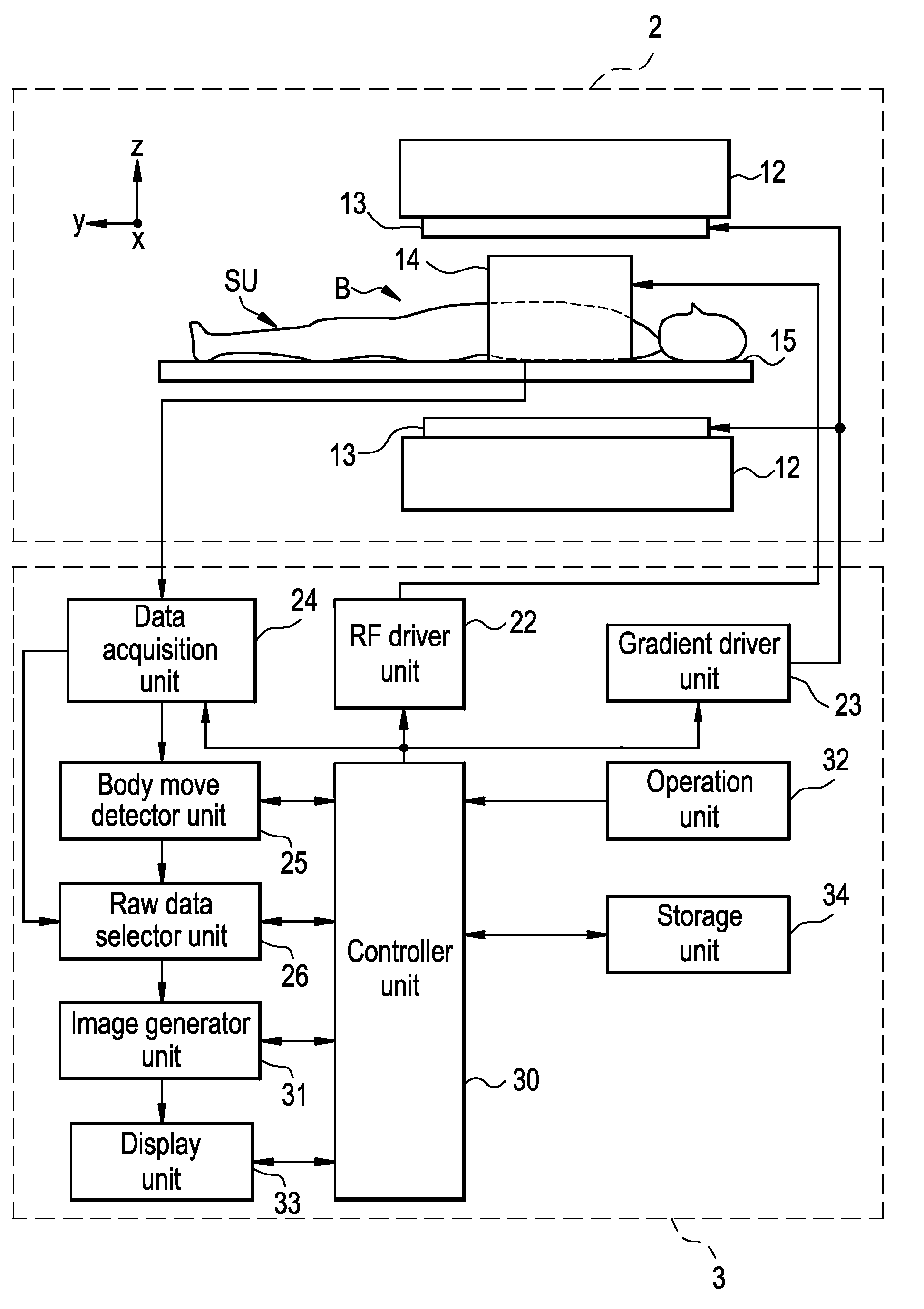
[0024] Now referring to FIG. 1, there is show a schematic block diagram illustrating the overview of a magnetic resonance imaging apparatus 1 in accordance with the preferred embodiment of the present invention.

[0025] As shown in FIG. 1, the magnetic resonance imaging apparatus 1 has a scan unit 2 and an operating console unit 3, and generates an image of an subject SU based on the magnetic resonance signals generated in the subject SU by emitting RF pulses to the subject in a static magnetic field space.

[0026] Now the scan unit 2 will be described in greater details.

[0027] The scan unit 2, as shown inFIG. 1, has a static field magnet unit 12, a gradient coil unit 13, an RF coil unit 14, and a cradle 15, and performs a scan to obtain the magnetic resonance signals developed in the subject SU, by emitting electromagnetic waves to the subject SU so as to e...

second embodiment

[0079] A second preferred embodiment in accordance with the present invention will be described herein below in greater details.

[0080] The present embodiment is similar to the preceding first preferred embodiment, except for the operation that the scan unit 2 performs the navigator sequence. The description of the similar operation to the first preferred embodiment will be omitted.

[0081] The operation of imaging the subject SU in this embodiment will be described in greater details.

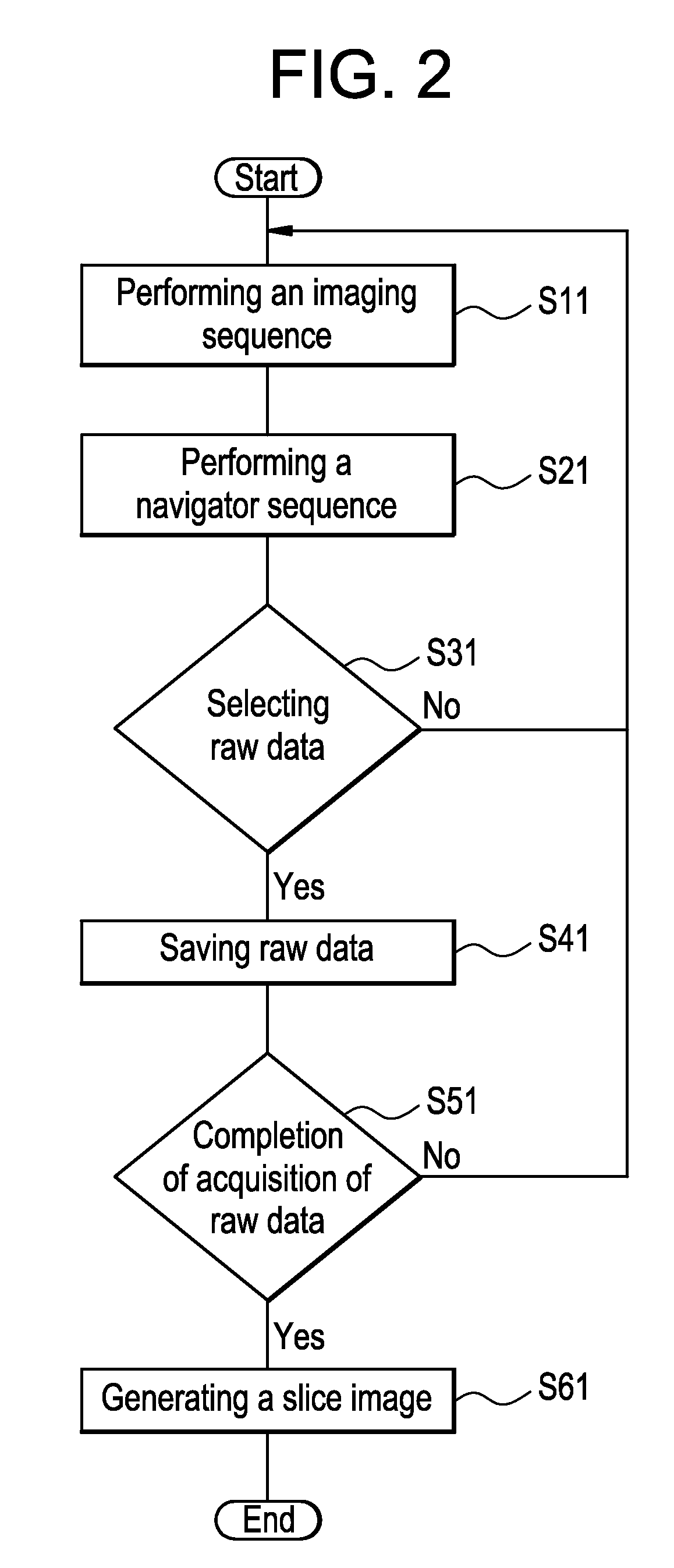
[0082] When the present embodiment performs the scan S, it performs the imaging sequence IS as similar to the preceding first embodiment, as shown in FIG. 2 and FIG. 3 (S11).

[0083] Next, as shown in FIG. 2 and FIG. 3, it performs the navigator sequence NS (S21).

[0084] To monitor the deglutition movement of the subject SU, the scan unit 2 performs the navigator sequence NS for exciting selectively the spin in the navigator area NA including the epiglottis to obtain the magnetic resonance signals as na...


PUM


Abstract

The present invention provides an MRI apparatus which prevents from developing body move artifacts on the image to improve the image quality. In the navigator sequence a scan unit emits first RF pulse so as to excite first slice plane area which is out of the imaging area and is intersecting the navigator area including the epiglottis, then it emits second RF pulse so as to excite second slice plane area which is out of the imaging area and is intersecting the first slice plane area in the navigator area including the epiglottis. Thereafter a body move detector detects the displacement of epiglottis due to the deglutition movement based on the navigator echo data, a raw data selector unit selects as raw data the imaging data thus obtained in the imaging sequence based on the displacement of epiglottis, then the image of the slice plane including the esophagus in the subject is generated from the imaging data thus selected.

Description

CROSS REFERENCE TO RELATED APPLICATIONS [0001] This application claims the benefit of Japanese Application No. 2005-339016 filed Nov. 24, 2005. BACKGROUND OF THE INVENTION [0002] The present invention relates to a magnetic resonance imaging (MRI) apparatus, more specifically to generation of the image of a subject based on the magnetic resonance signal generated in the subject by emitting RF pulses to the subject in a static magnetic field space. [0003] The MRI apparatus is widely used in many fields such as medical and industrial fields. [0004] The MRI apparatus emits RF pulses to a subject in a static magnetic field space to excite the spin of protons within the subject by the nuclear magnetic resonance (NMR) phenomenon, then scans to obtain the magnetic resonance (MR) signals generated by the excited spin. The MR signals thus obtained by the scan are served as the raw data of a slice image, to generate the slice image of the subject (e.g., patent reference 1). [0005] [patent refe...

Claims


Application Information

Patent Timeline
Patent Type & Authority Applications(United States)
IPC IPC(8): G01V3/00
CPCG01R33/56308G01R33/5676
Inventor IWADATE, YUJITSUKAMOTO, TETSUJI
Owner GE MEDICAL SYST GLOBAL TECH CO LLC
Why Eureka
Try Eureka