Methods and devices for anchoring to tissue

Active Publication Date: 2007-06-14
ADVENT MEDICAL
View PDF105 Cites 76 Cited by
  • Summary
  • Abstract
  • Description
  • Claims
  • Application Information

AI Technical Summary

Benefits of technology

[0009] Accordingly, an objective of this invention is to provide a method to reduce gastroesophageal reflux by endosonography-guided intervention. Specifically, endosonography is used to insert an anchoring element thr

Problems solved by technology

However this procedure can have a high incidence of postoperative complications and extended recovery times. Therefore endoscopic methods for enhancing the lower esophageal sphincter have been developed as an alternative to surgery.
Endoscopic treatment is often limited because the operator can only visualize the mucosal lining of the gastrointestinal wall th

Method used


Image

  • Methods and devices for anchoring to tissue
  • Methods and devices for anchoring to tissue
  • Methods and devices for anchoring to tissue

Examples


Embodiment Construction

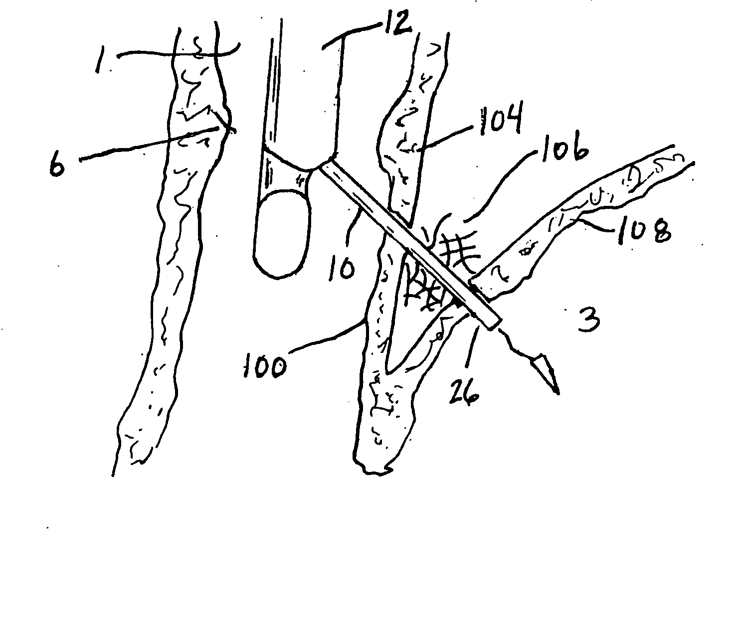
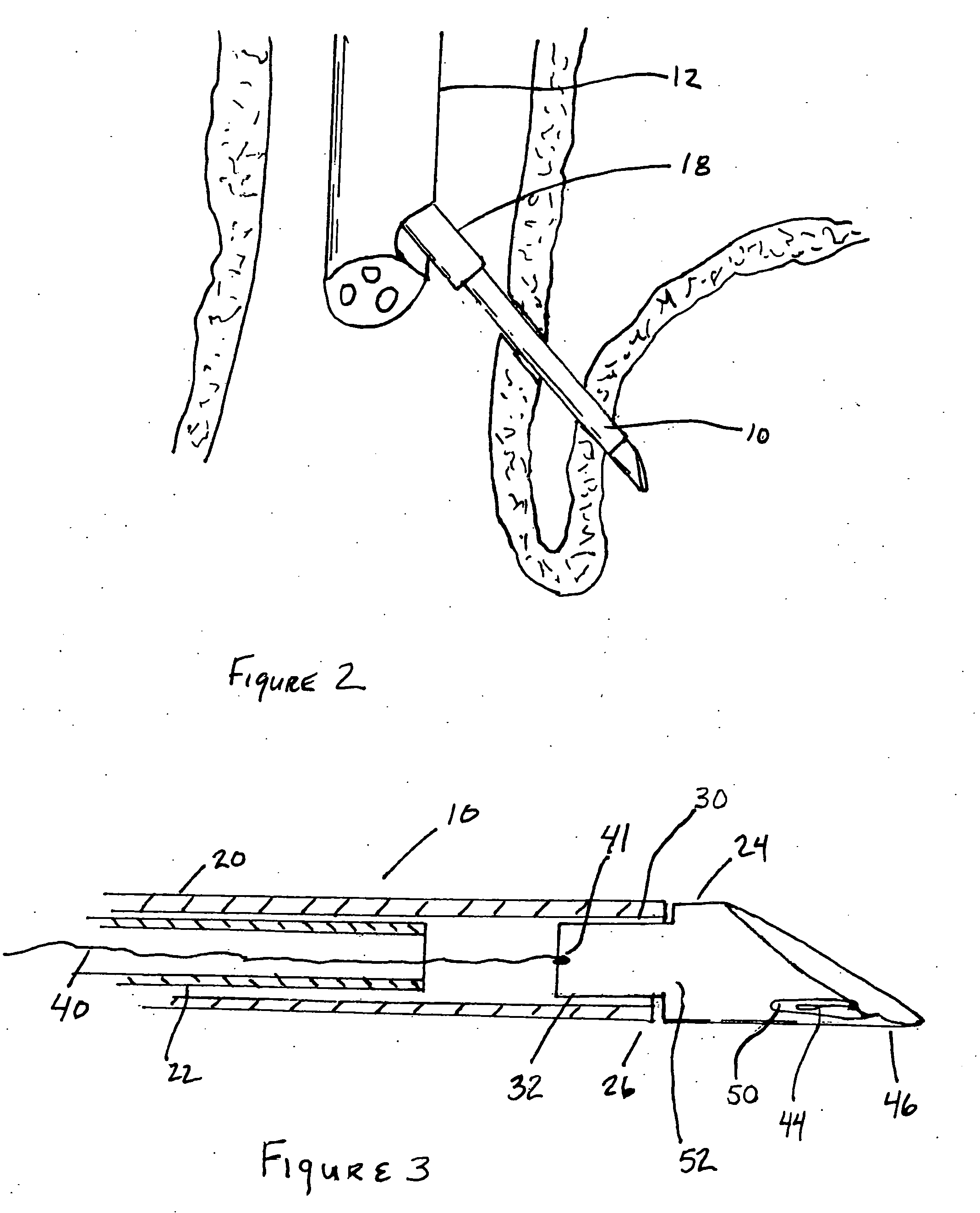
[0036] The system and method described herein may offer improvements over the techniques currently utilized to perform endoscopic procedures. This tissue securement system has several embodiments that are intended to work together to create a novel device and method for the treatment of GERD. However these embodiments also function independently and some of the embodiments of this system may be removed and the system may still achieve its desired function. Alternatively several of these embodiments may be useful as stand alone devices. The principle elements of this system are; an echoendoscope, a delivery needle, a stylet, a needle tip, a connecting element, an anchoring or ligating element and securement elements.

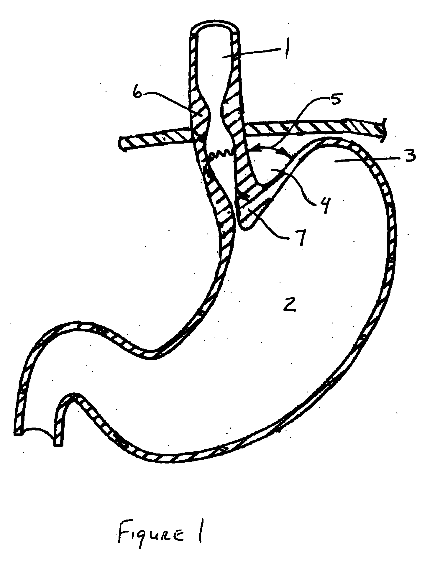
[0037]FIG. 1 is a cross sectional view of the esophageal-gastro-intestinal tract shown from the esophagus 1 to the stomach 2. The fundus 3 forms the superior portion of the stomach 2. The esophagus 1 enters the stomach 2 at a point below the fundus 3 forming the cardiac ...


PUM


Abstract

The present invention relates to a tissue securement system, device and method for endoscopy or endosonography-guided transluminal interventions whereby a ligation is placed and secured into tissue. An objective of this invention is to provide a stable securement platform for the coupling of secondary anchors to the gastro-intestinal tract. Specifically, endosonography is used to insert an anchoring element through the walls of adjacent body cavities. The ends of the anchoring element are coupled together to form a loop. This anchoring element can be used to secure other anchors or devices to the gastro intestinal tract.

Description

ClAIM OF PRIORITY [0001] This application is a continuation-in-part of co-pending U.S. patent application Ser. No. 11 / 449,365, filed Jun. 8, 2006, entitled “Methods and Devices for Endosonography-Guided Fundoplexy” which claims priority to U.S. Provisional Application No. 60 / 688837, filed Jun. 9, 2005, both of which are hereby expressly incorporated by reference.BACKGROUND [0002] 1. Field of the Invention [0003] The present invention relates to a tissue securement system, device and method for endoscopy or endosonography-guided transluminal interventions whereby a ligation or anchor is placed and secured into soft tissue. [0004] 2. Background of the Invention [0005] Gastroesophageal reflux disease (GERD) is a chronic condition caused by the failure of the anti-reflux barrier located at the gastroesophageal junction to keep the contents of the stomach from refluxing back into the esophagus. [0006] Surgical fundoplication is the gold standard for anatomic correction of the cardia in p...

Claims


Application Information

Patent Timeline
IPC IPC(8): A61B17/08
CPCA61B17/0401A61B17/0469A61B17/0482A61B17/0487A61B17/0625A61B17/12013A61B2017/003A61B2017/00827A61B2017/0409A61B2017/0454A61B2017/0464A61B2017/06176
Inventor BINMOELLER, KENNETH F.
Owner ADVENT MEDICAL
Why Eureka
Try Eureka