System and method for transcutaneous monitoring of endotracheal tube placement

a technology of endotracheal tube and transcutaneous monitoring, which is applied in the field of endotracheal tubes, can solve the problems of increasing operating time, difficult to properly align the tube into the trachea of patients, and blocking the airway of patients' airways

Inactive Publication Date: 2007-06-21
RUTGERS THE STATE UNIV
View PDF10 Cites 48 Cited by
  • Summary
  • Abstract
  • Description
  • Claims
  • Application Information

AI Technical Summary

Problems solved by technology

Certain medical conditions can cause a patient's airway to become blocked, thereby preventing air from passing to the lungs.
Once the endotracheal tube passes the larynx, it is difficult to properly align the tube into the trachea, because the inlets of the trachea and the esophagus are very close to each other, and the endotracheal tube is often inadvertently placed into the esophagus.
Such misalignment significantly increases operating time and reduces the efficiency of the medical procedure.
Such misalignment can also injure a patient by bruising the trachea and the esophagus tissues.
Although direct laryngoscopy may be the most commonly used intubation technique, this method is cumbersome and poses a serious risk to patients that have neck injury.

Method used


Image

  • System and method for transcutaneous monitoring of endotracheal tube placement
  • System and method for transcutaneous monitoring of endotracheal tube placement
  • System and method for transcutaneous monitoring of endotracheal tube placement

Examples


Embodiment Construction

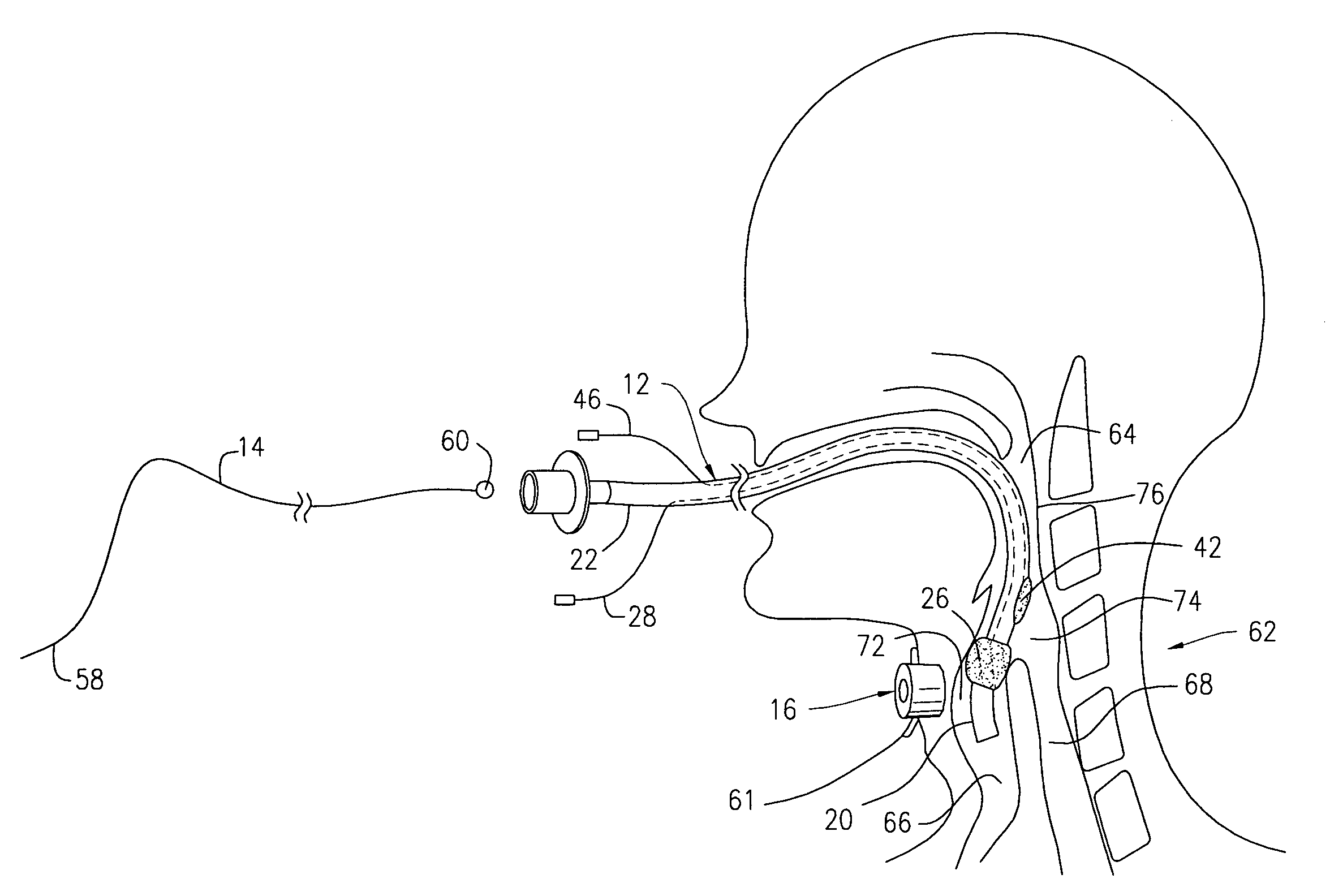
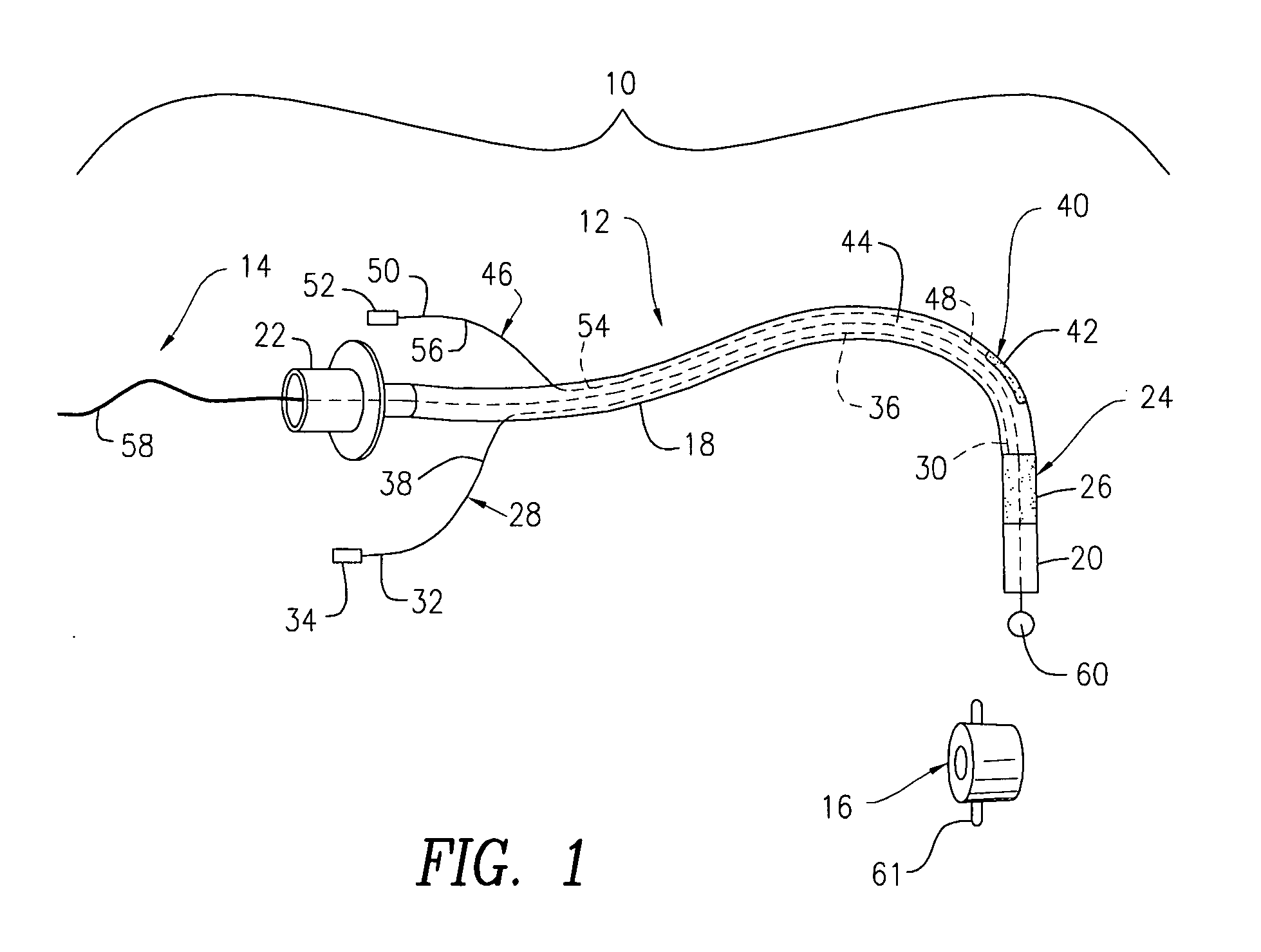
[0016]FIG. 1 illustrates a tracheal intubation device 10 that is used to facilitate ventilation in patients that have blocked airways. The tracheal intubation device 10 includes an endotracheal tube 12, a metal stillette 14, and an external guide system 16. The endotracheal tube 12 includes an elongate tubular body 18 that has a distal end 20 and a proximal end 22. The tubular body 18 can be made from any flexible material known in the art, such as plastic, silicon, and tygon.

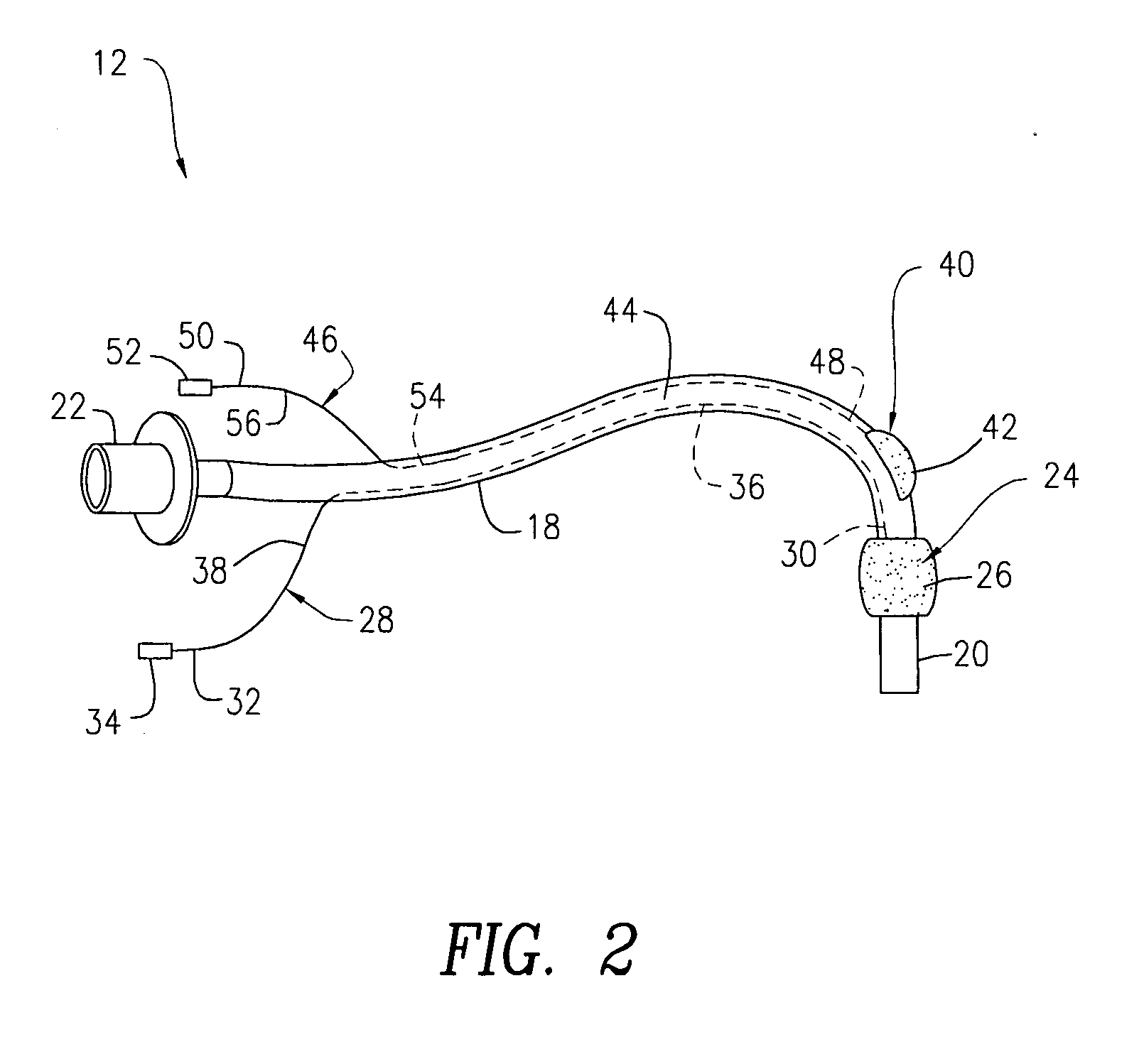
[0017]With reference to FIGS. 1 and 2, the endotracheal tube 12 further includes a primary cuff 24 attached to the tubular body 18 adjacent to the distal end 20 of the tubular body 18. The primary cuff 24 includes a first inflatable balloon 26, which extends about the entire circumference of the tubular body 18. For reasons to be discussed hereinafter, the first balloon 26 is sized and shaped so as to inflate into a fully expanded configuration as shown in FIG. 2 and to deflate into a fully collapsed configurat...


PUM


Abstract

The present invention relates to a tracheal intubation device and method for placing an endotracheal tube within a patient's trachea. More particularly, the endotracheal tube includes primary and secondary cuffs, in the form of inflatable balloons. A stillette is positioned within the endotracheal tube. The tracheal intubation device includes a guiding mechanism for guiding the stillette and the endotracheal tube within a patent's body. The guiding mechanism is positioned external to a patient and sized and shaped so as to transmit a signal and to receive a signal indicating the location of the stillette in a patient's body. After the endotracheal tube is positioned in the oropharynx, inflating the secondary cuff urges the endotracheal tube toward the trachea of a patient.

Description

FIELD OF THE INVENTION[0001]The present invention relates to endotracheal tubes, and more particularly, to a device and method for facilitating the placement of an endotracheal tube within the trachea.BACKGROUND OF THE INVENTION[0002]Certain medical conditions can cause a patient's airway to become blocked, thereby preventing air from passing to the lungs. A commonly used therapy to treat a blocked airway involves inserting an endotracheal tube into the patient's trachea in order to restore airway patency. The insertion of the endotracheal tube into a patient's trachea is referred to as tracheal intubation.[0003]In a tracheal intubation procedure, the endotracheal tube passes through a patient's mouth, through the larynx, and into the trachea. Once the endotracheal tube passes the larynx, it is difficult to properly align the tube into the trachea, because the inlets of the trachea and the esophagus are very close to each other, and the endotracheal tube is often inadvertently place...

Claims


Application Information

Patent Timeline
Patent Type & Authority Applications(United States)
IPC IPC(8): A62B9/06A61M16/00
CPCA61M16/04A61M16/0411A61M2205/32A61M16/0488A61M16/0445A61M16/0459A61M16/0486
Inventor QURESHI, ADNAN I.DIVANI, AFSHIN A.
Owner RUTGERS THE STATE UNIV
Why Eureka
Try Eureka