
Intrauterine ultrasound and method for use

a fibroids and ultrasonography technology, applied in the field of medical instruments and methods, can solve the problems of poor resolution, limited cardiac use, poor penetration rate of low frequency approaches, etc., and achieve the effect of good resolution of fibroids or other uterine structures

Inactive Publication Date: 2007-07-12
GYNESONICS
View PDF99 Cites 42 Cited by
  • Summary
  • Abstract
  • Description
  • Claims
  • Application Information

AI Technical Summary

Benefits of technology

[0008] The present invention provides improved, small-sized ultrasonic imaging apparatus intended for transcervical introduction into the uterus for imaging of the uterine wall. The apparatus and methods of the present invention will be particularly suitable for imaging fibroids disposed at virtually any depth within the uterine wall, typically being anywhere between the surface of the uterine wall to a depth of 6 cm or more. Advantageously, the present invention further provides for adjusting the imaging penetration of the ultrasonic array so that good resolution of the fibroids or other uterine structures can be obtained over the entire range of depths from 0-6 cm or more within the uterine wall. Typically, the imaging penetration is varied by changing the operational frequency of that transducer, typically over the range from 5 MHz to 12 MHz.

Problems solved by technology

These low frequency approaches generally had good penetration but poor resolution, i.e., ability to see fine images.
Such ultrasound arrays provide physicians with a clearer, more familiar image format but are generally limited to cardiac use.
The transducers, however, had not been optimized for use in these tissues.
This image is limited by the depth of penetration of only a few millimeters and has not found to be clinically useful.

Method used


Image

  • Intrauterine ultrasound and method for use
  • Intrauterine ultrasound and method for use
  • Intrauterine ultrasound and method for use

Examples


Embodiment Construction

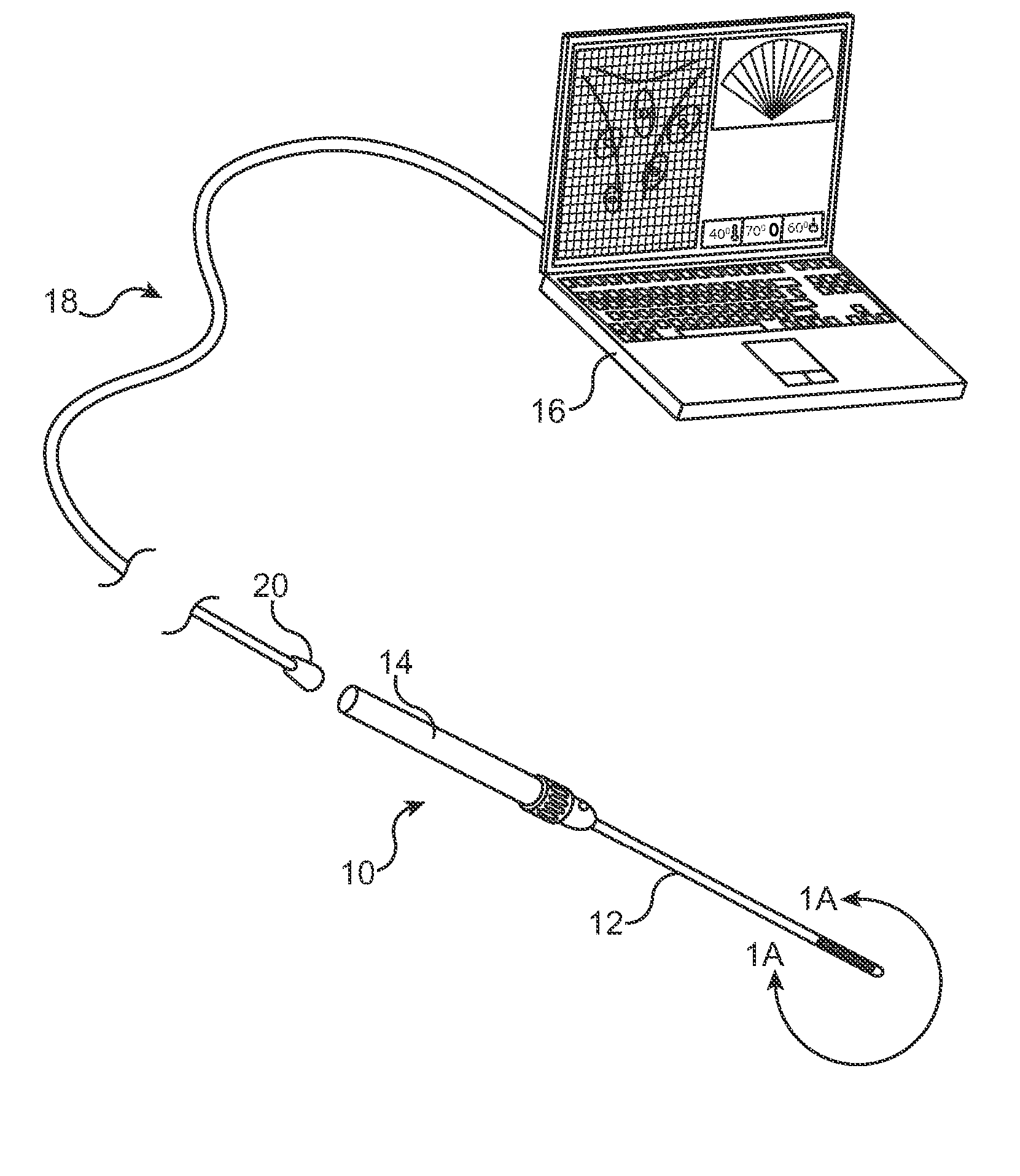
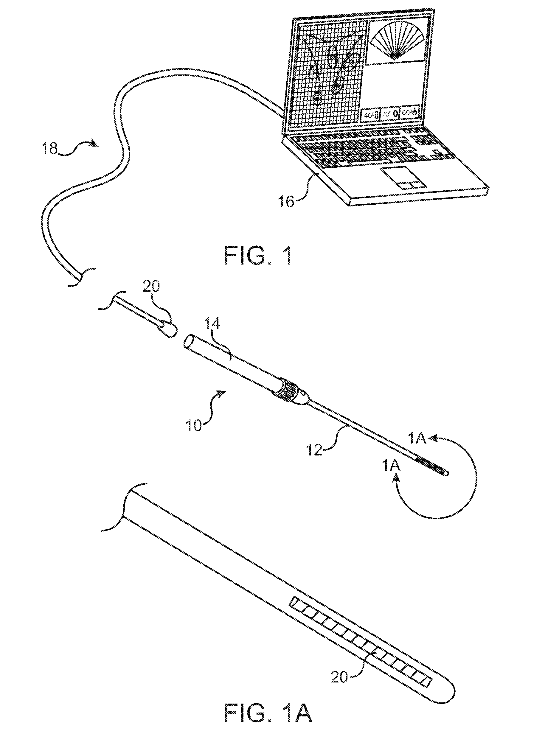
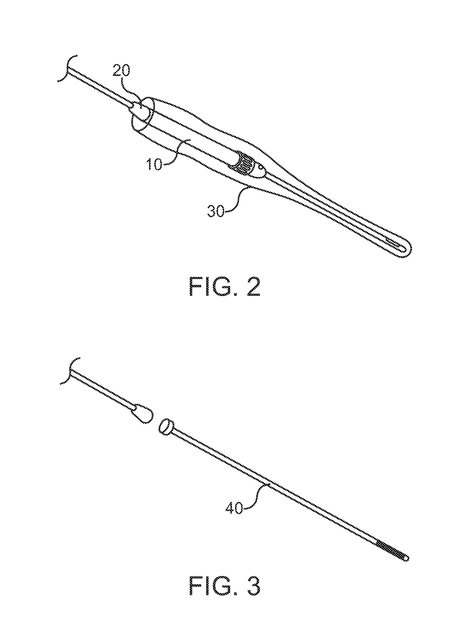
[0017] The present invention provides a very small diameter probe or catheter for access to the interior of the uterus with little or no dilatation of the cervix, typically having a width or diameter from 2 mm to 10 mm, usually from 3 mm to 8 mm. The exemplary probe includes a 64 element phased ultrasonic array with a 13 mm aperture, although as few as 32 elements or as many as 128 elements may be used as well. The aperture of the array may also be in the range from 6 mm to 14 mm. Increasing the aperture size is advantageous since the resolution of the image is improved. Electronic steering of the ultrasound beams (±90°, usually ±45° depending on the frequency of operation and the ultrasound element spacing) may also be provided, with the frequency of operation from 5 to 12 MHz. Depending on the target that is being imaged the frequency may be changed to change resolution and imaging penetration. For example, to image the endometrial cavity one may use a higher frequency and then sw...


PUM


Abstract

A method and apparatus for medical imaging is described. The apparatus applies specifically to accessing and targeting tissue in a small cavity or tightly enclosed space. The medical imaging apparatus or device uses ultrasound waves with elements that act as both a transmitter and receiver in order to image body tissues. The ultrasound is an array or plurality of arrays that may be arranged on the tip on a probe or catheter for insertion into a patient's body.

Description

CROSS-REFERENCES TO RELATED APPLICATIONS [0001] This application is claims priority to U.S. Provisional Application No. 60 / 758,727 (Attorney Docket No. 025676-000400US), filed on Jan. 12, 2006, and U.S. Provisional Application No. 60 / 821,009 (Attorney Docket No. 025676-000410US) filed on Aug. 1, 2006, the full disclosures of which are incorporated herein by reference.BACKGROUND OF THE INVENTION [0002] The present invention relates generally to medical apparatus and methods. More particularly, the present invention relates to methods and apparatus for ultrasonically imaging fibroids in the uterine cavity. [0003] Ultrasound medical imaging has been known for several decades. Medical ultrasound imaging began using low frequencies (2-5 MHz) for surface imaging of internal body structures. These low frequency approaches generally had good penetration but poor resolution, i.e., ability to see fine images. As technology advanced the ability to make smaller, higher frequency probes became p...

Claims


Application Information

Patent Timeline
IPC IPC(8): A61B8/14
CPCA61B8/12A61B8/445G01S15/8918G01S15/894G01S15/8952A61B8/4488
Inventor MUNROW, MICHAEL
Owner GYNESONICS
Learn More