System and Method For Labeling and Identifying Lymph Nodes In Medical Images

a lymph node and image technology, applied in image enhancement, medical/anatomical pattern recognition, instruments, etc., can solve the problems of time-consuming and sometimes inaccurate process, lymph node segmentation remains a challenging task, and affects the workload of radiologists

Active Publication Date: 2007-10-11
SIEMENS HEALTHCARE GMBH +1
View PDF3 Cites 53 Cited by
  • Summary
  • Abstract
  • Description
  • Claims
  • Application Information

AI Technical Summary

Problems solved by technology

Although these methods show promising results, lymph node segmentation still remains a challenging task.
Further, even with such tools available, radiologists must still manually provide anatomical labels for lymph nodes during staging and assessment....

Method used


Image

  • System and Method For Labeling and Identifying Lymph Nodes In Medical Images
  • System and Method For Labeling and Identifying Lymph Nodes In Medical Images
  • System and Method For Labeling and Identifying Lymph Nodes In Medical Images

Examples


Embodiment Construction

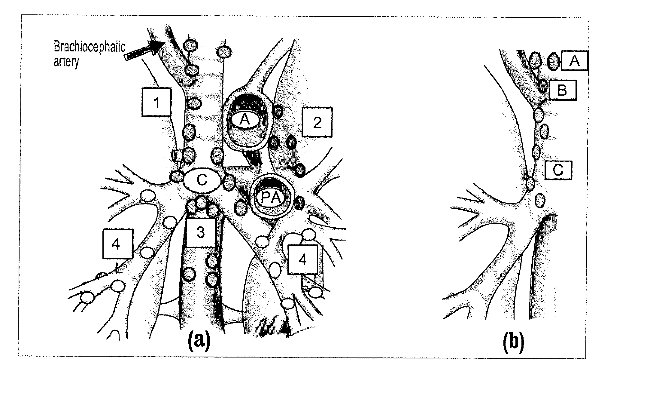
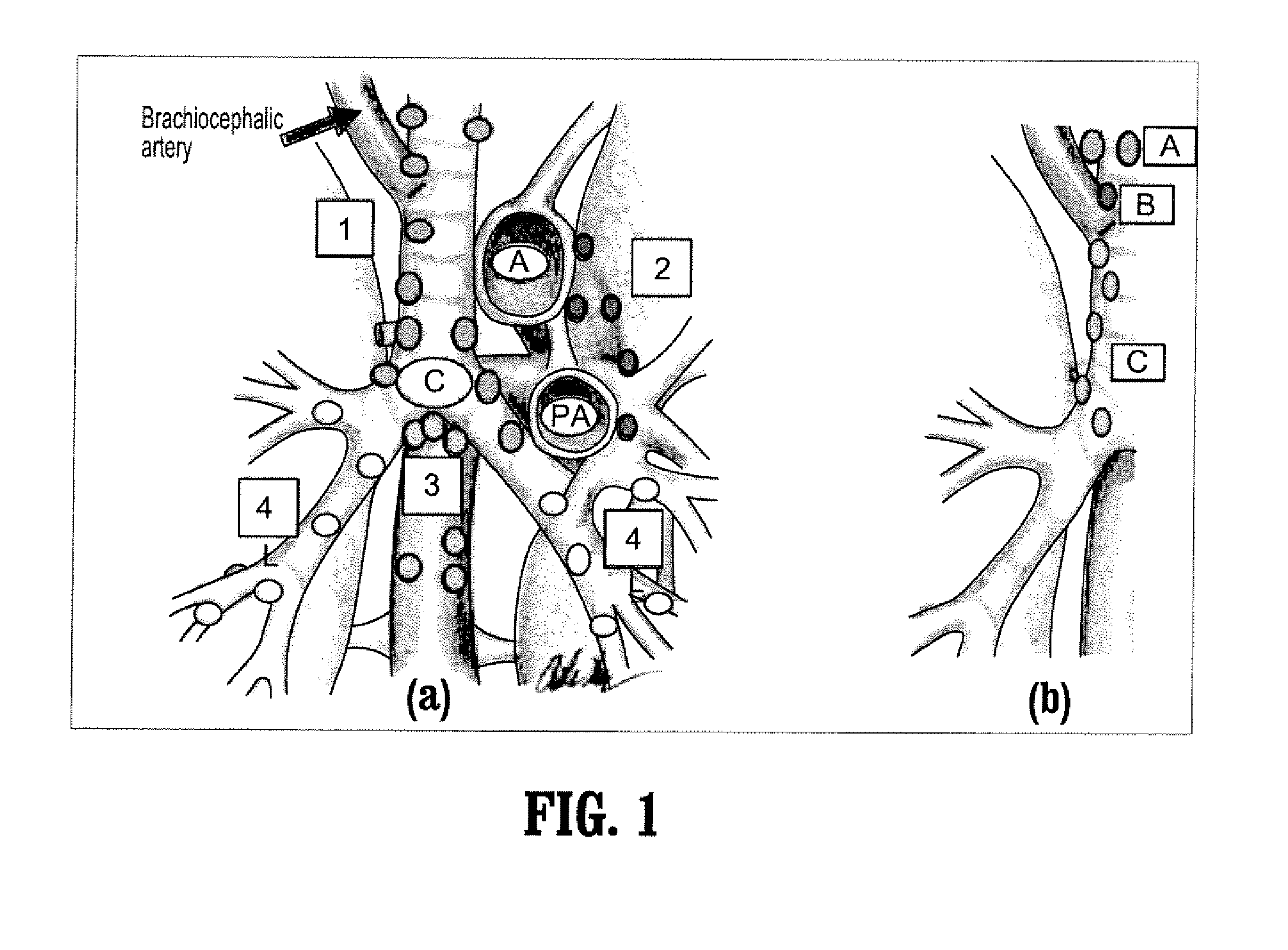
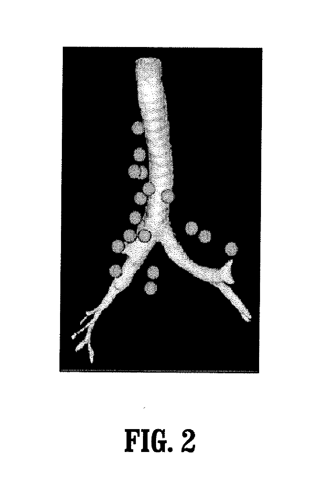
[0036] A system for labeling and identifying lymph nodes in medical images according to an exemplary embodiment of the present invention will now be described.

[0037]FIG. 4 illustrates a system 400 for labeling and identifying lymph nodes in medical images according to an exemplary embodiment of the present invention. As shown in FIG. 4, the system 400 includes an acquisition device 405, a personal computer (PC) 410 and an operator's console 415 connected over a wired or wireless network 420.

[0038] The acquisition device 405 may be a computed tomography (CT) imaging device or any other three-dimensional (3D) high-resolution imaging device such as a magnetic resonance (MR) scanner or ultrasound scanner.

[0039] The PC 410, which may be a portable or laptop computer, a medical diagnostic imaging system or a picture archiving communications system (PACS) data management station, includes a central processing unit (CPU) 425 and a memory 430 connected to an input device 450 and an output...


PUM


Abstract

A method for assigning a lymph node in a medical image with an anatomical name, the method including: identifying landmarks in a medical image; computing features relative to the landmarks given a location of a lymph node in the medical image; and assigning an anatomical name to the location of the lymph node by using a classifier that compares the computed features with classification rules.

Description

CROSS-REFERENCE TO RELATED APPLICATION [0001] This application claims the benefit of U.S. Provisional Application No. 60 / 762,029, filed Jan. 25, 2006, a copy of which is herein incorporated by reference.BACKGROUND OF THE INVENTION [0002] 1. Technical Field [0003] The present invention relates to anatomic object labeling and identification in medical images, and more particularly, to a system and method for labeling and identifying lymph nodes in medical images. [0004] 2. Discussion of the Related Art [0005] The identification and evaluation of lymph nodes for cancer staging forms a large portion of a radiologist's workflow. Examples of this are described in J. P. Ko, E. A. Drucker, J. A. Shepard, C. F. Mountain, C. Dresler, B. Sabloff, and T. C. McLoud, “CT depiction of regional nodal stations for lung cancer staging”, AJR Am J Roentgenol. 2000 March; 174(3):775-82, and S. Aquino, M. Harisinghani, and B. Branstetter, “Lymph Node Anatomy and Imaging for Cancer Staging”, RSNA 2005. Th...

Claims


Application Information

Patent Timeline
IPC IPC(8): G06K9/00A61B5/05
CPCG06K2209/05G06T7/0042G06T2207/30101G06T2207/10088G06T2207/10132G06T2207/10081G06T7/73G06V2201/03
Inventor KIRALY, ATILLA PETERZHANG, LINOVAK, CAROL L.GUNDEL, LUTZ
Owner SIEMENS HEALTHCARE GMBH
Why Eureka
Try Eureka