Mitral Valve Annuloplasty Device with Vena Cava Anchor

a technology of annuloplasty and mitral valve, which is applied in the field of mitral valve annuloplasty device with vena cava anchor, can solve the problems of reducing circulatory efficiency, rapid deterioration, and regurgitation of the mitral valv

Active Publication Date: 2007-10-11
CARDIAC DIMENSIONS
View PDF99 Cites 44 Cited by
  • Summary
  • Abstract
  • Description
  • Claims
  • Application Information

AI Technical Summary

Problems solved by technology

Mitral valve regurgitation is a serious, often rapidly deteriorating, condition that reduces circulatory efficiency.
Each of these procedures is highly invasive as access to the heart is gained through an opening in the patient's chest.
Patients with mitral valve regurgitation are often relatively frail which increases the risks associated with such an invasive operation.

Method used


Image

  • Mitral Valve Annuloplasty Device with Vena Cava Anchor
  • Mitral Valve Annuloplasty Device with Vena Cava Anchor
  • Mitral Valve Annuloplasty Device with Vena Cava Anchor

Examples


Embodiment Construction

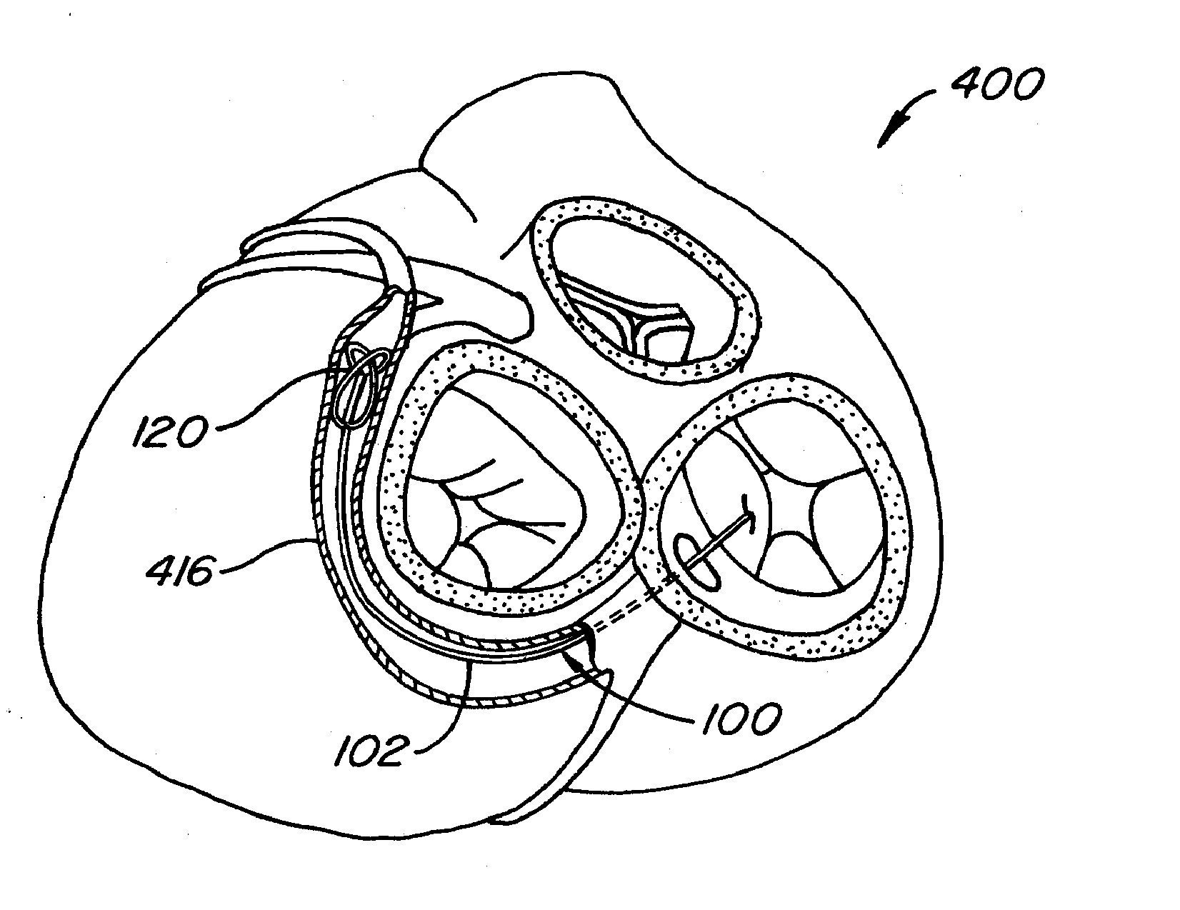
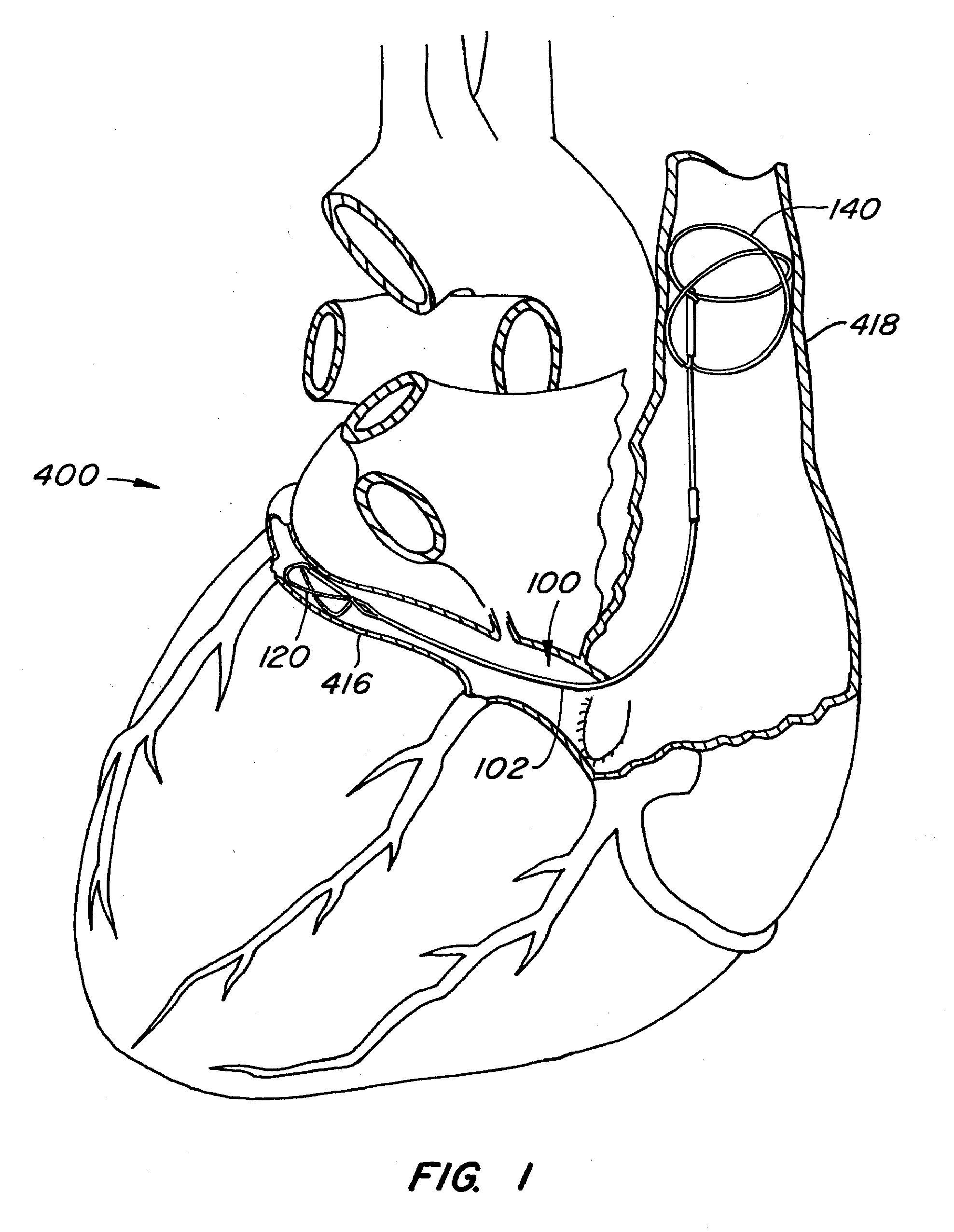
[0024] The present invention relates to a medical device and uses thereof that supports or changes the shape of tissue near a vessel in which the device is placed. The present invention is particularly useful in reducing mitral valve regurgitation by changing the shape of or supporting a mitral valve annulus. In preferred embodiments, the device comprises a distal anchor adapted to be anchored in the coronary sinus and a proximal anchor adapted to be anchored in the superior vena cava, with a support structure disposed between the anchors. The length and diameter of a coronary sinus differ from patient to patient, and it is desirable for an intravascular device to function effectively in all patients, regardless of these differences. By anchoring the proximal anchor in the superior vena cava, the distal anchor may be positioned in the coronary sinus at a desired location to reduce mitral valve regurgitation without producing other adverse consequences to the patient. The superior ve...


PUM


Abstract

The present invention relates to a medical device and uses thereof that supports or changes the shape of tissue near a vessel in which the device is placed. The present invention is particularly useful in reducing mitral valve regurgitation by changing the shape of or supporting a mitral valve annulus. The device includes a support structure, a proximal anchor adapted to be positioned in a superior vena cava, and a distal anchor adapted to be positioned in a coronary sinus. The support structure engages a vessel wall to change the shape of tissue adjacent the vessel in which the intravascular support is placed.

Description

BACKGROUND OF THE INVENTION [0001] The mitral valve is located between the chambers of the left atrium and the left ventricle. When the left ventricle contracts to pump blood throughout the body, the mitral valve closes to prevent blood from being pumped back into the left atrium. In patients suffering from mitral valve regurgitation the mitral valve fails to properly close upon each contraction due to, for example, genetic malformation, disease, or injury, and blood is pumped back into the atrium. Mitral valve regurgitation is a serious, often rapidly deteriorating, condition that reduces circulatory efficiency. [0002] Two of the more common techniques for restoring damaged mitral valve function are surgical replacement of the valve with a mechanical valve and implantation of a flexible ring around the native valve to support it. Each of these procedures is highly invasive as access to the heart is gained through an opening in the patient's chest. Patients with mitral valve regurgi...

Claims


Application Information

Patent Timeline
Patent Type & Authority Applications(United States)
IPC IPC(8): A61F2/24
CPCA61F2/2451
Inventor MATHIS, MARK L.NIEMINEN, GREGORYARONSON, NATHANBEGET, GARRETT
Owner CARDIAC DIMENSIONS
Why Eureka
Try Eureka