Automatic Positioning Quality Assessment for Digital Mammography

a digital mammography and positioning image technology, applied in the field of radiology and mammography, can solve the problems of inability of radiologists to evaluate the mammogram correctly, time-consuming, manual measurement, and thereby time-consuming, and achieve the goal of reducing the number of recalls, improving the overall mammography process, and facilitating a common standardization of mammography image quality

Inactive Publication Date: 2007-10-25
SECTRA IMTEC
View PDF8 Cites 22 Cited by
  • Summary
  • Abstract
  • Description
  • Claims
  • Application Information

AI Technical Summary

Benefits of technology

[0008] It is also a main object of the invention to improve the overall mammography process and reduce the number of recalls due to insufficiently positioned mammographic images...

Problems solved by technology

However, if the breast is improperly positioned the radiologist might not be able to evaluate the mammogram correctly.
However these kinds of measurements are done manually and thereby time consuming and are unlikely to be performed on a daily basis, especially if one considers the amount of time generally available for each mammography examination at a normal mammography unit.
Even more time consuming is the procedure with conventional film-based mammography where the images have to be developed and hanged on a...

Method used


Image

  • Automatic Positioning Quality Assessment for Digital Mammography
  • Automatic Positioning Quality Assessment for Digital Mammography
  • Automatic Positioning Quality Assessment for Digital Mammography

Examples


Embodiment Construction

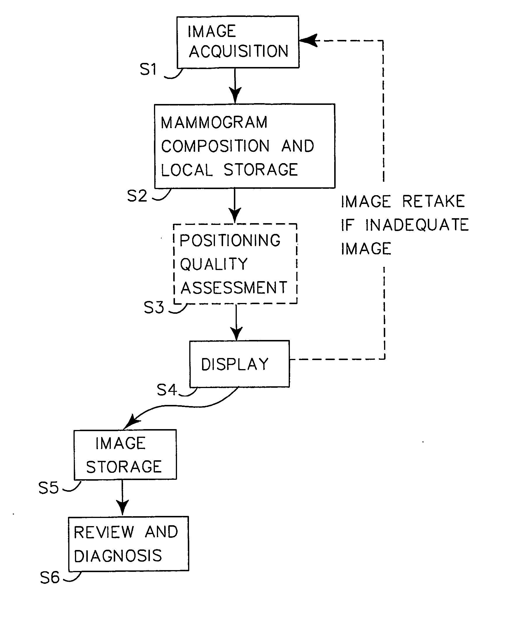
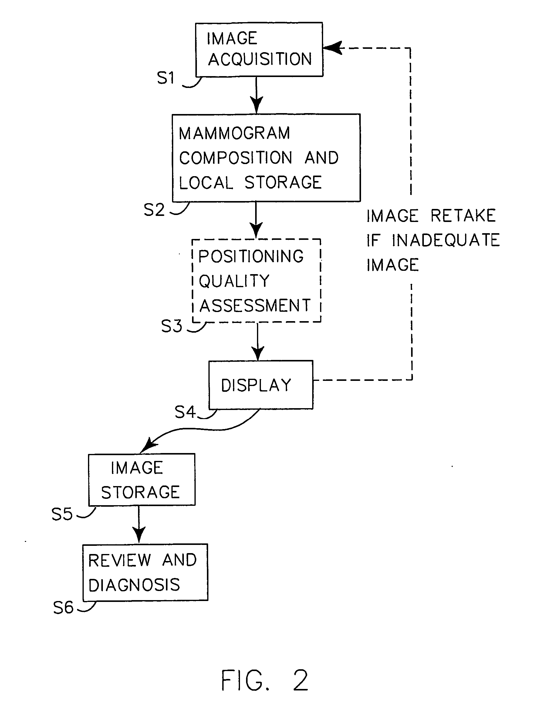
[0031] Throughout the drawings, the same reference characters will be used for corresponding or similar elements.

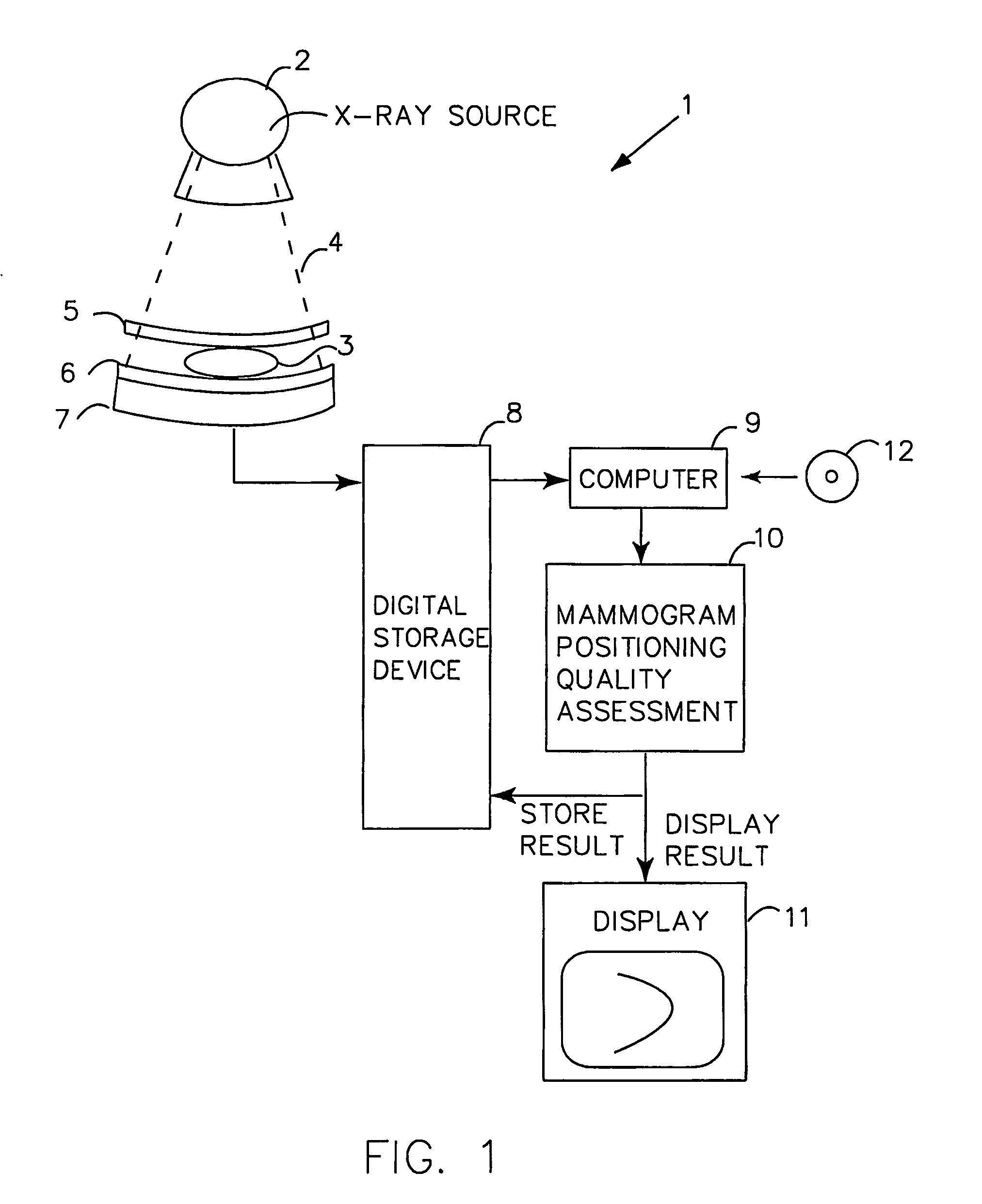
[0032] For a better understanding of the invention, it may be useful to begin with a general system overview of an exemplary digital mammography system, referring to FIG. 1. The system 1 includes an X-ray source 2 directed to expose a patient's breast 3 with X-ray beams 4. The breast is generally compressed, using a predefined compression force, between two compression plates 5, 6. Below the lower compression plate, some detector means 7 is arranged. The detector 7 is usually in the form of a two-dimensional array of radiation sensitive elements, the outputs of which are mapped into a corresponding array of digital pixels representing the digital mammogram. The digital pixel information is stored as an image file in a digital storage device 8, and the digital mammogram is then generated in a computer-based acquisition workstation, for example the Sectra Acquisition works...


PUM


Abstract

The invention presents a way to automatically assess the quality of acquired (S1, S2) digital mammographic images with respect to the image positioning of a patient's breast. The automated digital quality assessment (S3) is executed in real time and preferably notifies (S4) the technologist instantly if the image positioning quality of a mammographic image is inadequate. This makes it possible to retake the image while the patient is still present at the examination facility. The quality assessment notification includes information to the technologist, preferably both visually and statistically of land-mark positioning measurements. Alternatively, the digital quality assessment is accompanied by a computerized decision of whether the mammogram needs to be retaken, requiring a minimum of involvement, if at all, from the technologist. The invention hence provides a set of quality-assured mammographic images that can be stored (S5) and later accessed by a radiologist for review and diagnosis (S6).

Description

TECHNICAL FIELD OF THE INVENTION [0001] The present invention generally relates to the field of radiology and mammography, and more specifically to assessment of positioning image quality in digital mammography. BACKGROUND OF THE INVENTION [0002] Mammography is today the most important and accurate method for diagnosing breast cancer. The diagnostic value of mammography images is highly dependent on image quality, which, in turn, depends on a number of technical and image processing related factors such as proper exposure, compression, sharpness and contrast. However, if the breast is improperly positioned the radiologist might not be able to evaluate the mammogram correctly. With digital mammography, parameters such as brightness, contrast, magnification etc. can be altered in real time after the mammography examination is completed to improve recognition of suspicious lesions in the breast. Different types of segmentations can also be applied to distinguish certain areas such as t...

Claims


Application Information

Patent Timeline
IPC IPC(8): A61B6/04G06T7/00
CPCA61B6/0414A61B6/502G06T2207/30068G06T7/0042G06T7/0012G06T7/73
Inventor SELSE, EMILPETTERSSON, KRISTINA
Owner SECTRA IMTEC
Why Eureka
Try Eureka