Automatic analysis in virtual endoscopy

a virtual endoscope and automatic analysis technology, applied in the field of automatic analysis of virtual endoscopes, can solve the problems of exposing patients to the risk of bowel perforation, affecting the quality of life of patients, and 1.5 million fiberoptic colonoscopy procedures performed

Inactive Publication Date: 2007-11-29
WAKE FOREST UNIV HEALTH SCI INC
View PDF31 Cites 27 Cited by
  • Summary
  • Abstract
  • Description
  • Claims
  • Application Information

AI Technical Summary

Problems solved by technology

However, only about 1.5 million fiberoptic colonoscopies are performed each year.
Another problem is that conventional colonoscopies fail to provide access to the entire colon in approximately 15% of cases.
Colonoscopies also expose patients to the risk of bowel perforation.
Such human inspection is time consuming, tedious, expensive, and under certain circumstances prone to error of interpretation.

Method used


Image

  • Automatic analysis in virtual endoscopy
  • Automatic analysis in virtual endoscopy
  • Automatic analysis in virtual endoscopy

Examples


Embodiment Construction

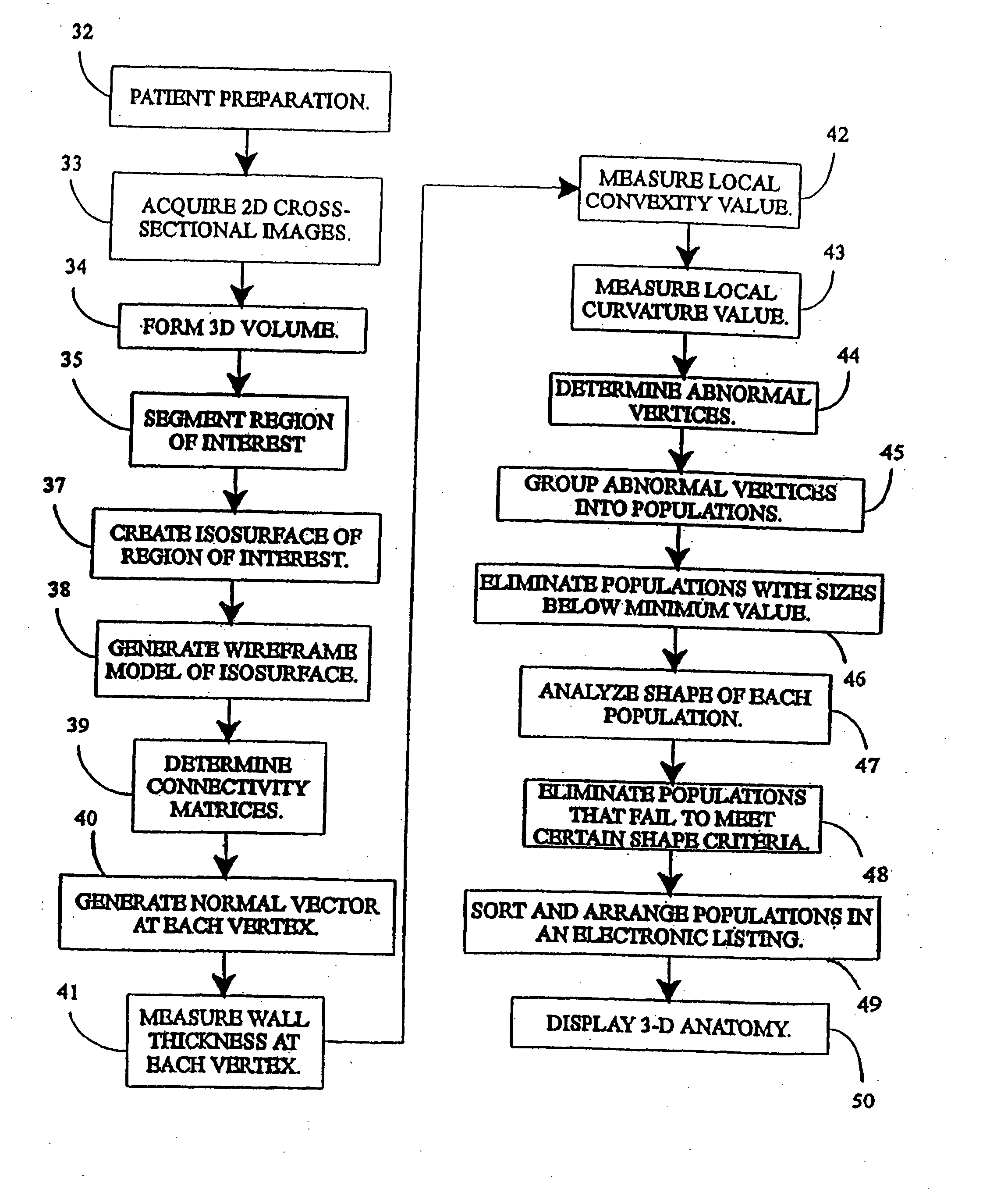
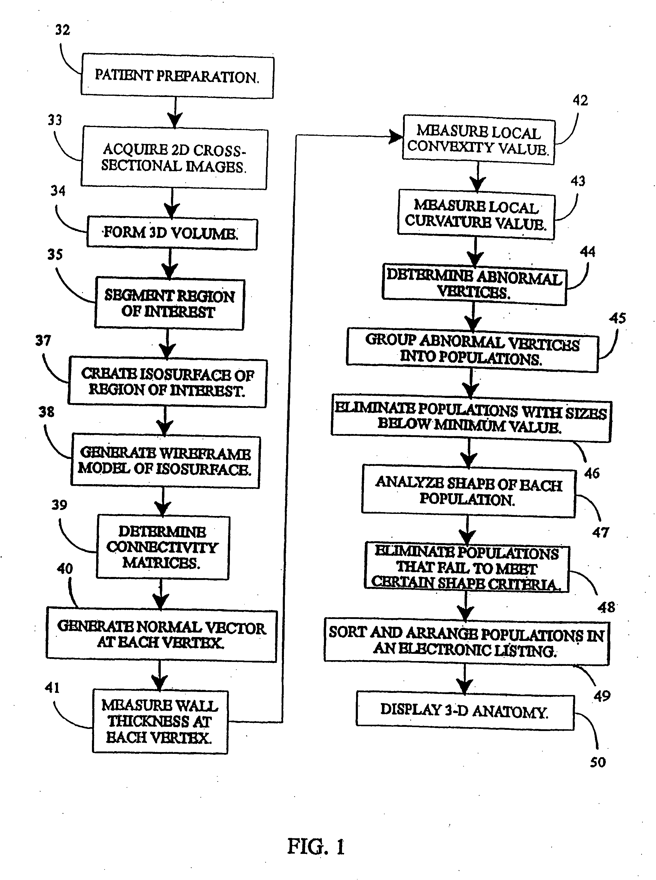
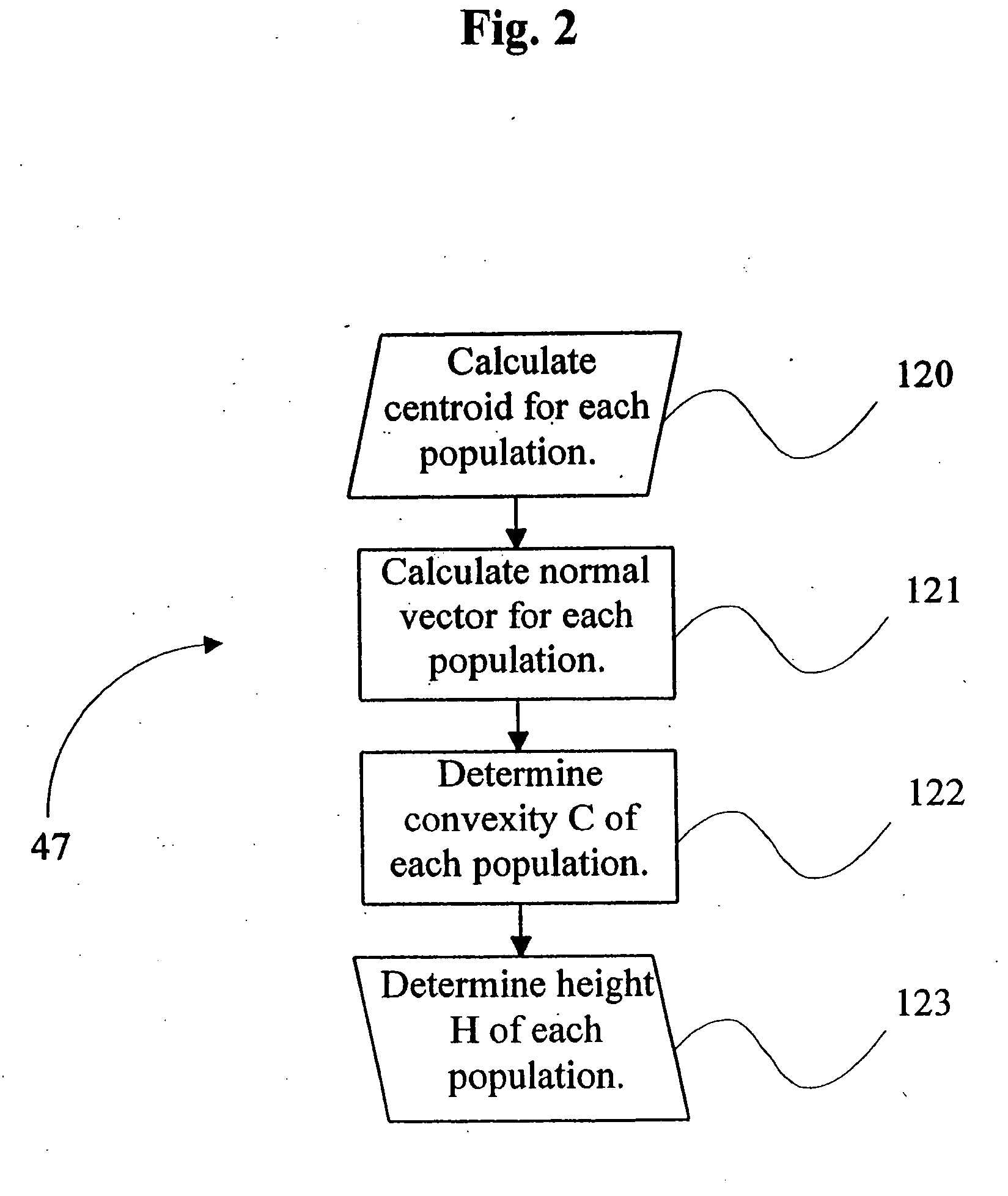
[0021] The present invention generally relates to a method and system, as schematically represented in FIGS. 1-4, for generating interactive, three-dimensional renderings of three-dimensional structures generally having a lumen. The structures are usually in the general form of selected regions of a body and in particular, human or animal body organs which have hollow lumens such as colons, blood vessels, and airways. In accordance with the present invention, the interactive three-dimensional renderings are generated in a computer-controlled process from a series of two-dimensional, cross-sectional images of the selected body organ acquired, for example, from a helical computed tomography (CT) Scan. The three-dimensional renderings are interactive in that such renderings can be manipulated by a user on a visual display of a computer system, such as a computer monitor, to enable movement in, around and through the three-dimensional structure while simultaneously displaying multiplana...


PUM


Abstract

A computer system and a computer-implemented method are provided for interactively displaying a three-dimensional rendering of a structure having a lumen and for indicating regions of abnormal wall structure. A three-dimensional volume of data is formed from a series of two-dimensional images representing at least one physical property associated with the three-dimensional structure. An isosurface of a selected region of interest is created by a computer from the volume of data based on a selected value or values of a physical property representing the selected region of interest. A wireframe model of the isosurface is generated by the computer wherein the wireframe model includes a plurality of vertices. The vertices are then grouped into populations of contiguous vertices having a characteristic indicating abnormal wall structure by the computer. The wireframe model is then rendered by the computer in an interactive three-dimensional display to indicate the populations of abnormal wall structure.

Description

RELATED APPLICATION [0001] This application is a continuation of co-pending application Ser. No. 10 / 109,547, entitled, “Automatic Analysis in Virtual Endoscopy”, filed on Mar. 28, 2002, now issued as U.S. Pat. No. 7,149,564, which in turn is a continuation of application Ser. No. 09 / 299,061, entitled “Automatic Analysis in Virtual Endoscopy”, filed on Apr. 23, 1999, now issued as U.S. Pat. No. 6,366,800, which in turn is a continuation of application Ser. No. 08 / 805,584, entitled “Automatic Analysis in Virtual Endoscopy”, filed on Feb. 25, 1997, now issued as U.S. Pat. No. 5,920,319, which in turn is a continuation-in-part of application Ser. No. 08 / 331,352, filed on Oct. 27, 1994, now issued as U.S. Pat. No. 5,782,762, which are each incorporated herein by reference.FIELD OF THE INVENTION [0002] The present invention relates to a system and method for interactively displaying a three-dimensional rendering of a structure having a lumen and, more particularly, to a system and method ...

Claims


Application Information

Patent Timeline
Patent Type & Authority Applications(United States)
IPC IPC(8): A61B5/05G06T17/00A61B1/04A61B6/03A61B5/107G06T1/00G06T5/00G06T15/08G06T17/20G09B23/28
CPCA61B5/1076G06T7/0012G06T7/0081G06T7/0091G06T15/08G06T17/00Y10S128/92G06T2207/10081G06T2207/20012G06T2207/30032G06T2207/30064G09B23/285G06T17/20G06T7/11G06T7/155Y02A90/10
Inventor VINING, DAVID J.HUNT, GORDON W.AHN, DAVID K.STELTS, DAVID R.GE, YAORONGHEMLER, PAUL F.SALIDO, TIFFANY W.
Owner WAKE FOREST UNIV HEALTH SCI INC
Why Eureka
Try Eureka