Quantifying Blood Volume Using Magnetization Transfer Magnetic Resonance Imaging

a magnetic resonance imaging and blood volume technology, applied in the field of non-invasive methods for quantifying blood volume, can solve the problems of unresolved need for improving non-invasive blood volume quantifying techniques

Inactive Publication Date: 2008-01-24
VAN ZIJL PETER C M +1
View PDF0 Cites 19 Cited by
  • Summary
  • Abstract
  • Description
  • Claims
  • Application Information

AI Technical Summary

Benefits of technology

[0006] One advantage includes determining blood volume.
[0007] Another advantage resides in determining parenchymal blood volume through magnetization transfer ratios (MTRs).
[0008] Another advantage resides in determining blood volume through MRI scans

Problems solved by technology

In view of the aforementioned deficiencies of conventional blood volume quantifying technique

Method used


Image

  • Quantifying Blood Volume Using Magnetization Transfer Magnetic Resonance Imaging
  • Quantifying Blood Volume Using Magnetization Transfer Magnetic Resonance Imaging
  • Quantifying Blood Volume Using Magnetization Transfer Magnetic Resonance Imaging

Examples


Embodiment Construction

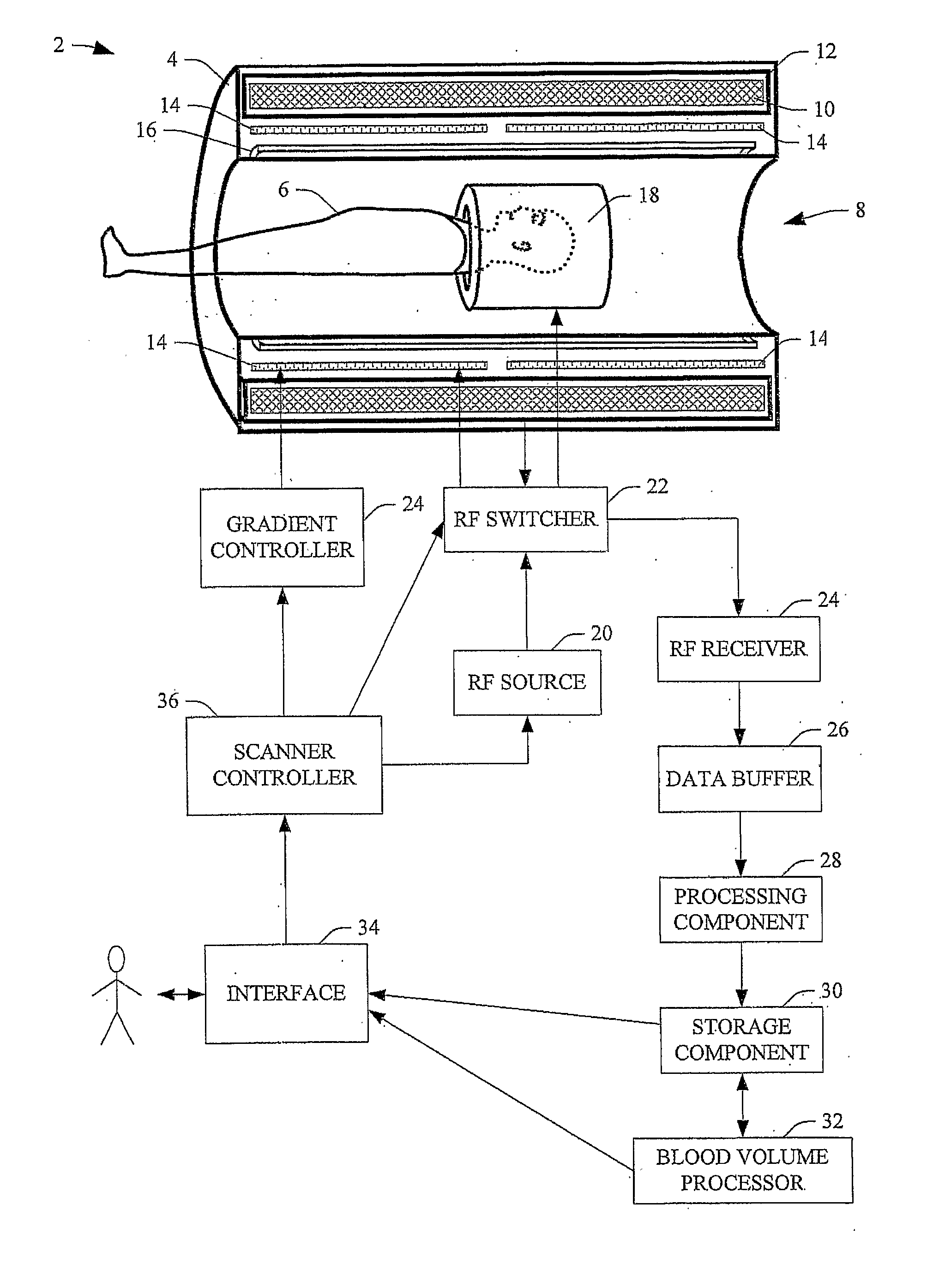
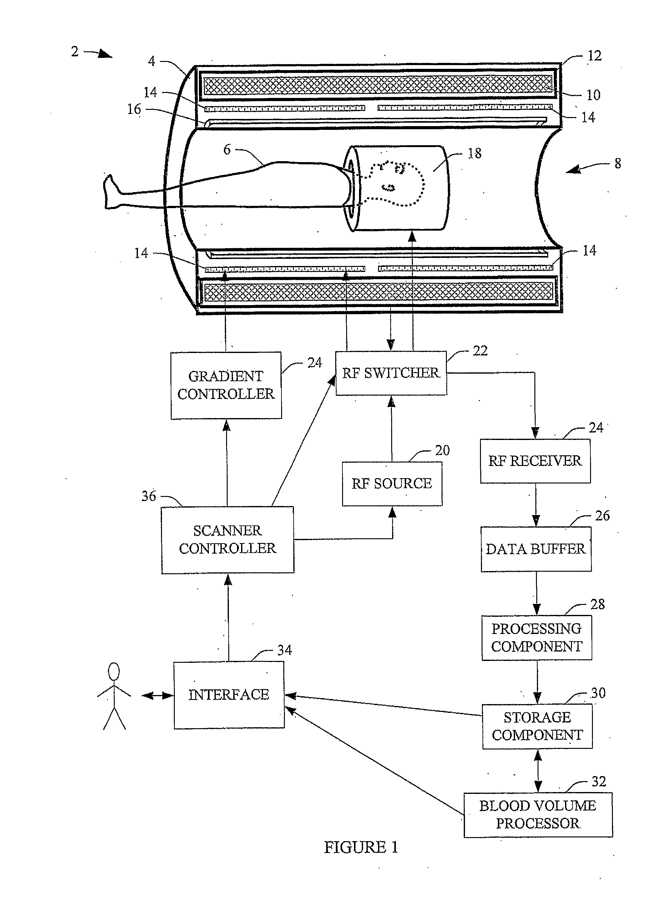
[0026]FIG. 1 illustrates a magnetic resonance imaging (MRI) scanner 2 used to facilitate determining blood volume through magnetization transfer (MT) properties. An absolute blood volume map can be obtained by combining conventional MRI scanning techniques and using large MT-based reduction of tissue signal with respect to blood signal for non-invasive determination of absolute and relative blood volume effects in all tissues.

[0027] The scanner 2 includes a scanner housing 4. A subject 6 (or other object) is at least partially disposed within a bore 8 of the housing 4 for one or more scanning procedures. A magnet 10 resides in the scanner housing 4. Typically, the magnet 10 is a persistent superconducting magnet surrounded by cryoshrouding 12. However, other known magnets can be employed. The magnet 10 generates a magnetic field (B0) in the subject 6. Typical magnetic fields strengths are about 0.5 Tesla, 1.0 Tesla, 1.5 Tesla, 3 Tesla or higher (e.g., about 7 Tesla).

[0028] Magneti...


PUM


Abstract

A magnetic resonance method for imaging blood volume in parenchyma via magnetic transfer (MT) includes: determining a MT effect of parenchyma; determining a MT effect of tissue; and quantifying the parenchymal blood volume using the difference between the MT effect of parenchyma and the MT effect of tissue. In one embodiment, the parenchymal blood volume is quantified through the following: MTRpar=MTRtissue(1−BV/Vpar), where MTRpar is the magnetization transfer ratio of parenchyma, MTRtissue is the magnetization transfer ratio of tissue, BV is the blood volume, and Vpar is a total parenchymal water volume.

Description

BACKGROUND [0001] The following relates to non-invasive techniques for quantifying blood volume. It finds particular application to quantifying microvascular blood volume through magnetic resonance imaging techniques. [0002] Conventionally, invasive and non-invasive techniques are used to measure blood volume. Invasive techniques include positron emission tomography (PET), dynamic contrast magnetic resonance imaging (DC MRI), dynamic susceptibility contrast MRI (DSC MRI), near infrared spectroscopy (NIRS), and optical imaging. Typical PET techniques are based on the administration of radioactive compounds, for example, through an arterial line or inhalation of C-11 or O-15 carbon monoxide. MRI techniques usually employ exogenous contrast agents (e.g., Gd-DTPA, MION) and a venous catheter for contrast agent injection / infusion. [0003] One non-invasive approach uses nulling of the blood signal to measure blood volume changes. This approach is referred to as vascular space occupancy (VA...

Claims


Application Information

Patent Timeline
IPC IPC(8): A61B5/055
CPCA61B5/055G01R33/563G01R33/5605
Inventor VAN ZIJL, PETER C.M.ZHOU, JINYUAN
Owner VAN ZIJL PETER C M
Why Eureka
Try Eureka