Minimally-invasive devices and methods for treatment of congestive heart failure

a congestive heart failure, minimally invasive technology, applied in the direction of surgical forceps, catheters, applications, etc., can solve the problems of inability to pump at a sufficient rate to maintain adequate blood circulation throughout the body, inability to perform highly invasive and traumatic procedures, and shortening of breath, so as to facilitate the reduction of left ventricular volume, reduce pain and trauma to the patient, and reduce the risk of infection and neurological events.

Inactive Publication Date: 2008-02-07
ETHICON INC
View PDF96 Cites 19 Cited by
  • Summary
  • Abstract
  • Description
  • Claims
  • Application Information

AI Technical Summary

Benefits of technology

[0008] The invention provides devices and methods for treating CHF, as well as other diseases resulting in an enlarged heart, that not only significantly reduce the pain and trauma to the patient, but that may reduce the risk of infection and the risk of neurological events associated with the Batista Operation. The invention facilitates the reduction of left ventricular volume by removing a section of the heart wall or otherwise reshaping the ventricle without requiring a median sternotomy or gross thoracotomy. The invention further allows the procedure to be performed on cardiopulmonary bypass with the heart isolated and arrested, yet without the gross thoracic incision required by, or the risk of embolism produced by, conventional aortic cross-clamps. Moreover, the invention may significantly reduce the risk that air will be introduced into the bloodstream and embolized to the brain during or after the procedure.
[0015] reshaping an outer wall of the patient's heart while the heart is arrested so as to reduce the transverse dimension of the left ventricle.
[0017] By obviating the need for an aortic cross-clamp, the need for the median sternotomy through which such a cross-clamp is placed is also eliminated. The left ventricle may then be reshaped and volumetrically reduced using thoracoscopic instruments positioned through small incisions, punctures or ports located in the intercostal spaces between the ribs.
[0021] manipulating the tissue attaching device to attach the first location to a second location on a wall of the heart so as to reduce the transverse dimension of the left ventricle, the user's hands remaining outside the patient's chest when manipulating the tissue attaching device.
[0025] In still other embodiments, a restrictive girdle or band is placed around the outside of the heart to restrict the left ventricle to the desired diameter or volume. The band or girdle is preferably elastic so as to expand and contract with the heart as it pumps. Preferably, the girdle or band is applied to the heart using specialized thoracoscopic instruments placed through intercostal spaces in the rib cage while generally maintaining the user's hands outside the chest, thereby eliminating the need for a gross thoracotomy.
[0026] Because the chest is not grossly opened, the heart is isolated from the rest of the circulatory system, and in some embodiments, even the ventricle itself is not opened, the methods of the invention may reduce the risk that air will pass through the ventricular incision and into the bloodstream. To reduce this risk even further, the invention also allows the chest to be flooded with carbon dioxide or other suitable gas during the procedure to maintain the chest cavity free of air. A tube may be placed through one of the intercostal ports and gas delivered through the tube into the chest at a pressure suitable to ensure that air cannot enter the chest cavity. Additionally, trocar sleeves or tubular ports may be used which have internal seals like those used for gaseous insufflation in laparoscopic procedures, thereby preventing the unwanted introduction of air into the chest. Further, where some risk of air embolism is present due to the opening of the left ventricle, following closure the left ventricle and aorta may be flushed with saline and then vented through a lumen in the aortic occlusion catheter while maintaining aortic occlusion, thereby removing any trapped air that may be present.

Problems solved by technology

In congestive heart failure or CHF, the heart has become so enlarged as a result of viral infection, myocardial infarction or other disease that it is unable to pump at a sufficient rate to maintain adequate circulation of blood throughout the body.
As a result, blood backs up into the lungs, causing shortness of breath and other symptoms, and, if left untreated, the disease can lead to death.
Unfortunately, due to a donor shortage, of the 40,000 patients who may require a transplant each year, only 2500 actually get one, with up to 15-20% of patients dying while on the waiting list for a donor heart.
Although the Batista Operation can extend the life of a patient who would otherwise die without a transplant, it is a highly invasive and traumatic procedure.
In order to expose the heart, the chest must be opened widely by sawing the sternum in half and spreading apart the rib cage, known as a median sternotomy, producing a great deal of pain, risk of infection, and long recovery time.
For elderly or extremely ill patients, the trauma produced by the operation could contribute significantly to the mortality and morbidity associated with the procedure.
Moreover, the Batista Operation has typically been performed while the heart is beating, causing a great deal of blood loss through the ventricular incision, and risking the introduction of air into the bloodstream, potentially causing stroke or other neurological problems.
However, because such cross-clamps crush the walls of the aorta together in order to occlude the vessel, cross-clamps may produce the added risk of releasing calcific particles from the inner walls of the aorta, which may embolize in the bloodstream and produce neurological events such as stroke.
Moreover, the risk remains that air will become trapped in the ventricle after it has been closed, allowing the air to migrate to the brain as soon as the cross-clamp is removed.

Method used


Image

  • Minimally-invasive devices and methods for treatment of congestive heart failure
  • Minimally-invasive devices and methods for treatment of congestive heart failure
  • Minimally-invasive devices and methods for treatment of congestive heart failure

Examples


Embodiment Construction

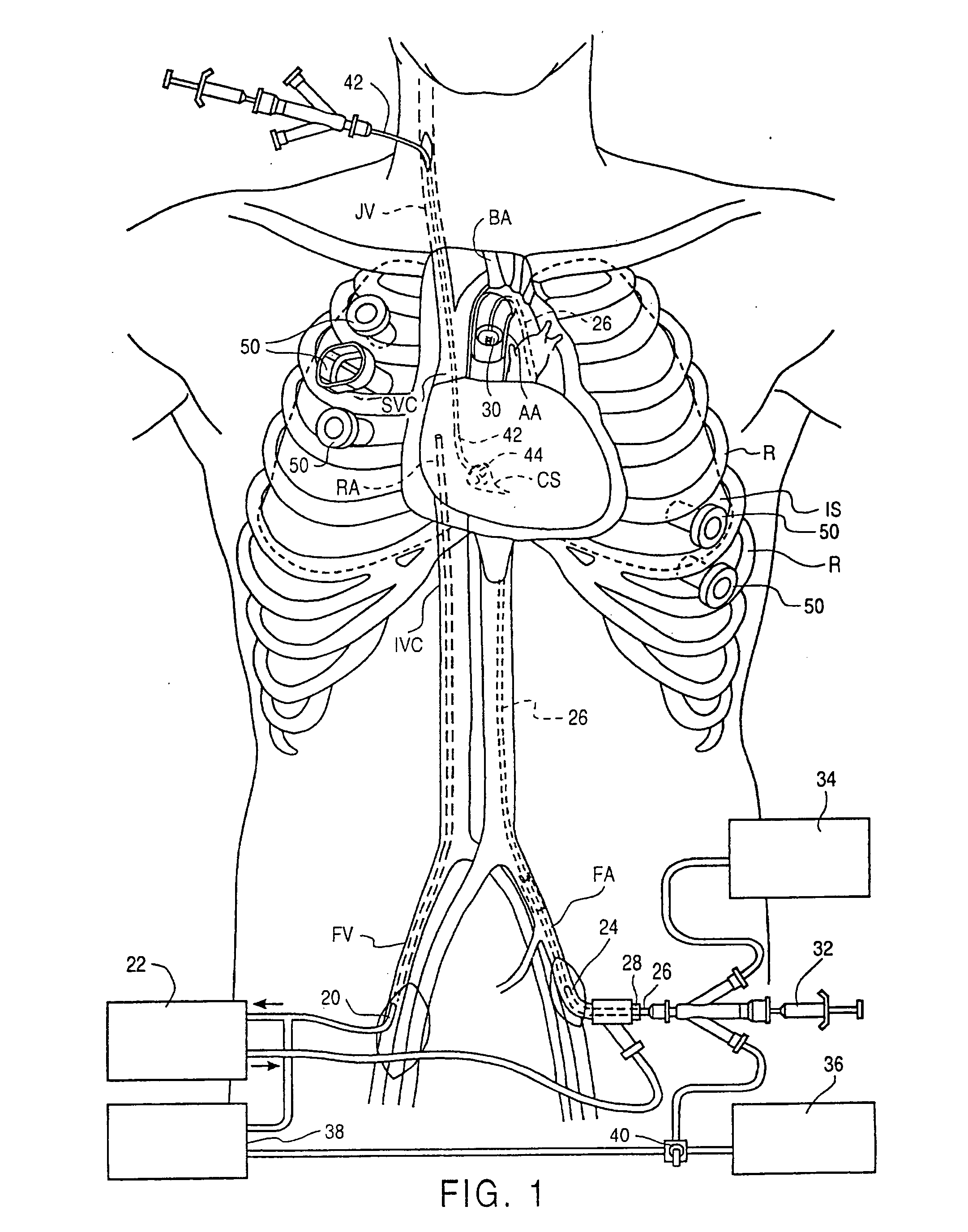
[0042] Referring to FIG. 1, an endovascular cardiopulmonary bypass (CPB) system useful in the method of the invention is illustrated as it is used in a patient. Additional aspects of such endovascular CPB systems suitable for use in the methods of the invention are described in the following patent applications, which are incorporated herein by reference: Ser. No. 08 / 282,192, filed Jul. 28, 1994, now U.S. Pat. No. 5,584,803, Ser. No. 08 / 612,341, filed Mar. 7, 1996, and Ser. No. 08 / 486,216, filed Jun. 7, 1995, now U.S. Pat. No. 5,766,151. The system includes a venous drainage cannula 20 placed into a femoral vein FV (or other suitable peripheral vein) and preferably having sufficient length to extend into the inferior vena cava IVC, the right atrium RA or the superior vena cava SVC. Venous drainage cannula 20 is connected to an extracorporeal CPB system 22, which filters and oxygenates the blood withdrawn from the patient. The system further includes an arterial return cannula 24 pla...


PUM


Abstract

A method of treatment of congestive heart failure comprises the steps of introducing an aortic occlusion catheter through a patient's peripheral artery, the aortic occlusion catheter having an occluding member movable from a collapsed position to an expanded position; positioning the occluding member in the patient's ascending aorta; moving the occluding member from the collapsed shape to the expanded shape after the positioning step; introducing cardioplegic fluid into the patient's coronary blood vessels to arrest the patient's heart; maintaining circulation of oxygenated blood through the patient's arterial system; and reshaping an outer wall of the patient's heart while the heart is arrested so as to reduce the transverse dimension of the left ventricle. The ascending aorta may be occluded and cardioplegic fluid delivered by means of an occlusion balloon attached to the distal end of an elongated catheter positioned transluminally in the aorta from a femoral, subclavian, or other appropriate peripheral artery.

Description

CROSS-REFERENCES TO RELATED APPLICATIONS [0001] This application is a Continuation of co-pending U.S. patent application Ser. No. 08 / 685,262, filed Jul. 23, 1996, which is a Continuation-In-Part of application Ser. No. 08 / 485,600, filed Jun. 7, 1995, which is a Continuation-In-Part of application Ser. No. 08 / 281,962, filed Jul. 28, 1994, which is a Continuation-In-Part of application Ser. No. 08 / 163,241, filed Dec. 6, 1993, now U.S. Pat. No. 5,571,2215, which is a Continuation-In-Part of application Ser. No. 08 / 023,778, filed Feb. 22, 1993, now issued as U.S. Pat. No. 5,452,733, the complete disclosures of which are hereby incorporated herein by reference for all purposes.BACKGROUND OF THE INVENTION [0002] In congestive heart failure or CHF, the heart has become so enlarged as a result of viral infection, myocardial infarction or other disease that it is unable to pump at a sufficient rate to maintain adequate circulation of blood throughout the body. As a result, blood backs up int...

Claims


Application Information

Patent Timeline
Patent Type & Authority Applications(United States)
IPC IPC(8): A61B19/00A61B17/00A61B17/02A61B17/04A61B17/06A61B17/122A61B17/28A61B17/30A61B17/34A61B18/00A61B18/14A61F2/24A61F2/958A61F7/00A61F7/10A61M1/00A61M1/36
CPCA61B17/00234A61M2205/366A61B17/0218A61B17/0293A61B17/0467A61B17/0469A61B17/06061A61B17/122A61B17/29A61B17/2909A61B17/3421A61B18/1492A61B19/26A61B19/52A61B2017/00053A61B2017/00243A61B2017/00247A61B2017/00575A61B2017/00632A61B2017/00867A61B2017/00907A61B2017/00946A61B2017/0237A61B2017/0243A61B2017/047A61B2017/0472A61B2017/0474A61B2017/0475A61B2017/0496A61B2017/2912A61B2017/2926A61B2017/2927A61B2017/2932A61B2017/2943A61B2017/306A61B2017/308A61B2017/3405A61B2017/3492A61B2018/00214A61B2018/00232A61B2018/00261A61B2018/00291A61B2018/00363A61B2018/00392A61B2018/00577A61B2018/00982A61B2019/461A61B2019/5206A61F2/2442A61F2/2445A61F7/10A61F2007/0001A61F2007/0054A61F2007/101A61M1/3621A61M25/1011A61M39/0247A61M2025/028A61M2039/027A61M2039/0279A61M2202/047A61M2205/3344A61M2205/3355A61B17/0057A61B90/36A61B90/50A61B2090/061A61B2090/306
Inventor STEVENS, JOHN H.BOLDUC, LEE R.BOYD, STEPHEN W.DONLON, BRIAN S.GIFFORD, HANSON S. III
Owner ETHICON INC
Why Eureka
Try Eureka