
Remotely enabled pacemaker and implantable subcutaneous cardioverter/defibrillator system

a technology of remote enabled pacemakers and implantable cardioverters, which is applied in the field of implantable medical devices, can solve the problems of prohibitively expensive implanting of currently available imds in all such patients, difficult to implant in some patients, and high cost of imds and implant procedures, so as to reduce pain and discomfort, and achieve safe and effective operations

Active Publication Date: 2006-10-26
MEDTRONIC INC
View PDF59 Cites 253 Cited by
  • Summary
  • Abstract
  • Description
  • Claims
  • Application Information

AI Technical Summary

Benefits of technology

The present invention provides a system that allows different medical devices to communicate with each other to coordinate the delivery of therapy. This can include an external device (AED), a subcutaneous device (SubQ ICD), and an implantable device (IMD). The system can continuously communicate and automatically initialize the devices to detect arrhythmic episodes and deliver coordinated therapy as needed. Additionally, the invention includes a communication system between a pain-suppressing device and a device that releases a bolus of pain suppression drug to reduce pain and discomfort associated with high voltage stimulation. The control system and computer-implemented software coordinate monitoring and communications between the external, subcutaneous, and implanted devices, ensuring safe and effective operations.

Problems solved by technology

Such devices are often difficult to implant in some patients (particularly children and thin, elderly patients) and typically require the sacrifice of 1 or 2 veins to implant the lead system because leads associated with implantation of an IMD utilize a transvenous approach for cardiac electrodes and lead wires.
Although IMDs and implant procedures are very expensive, most patients who are implanted have experienced and survived a sudden cardiac death episode because of interventional therapies delivered by the IMDs.
However, implanting currently available IMDs in all such patients would be prohibitively expensive.
Further, even if the cost factor is eliminated there is shortage of trained personnel and implanting resources.
Further, endocardial lead placement is not possible with patients who have a mechanical heart valve implant and is not generally recommended for pediatric cardiac patients.
There are technical challenges associated with the implantation of a SubQ ICD.
This is particularly because the SubQ ICD is limited to far-field sensing since there are no intracardial or epicardial electrodes in a subcutaneous system.
Further, sensing of atrial activation from subcutaneous electrodes is limited since the atria represent a small muscle mass and the atrial signals are not sufficiently detectable transthoracically.
Accordingly, the design of a SubQ ICD is a difficult proposition given the technical challenges to sense and detect arrhythmias.
Yet another challenge could be combining a SubQ ICD with an existing pacemaker (IPG) in a patient.
While this may be desirable in a case where an IPG patient may need a defibrillator, a combination implant of SubQ ICD and IPG may result in inappropriate therapy by the SubQ ICD, which may pace or shock based on spikes from the IPG.
The result can be over-counting beats from the atrium, ventricles or both; or, because of the larger pacing spikes, sensing of arrhythmic signals (which are typically much smaller in amplitude) may be compromised.
Additionally, the implanting of two or more devices in a patient can be challenging with respect to programming and coordinating therapies delivered by the devices.
Further, monitoring of the patient including the devices by use of conventional telemetry and diagnostics may pose additional burden on patient and device management resources.
Specifically, the operations of one device may be suspended when one another device is providing a life support therapy.

Method used


Image

  • Remotely enabled pacemaker and implantable subcutaneous cardioverter/defibrillator system
  • Remotely enabled pacemaker and implantable subcutaneous cardioverter/defibrillator system
  • Remotely enabled pacemaker and implantable subcutaneous cardioverter/defibrillator system

Examples


first embodiment

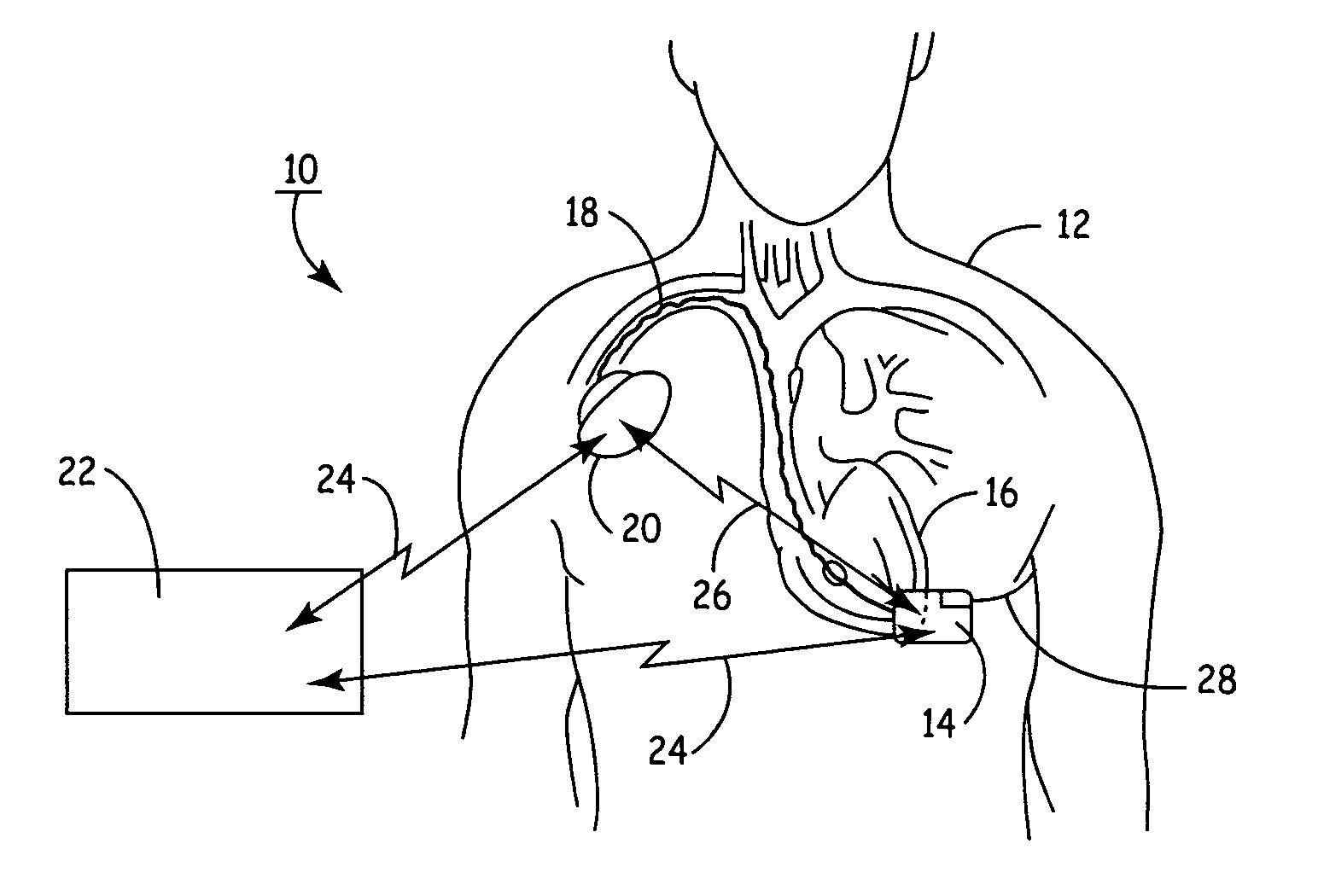
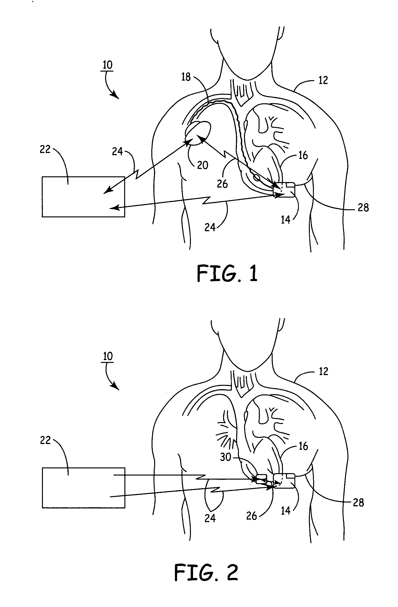
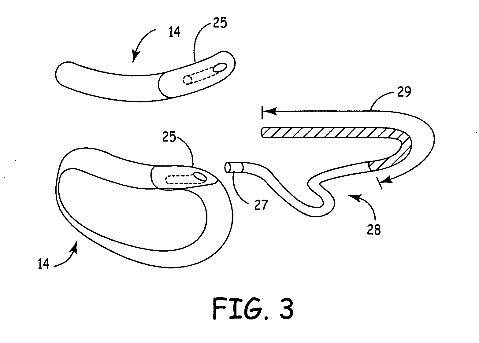
[0033]FIG. 1 illustrates the present invention, having a SubQ ICD 14 and IMD 20 implanted in patient 12. The SubQ ICD 14 is subcutaneously implanted outside the ribcage of patient 12 anterior to the cardiac notch. SubQ ICD 14 is shown coupled to subcutaneous lead 28. Lead 28 includes an electrode for subcutaneous sensing and cardioversion / defibrillation therapy delivery and is located transthoracically in relation to heart 16. Lead 28 is tunneled subcutaneously from the median implant pocket of SubQ ICD 14 laterally and posterially to the patient's back to a location opposite the heart such that the heart 16 is disposed between the SubQ ICD 14 and the distal electrode coil 29 (see FIG. 3). The implant location of device 14 and lead 28 is typically between the 3rd and 8th ribs.

[0034] IMD 20 is shown implanted pectorially in patient 12 and may take the form of any type of pacemaker / stimulator such as, but not limited to, a single chamber atrial pacemaker, a single chamber ventricular ...

second embodiment

[0037]FIG. 2 illustrates the present invention having SubQ ICD 14 and IMD 30 implanted in patient 12. The SubQ ICD 14 is subcutaneously implanted outside a patient's 12 ribcage similar to the description hereinabove.

[0038] IMD 30 is shown implanted epicardially in patient 12 and may take the form of any type of pacemaker / stimulator such as, but not limited to, a single chamber atrial pacemaker, a single chamber ventricular pacemaker, a dual chamber atrial / ventricular pacemaker, a bi-atrial pacemaker, a bi-ventricular pacemaker and the like. Cardiac leads / electrodes (not shown in FIG. 2) connect IMD 30 to the patient's heart 16 and may take the form of any typical lead configuration as is known in the art, such as, without limitation, epicardial right ventricular (RV) pacing leads, right atrial (RA) pacing leads, left ventricular pacing leads, unipolar or bipolar lead / electrode configurations, or any combinations of the above lead systems. Epicardial lead / electrode attachment to the ...


PUM


Abstract

Subcutaneous Implantable cardioverter-defibrillators (SubQ ICDS) are disclosed that are entirely implantable subcutaneously with minimal surgical intrusion into the body of the patient and provide distributed cardioversion-defibrillation sense and stimulation electrodes for delivery of cardioversion-defibrillation shock and pacing therapies across the heart when necessary. The SubQ ICD is implemented with other implantable and external medical devices and communicates to provide drugs and therapy in a coordinated and synergistic manner.

Description

FIELD OF THE INVENTION [0001] The present invention generally relates to an implantable medical device system, particularly comprising a subcutaneous implantable cardioverter-defibrillator (SubQ ICD) that is entirely implanted subcutaneously with no endovascular epicardial leads / electrodes. More specifically, the SCD is in bi-directional data communication and functional synergy with one or more implantable and / or externally mounted devices and, optionally to a transdermal drug delivery apparatus. BACKGROUND OF THE INVENTION [0002] Many types of implantable medical devices (IMDs) have been clinically implanted over the last twenty years that deliver relatively high-energy cardioversion and / or defibrillation shocks to a patient's heart when a malignant tachyarrhythmia, e.g., atrial or ventricular fibrillation, is detected. Cardioversion shocks are typically delivered in synchrony with a detected R-wave when fibrillation detection criteria are met, whereas defibrillation shocks are ty...

Claims


Application Information

Patent Timeline
Patent Type & Authority Applications(United States)
IPC IPC(8): A61N1/39
CPCA61N1/025A61N1/37211A61N1/3962A61N1/37288A61N1/37229A61N1/39622
Inventor MARKOWITZ, H. TOBYHETTRICK, DOUGLAS A.COMBS, WILLIAM J.SHELDON, TODD J.
Owner MEDTRONIC INC
Learn More