Four-dimensional (4D) image verification in respiratory gated radiation therapy

a technology of image verification and applied in the field of four-dimensional (4d) image verification in respiratory gated radiation therapy, can solve the problems of patient compliance, difficult identification of the location of the tumor, and inability to deliver the dos

Active Publication Date: 2008-02-07
SIEMENS HEALTHCARE GMBH +1
View PDF9 Cites 106 Cited by
  • Summary
  • Abstract
  • Description
  • Claims
  • Application Information

AI Technical Summary

Benefits of technology

[0014] Determining an optimal time point synchronization between the DRRs and the fluoroscopic images comprises: establishing a correspondence map between the DRRs and the fluoroscopic images; and minimizing an energy function to find an optimal mapping between the DRRs and the fluoroscopic images, the optimal mapping representing the optimal time point synchronization.
[0024] Determining an optimal time point synchronization between the DRRs and the fluoroscopic images comprises: establishing a correspondence map between the DRRs and the fluoroscopic images; and minimizing an energy function to find an optimal mapping between the DRRs and the fluoroscopic images, the optimal mapping representing the optimal time point synchronization.

Problems solved by technology

Notwithstanding these recent advances, miss-delivery of the dose can occur because of inter-fraction and intra-fraction target motions.
The variation in the size of the motion as well as the pattern of the motion can make the identification of the location of a tumor difficult.
A drawback of this method is that it requires patient compliance.
It is technically challenging to implement linear accelerator (LINAC)-based tracking using a dynamic multi-leaf collimator mainly due to quality assurance issues.
However, a challenge of respiratory gated treatment is to adapt the gating intervals based on current breathing patterns of the patient.
However, having an external signal is not equivalent to knowing the location of the tumor at all times. In addition, exploiting the external signal blindly can cause significant errors.
Thus, a drawback of this method is the variability of the correlation between the amplitude of the external signal (e.g., marker position) and the position of the internal target (e.g., tumor).

Method used


Image

  • Four-dimensional (4D) image verification in respiratory gated radiation therapy
  • Four-dimensional (4D) image verification in respiratory gated radiation therapy
  • Four-dimensional (4D) image verification in respiratory gated radiation therapy

Examples


Embodiment Construction

[0039] In the following description, we will outline a four-dimensional (4D) image verification method according to an exemplary embodiment of the present invention that addresses the variability of the correlation between the amplitude of an external signal (e.g., a respiratory belt around a patient's abdominal or thoracic area or marker position placed on the patient's thorax) and the position of an internal target. The method is divided into four phases, which are described hereinafter with reference to the accompanying figures.

Planning Phase

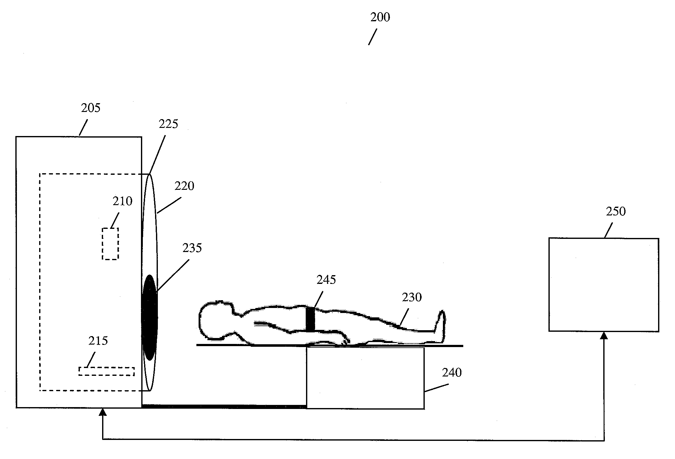
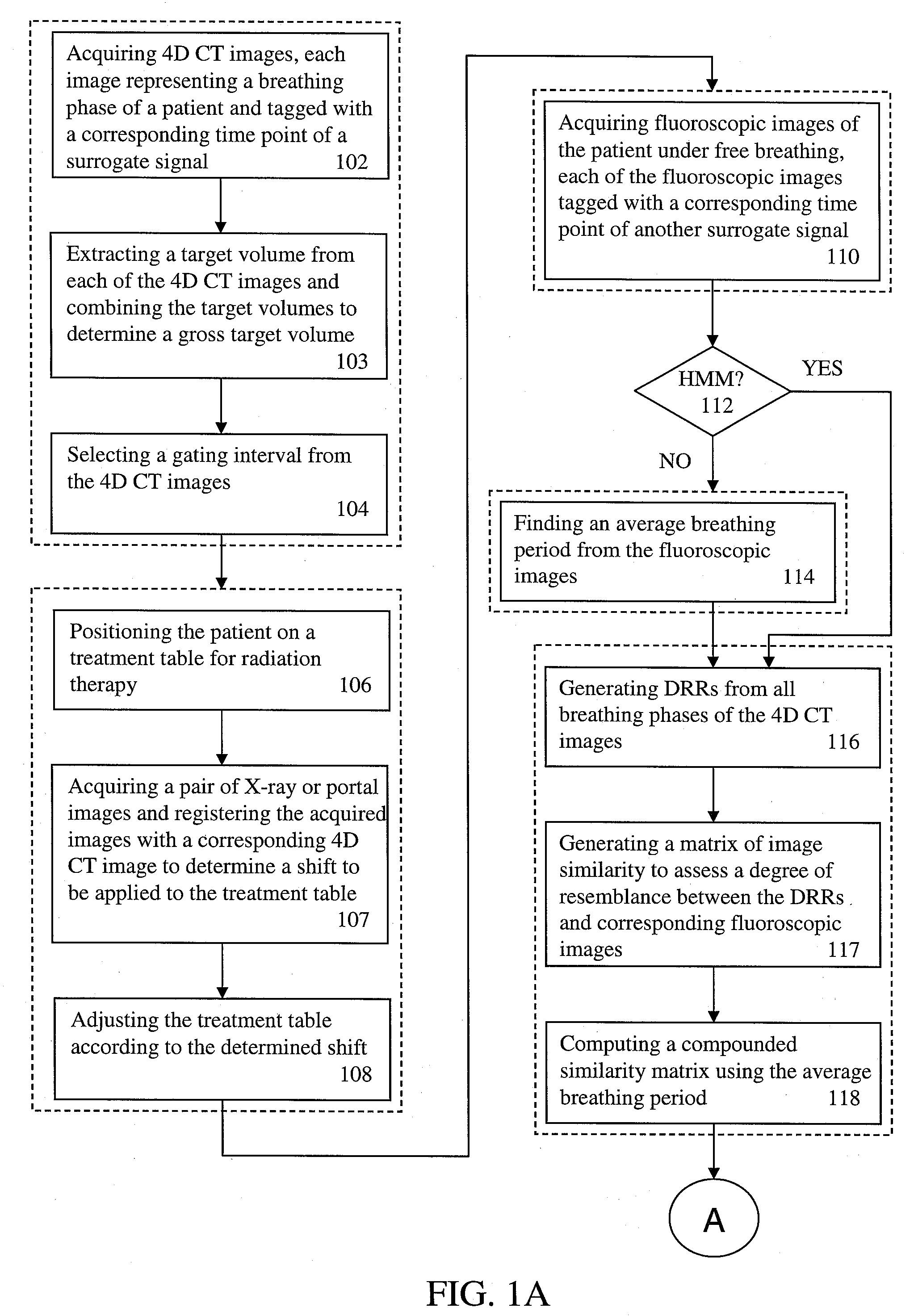
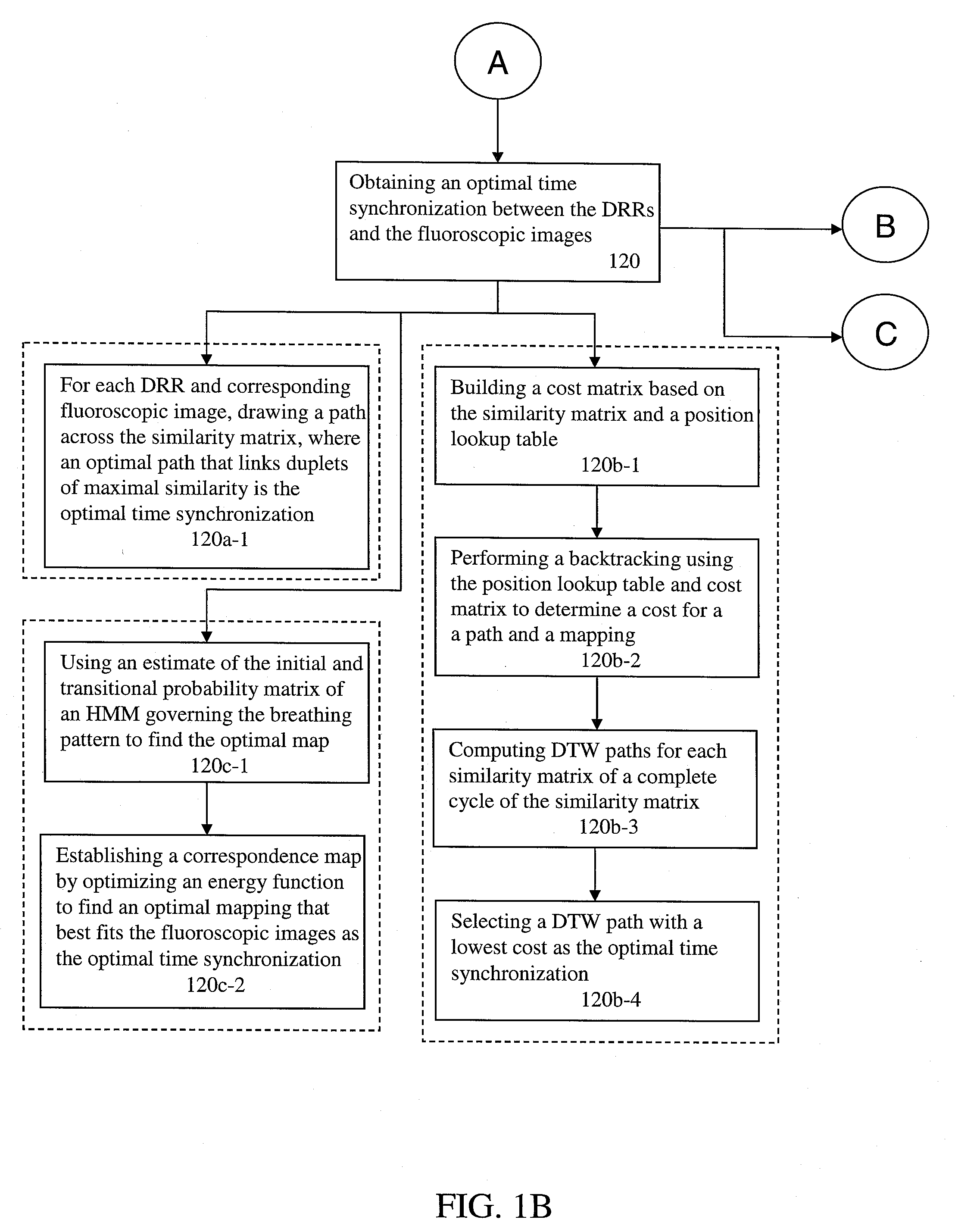
[0040] As shown in FIG. 1A, during the planning phase, a 4D computed tomography (CT) acquisition of a patient is made (102). The acquisition is made under a protocol that produces time resolved volumetric images at various phases of breathing (e.g., 8-12 phases). In other words, each image represents a breathing phase during a single breath of the patient. During this time, a surrogate signal is acquired, the surrogate signal being the acq...


PUM


Abstract

A method for four-dimensional (4D) image verification in respiratory gated radiation therapy, includes: acquiring 4D computed tomography (CT) images, each of the 4D CT images representing a breathing phase of a patient and tagged with a corresponding time point of a first surrogate signal; acquiring fluoroscopic images of the patient under free breathing, each of the fluoroscopic images tagged with a corresponding time point of a second surrogate signal; generating digitally reconstructed radiographs (DRRs) for each breathing phase represented by the 4D CT images; generating a similarity matrix to assess a degree of resemblance in a region of interest between the DRRs and the fluoroscopic images; computing a compounded similarity matrix by averaging values of the similarity matrix across different time points of the breathing phase during a breathing period of the patient; determining an optimal time point synchronization between the DRRs and the fluoroscopic images by using the compounded similarity matrix; and acquiring a third surrogate signal and turning a treatment beam on or off according to the optimal time point synchronization.

Description

CROSS-REFERENCE TO RELATED APPLICATION [0001] This application claims the benefit of U.S. Provisional Application No. 60 / 835,528, filed Aug. 4, 2006, the disclosure of which is incorporated by reference herein in its entirety.BACKGROUND OF THE INVENTION [0002] 1. Technical Field [0003] The present invention relates to four-dimensional (4D) image verification in respiratory gated radiation therapy. [0004] 2. Discussion of the Related Art [0005] Radiation therapy is a treatment modality with the primary goal of precisely delivering a prescribed dose of radiation to a tumor while minimizing an amount of the dose delivered to surrounding healthy tissues and critical structures. Recent technological advances in radiation therapy have enabled the delivery of a highly conformal radiation dose distribution to a morphologically complex target volume. Notwithstanding these recent advances, miss-delivery of the dose can occur because of inter-fraction and intra-fraction target motions. [0006] ...

Claims


Application Information

Patent Timeline
Patent Type & Authority Applications(United States)
IPC IPC(8): A61B6/00
CPCA61B5/113A61B5/1135A61B6/032A61B6/541A61N5/1037A61N2005/1062A61N5/1064A61N5/1068A61N2005/1059A61N2005/1061A61N5/1049
Inventor KHAMENE, ALIFLORIN, CHARLES HENRICELI, JUAN CARLOSOFSTAD, BARBARAROUSSON, MIKAELSAUER, FRANKSCHALLER, CHRISTIAN
Owner SIEMENS HEALTHCARE GMBH
Why Eureka
Try Eureka