Image processing method, apparatus, and program

a tumor contour and image processing technology, applied in image analysis, image enhancement, instruments, etc., can solve the problems of degrading the detection capability of contours, difficult tumor area detection through binarization process, and easy local change of contour extraction, so as to enhance the detection capability of tumor contours

Active Publication Date: 2008-02-21
FUJIFILM CORP
View PDF12 Cites 57 Cited by
  • Summary
  • Abstract
  • Description
  • Claims
  • Application Information

AI Technical Summary

Benefits of technology

[0013] The present invention has been developed in view of the circumstances described above, and it is an object of the present invention to provide an image processing method, apparatus, and program capable of enhancing detection capabilities for tumor contours.
[0014] It is a further object of the present invention to provide an image processing method, apparatus, and program capable of enhancing detection capabilities in contour detection of particular regions of different types of images to which the technology of the present invention is generally applicable.

Problems solved by technology

Thus, the method proposed in Patent Document 2, in which a tumor area is determined from the regions extracted through binarization process, may not correctly detect tumor areas in many occasions. FIGS. 2A to 2D and FIGS. 3A to 3D illustrate one-dimensional intensity profiles on straight lines passing through liver tumors.
However, in FIGS. 2D, 3B, and 3D, the distinction between the inside and outside of the tumor may not be made only by the intensity, so that detection of the tumor area through binarization process is difficult.
The method for obtaining a contour of a target region based on the local contrast (edge information) of an image disclosed in Patent Document 3 or Non-patent Document 2 has a problem that the contour extraction is susceptible to changes in local contrast, and as the contrast between the target region and background area becomes small, the contour detection capability is degraded.
Further, the contour is obtained as an ellipse, so that an accurate contour may not be obtained for a tumor having an irregular shape, thereby the accuracy of the image diagnosis may be degraded.

Method used


Image

  • Image processing method, apparatus, and program
  • Image processing method, apparatus, and program
  • Image processing method, apparatus, and program

Examples


first embodiment

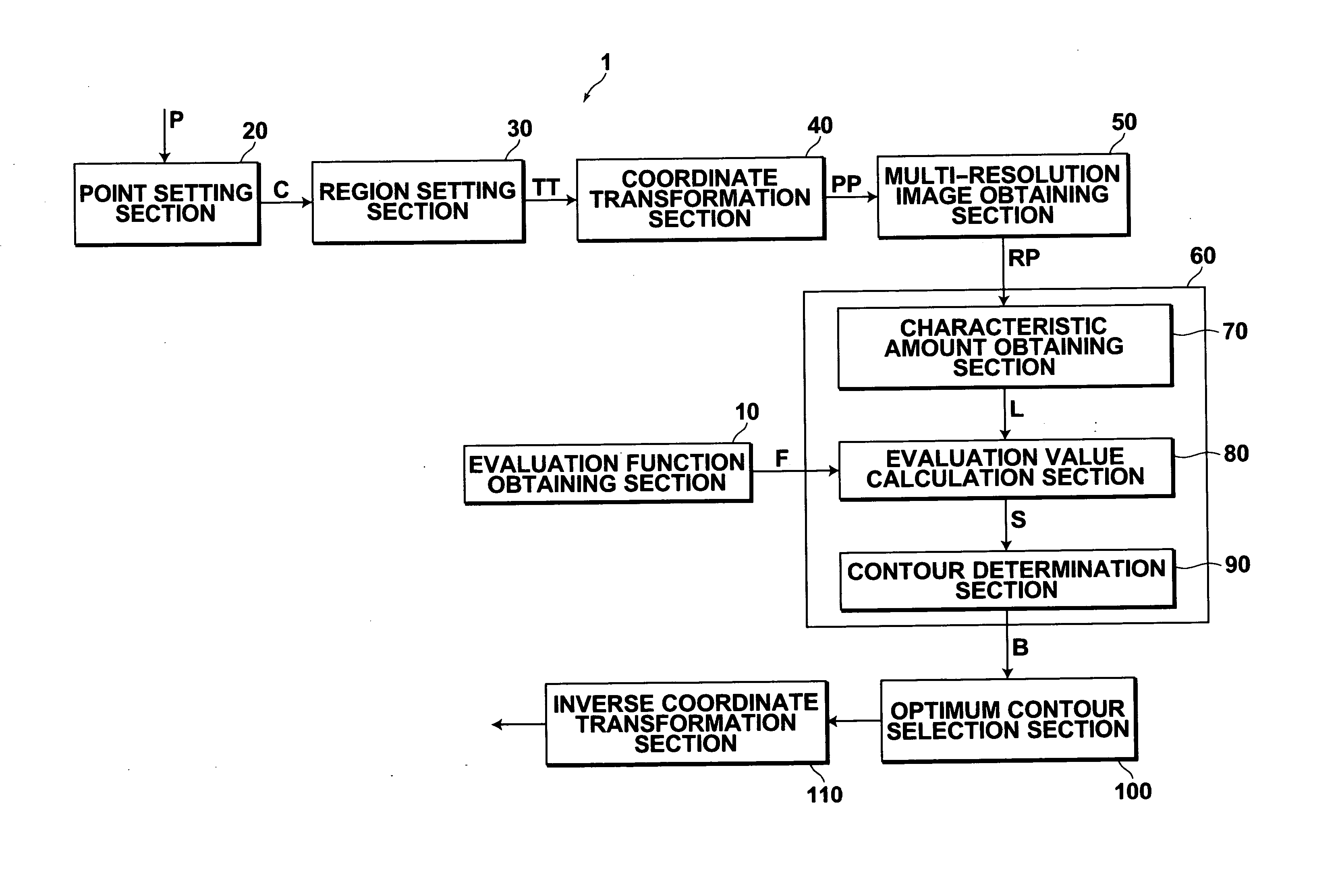
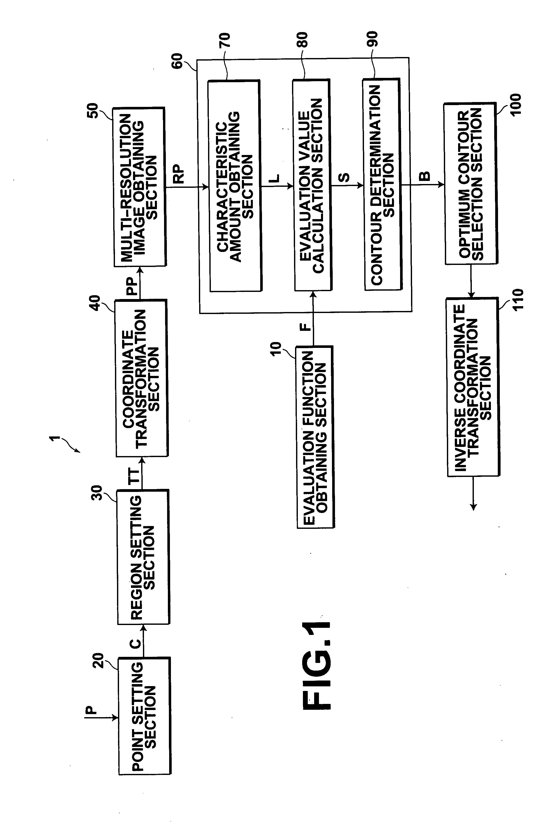
[0065] Hereinafter, the image processing apparatus of the present invention in which the apparatus is applied to determining a contour of a tumor area in a medical image, will be described with reference to the accompanying drawings. The configuration of an image processing apparatus 1 illustrated in FIG. 1 is realized by executing an image processing apparatus program, which is stored in an auxiliary storage, on a computer (e.g., personal computer, or the like). The program for realizing the image processing apparatus is recorded on an information recording medium, such as a CD-ROM, or distributed through a network, such as the Internet, and installed on the computer.

[0066] The image processing apparatus 1 is an apparatus for determining a contour of a tumor area in a medical image P obtained by a CT system or the like. As illustrated in FIG. 1, the image processing apparatus 1 includes: an evaluation function obtaining section 10 for performing machine learning in advance for lear...

second embodiment

[0113] When determining the contour of a tumor area in a three-dimensional medical image using the image processing apparatus described above, machine learning for learning a characteristic amount of each voxel in a plurality of three-dimensional sample images, each including a tumor area with a known contour, is performed by the evaluation function obtaining section 210 to obtain an evaluation function capable of evaluating whether or not each voxel is a voxel representing the contour based on the characteristic amount thereof. Here, as for the characteristic amount, intensity information of an adjacent region to a target voxel, for example, combinations of intensity values of a plurality of different voxels within a cubic region of five pixels in x, y and z axis directions centered on the target voxel may be used. Then, an arbitrary point C2 in a three-dimensional coordinate system is determined within the tumor area in the three-dimensional medical image by the point setting sec...


PUM


Abstract

Machine learning for learning a characteristic amount of each pixel in a plurality of sample images, each including a particular region with a known contour, is performed in advance to obtain an evaluation function capable of evaluating whether or not each pixel is a pixel representing the contour based on the characteristic amount thereof. An arbitrary point is set within a particular region in an input image, and a discrimination region including the particular region is set in the input image with reference to the arbitrary point. A characteristic amount is obtained from each pixel within the discrimination region, then based on the characteristic amount, an evaluation value indicating whether or not each pixel within the discrimination region is a pixel representing the contour is calculated using the evaluation function, and the contour of the particular region in the input image is determined based on the evaluation value.

Description

BACKGROUND OF THE INVENTION [0001] 1. Field of the Invention [0002] The present invention relates to a method, apparatus, and program for detecting a contour of a particular region in an image. More specifically, the present invention relates to an image processing method, apparatus, and program for detecting a contour of a tumor area in a medical image. [0003] 2. Description of the Related Art [0004] In the medical field, computer aided diagnosis (CAD), in which an image analysis is performed on a digital medical image (hereinafter, simply referred to as “medical image”) for diagnosis, is used in order to improve diagnostic accuracy and to reduce the burden on the medical staff, including doctors, who performs radiological image reading. [0005] One of the known CAD systems, for example, automatically analyzes a malignancy grade of a tumor based on radiological information, such as the size of a lesion in a medical image. U.S. Pat. No. 6,738,499 (Patent Document 1) proposes a method...

Claims


Application Information

Patent Timeline
Patent Type & Authority Applications(United States)
IPC IPC(8): G06K9/62G06V10/26G06V10/50G06V10/774
CPCG06K9/34G06K9/4609G06K9/4642G06T2207/30056G06K2209/05G06T7/0012G06K9/6256G06V10/26G06V10/443G06V10/50G06V2201/03G06V10/774G06F18/214
Inventor LI, YUANZHONG
Owner FUJIFILM CORP
Why Eureka
Try Eureka