Compact gynecological observation system for examination, imaging analysis and treatment

Inactive Publication Date: 2008-02-21
O D F MEDICAL
View PDF10 Cites 16 Cited by
  • Summary
  • Abstract
  • Description
  • Claims
  • Application Information

AI Technical Summary

Benefits of technology

[0025] g. a front window, located on the cover, through which the imaging assembly is able to ca

Problems solved by technology

Medical practice of the prior art has failed to provide a comfortable way to carry out gynecological examinations.
This method of examination is not only uncomfortable, but is also considered by some as humiliating and many patients avoid their periodic gy

Method used


Image

  • Compact gynecological observation system for examination, imaging analysis and treatment
  • Compact gynecological observation system for examination, imaging analysis and treatment
  • Compact gynecological observation system for examination, imaging analysis and treatment

Examples


Embodiment Construction

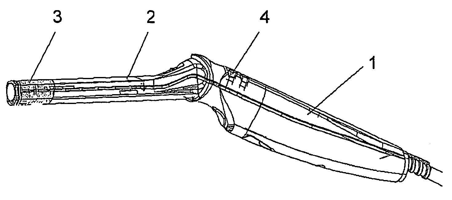
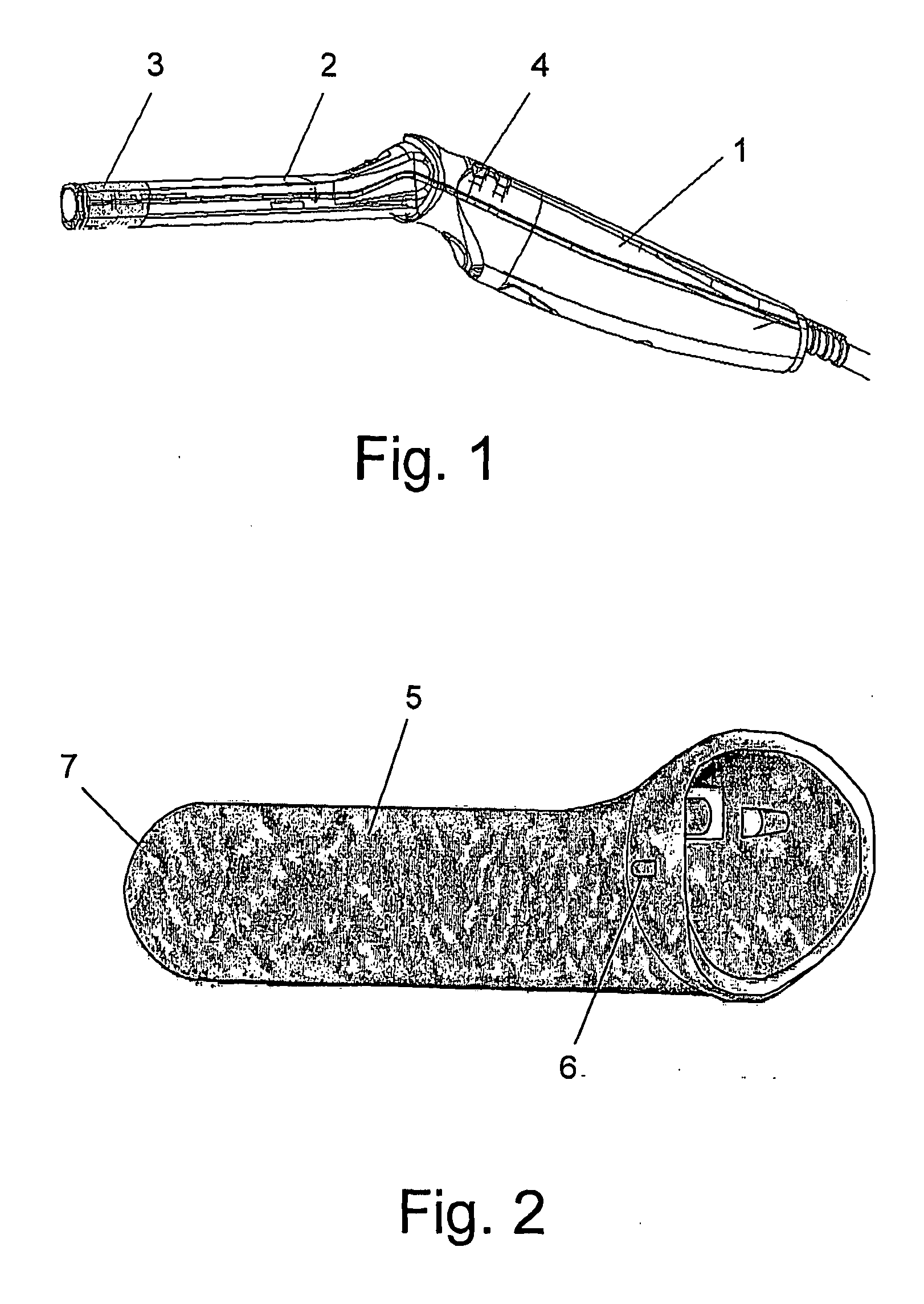
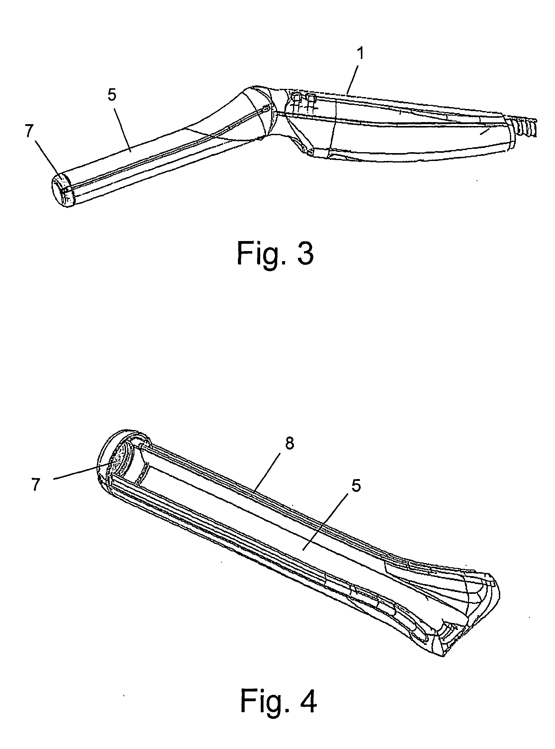
[0069] For a better understanding of the invention and to show how it may be carried into effect, reference will now be made, purely by way of example, to the accompanying drawings. With specific reference to the drawings in detail, it is stressed that the particulars shown are by way of example and for purposes of illustrative discussion of preferred embodiments of the present invention only, and are presented for the purpose of providing what is believed to be the most useful and readily understood description of the principles and conceptual aspects of the invention. In this regard, no attempt is made to show structural details of the invention in more detail than is necessary for a fundamental understanding of the invention. From the description taken together with the drawings it will be apparent to those skilled in the art how the several forms of the invention may be embodied in practice.

[0070] The device of the invention is designed to be introduced into the vagina for rout...


PUM


Abstract

The invention is a medical imaging device designed for insertion into the vagina for medical examination, imaging, image analysis and treatment of the interior of the vagina. The device comprises a front assembly, optimally shaped to match the shape of the vaginal canal; imaging and illumination assemblies; a handle; and an optimally shaped cover designed to fit tightly over and hermetically isolate at least that part of the front assembly that is inserted into the vaginal canal from the surrounding tissue and body fluids. The cover of the device of the invention can comprise one or more passages extending through it from its proximal end to its distal end enabling passage of one or more surgical tools and/or liquids and/or gases, from outside of the vaginal canal to the location of the scene imaged by the imaging assembly. The invention is also a medical imaging system comprising a medical imaging device of the invention, a computer and/or display station, and communication components. The medical imaging system may comprise operation software and image processing software.

Description

FIELD OF THE INVENTION [0001] The present invention relates to the field of medical devices. Specifically, the invention relates to the field of medical devices for performing imaging, image analysis, and treatment. More specifically, the invention relates to a medical imaging device intended to be introduced into the vagina for medical examination, imaging, image analysis, and treatment. BACKGROUND OF THE INVENTION [0002] Examination of the vaginal canal and cervix is a common procedure conducted by gynecologists world-wide on women of almost all ages. This procedure is conducted for detecting abnormalities in the vagina or cervix, including symptoms of cervical cancer and its pre-cancerous stages. [0003] Cervical cancer is one significant abnormality in the vagina and cervix which requires detection since early detection of cervical cancer can bring to early treatment, which may result in healing of the cancer and saving the patient's life. [0004] Additional abnormalities can be d...

Claims


Application Information

Patent Timeline
IPC IPC(8): A61B1/303
CPCA61B1/00082A61B1/00135A61B1/31A61B1/018A61B1/303A61B1/00142
Inventor GAL, EHUDBERINSKY-LITIOUGA, GENNADIYPINTEL, OFER
Owner O D F MEDICAL
Why Eureka
Try Eureka