Method of automated image color calibration

a color calibration and image technology, applied in the field of medical imaging and image processing, can solve the problems of difficult to extract the color property of acetowhite lesions properly, difficult to accurately assess the color information, and difficult to achieve the effect of correct extraction of the color property of acetowhite lesions,

Inactive Publication Date: 2008-10-02
STI MEDICAL SYST
View PDF12 Cites 45 Cited by
  • Summary
  • Abstract
  • Description
  • Claims
  • Application Information

AI Technical Summary

Problems solved by technology

Consequently, the color of the cervical epithelium may look very different (including normal and abnormal findings) in cervical images acquired with different instruments or at different times. This makes the assessment of the color information very challenging, even for an expert.
On the other hand, lack of color calibration makes it very difficult to extract the color property of the acetowhite lesions properly.
Non-uniform illumination and light distribution also has been a major obstacle in extracting lesion margins and blood vessel structures compared to the colposcopic annotations.

Method used


Image

  • Method of automated image color calibration
  • Method of automated image color calibration
  • Method of automated image color calibration

Examples


Embodiment Construction

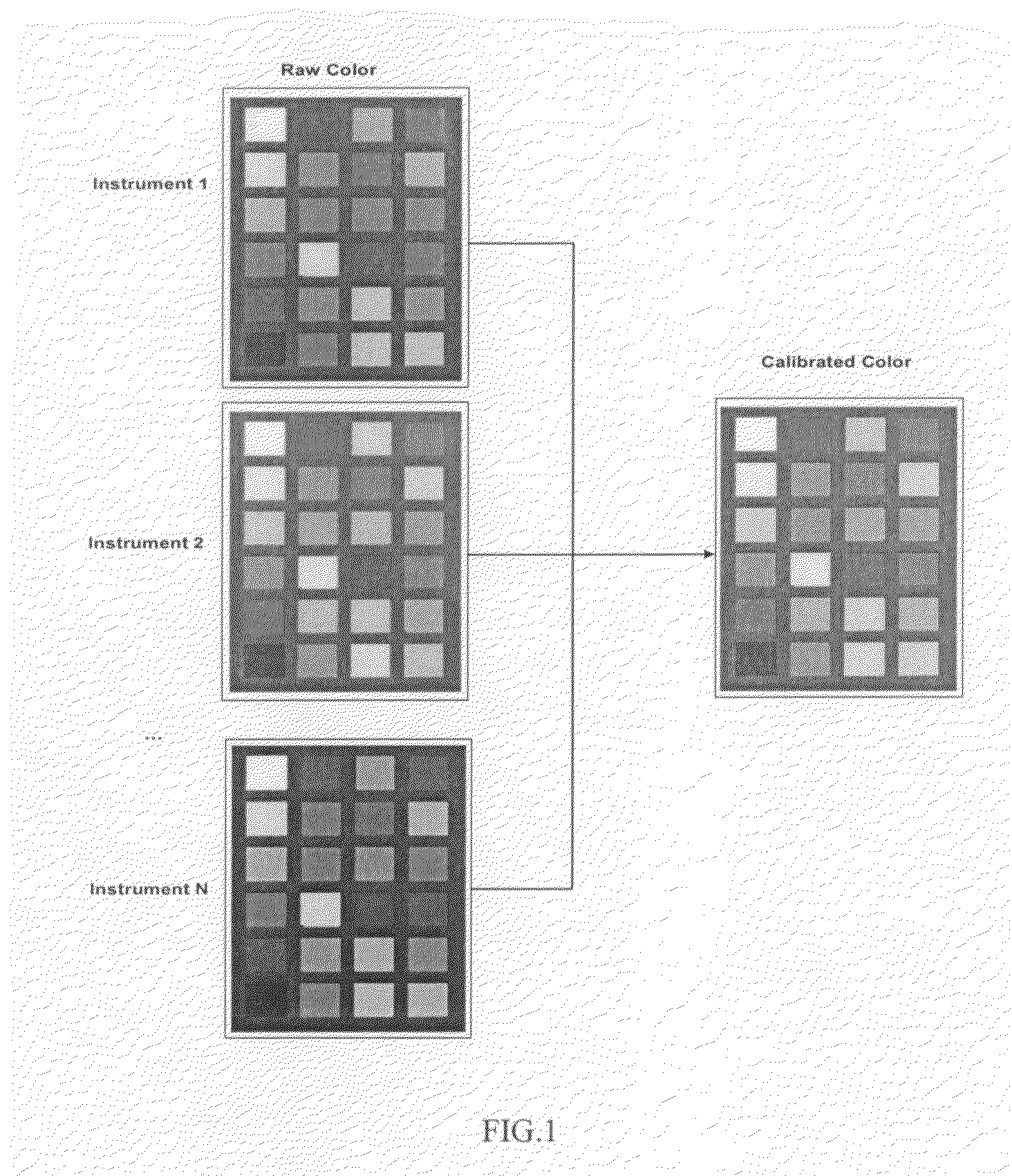
[0028]The presently preferred embodiment of the invention provides a method and an apparatus for mapping the color appearance of the images taken with different instruments at different times and locations and transforms them into a standard color space with normalized light distribution. FIG. 1 shows the concept of color calibration: mapping the raw color space of different instruments into a standard color space. The method preferably is performed in the following steps described in more detail below:[0029]1. Collecting raw cervical images and calibration data;[0030]2. Applying gray balancing to both the raw cervical images and calibration data using the gray balance algorithm;[0031]3. Performing color transformation to correct for inaccurate color in the gray balanced raw cervical image using a color correction matrix calculated from the gray balanced calibration data.

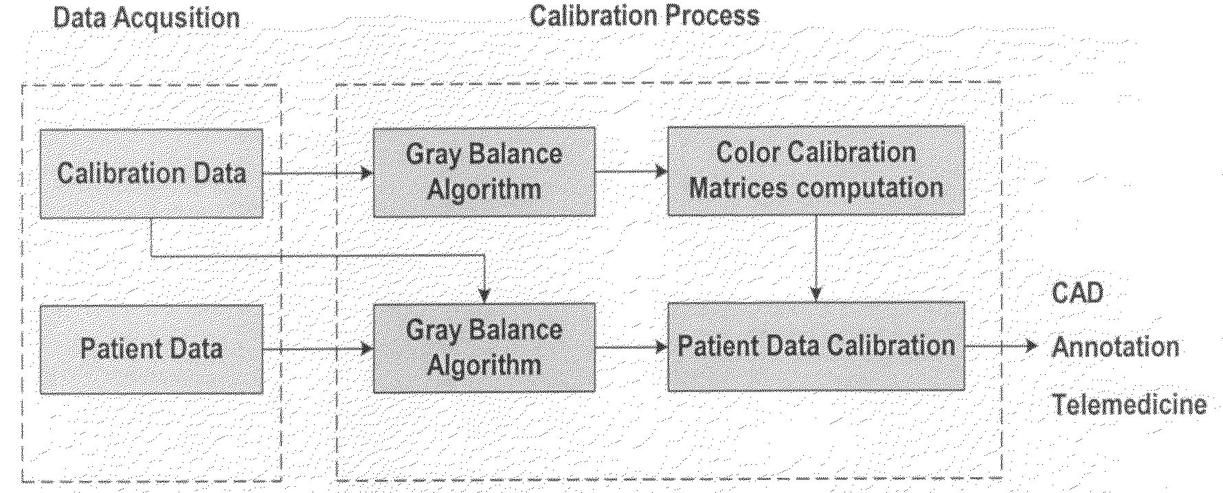
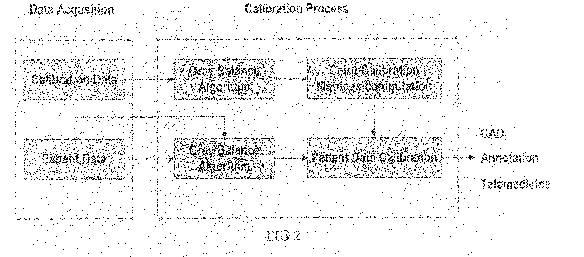
[0032]FIG. 2 shows the entire calibration procedure proposed for the colposcopic image calibration.

(1) Collection...


PUM


Abstract

A method of automated image calibration that corrects for non-uniform illumination and calibrates color that is simple, fast, automated, accurate and reliable. A gray balance algorithm is applied to correct for non-uniform illumination and a color calibration algorithm is then applied to calibrate the human subject data. The system has been applied in multiple clinical sites with different instruments.

Description

[0001]This application claims priority to U.S. provisional patent application 60 / 919,255 filed on Mar. 19, 2007.TECHNICAL FIELD[0002]This invention generally relates to medical imaging and image processing. The invention relates more specifically to correction for non-uniform illumination and color calibration of medical images for use in Computer-Aided-Diagnosis / Detection (CAD) systems.BACKGROUND ART[0003]Although this invention is being disclosed in connection with cervical cancer, it is applicable to many other areas of medicine. Uterine cervical cancer is the second most common cancer in women worldwide, with nearly 500,000 new cases and over 270,000 deaths annually (IARC, “Globocan 2002 database,” International agency for research in cancer, 2002, incorporated herein by reference). Because invasive disease is preceded by pre-malignant Cervical Intraepithelial Neoplasia (CIN), if detected early and treated adequately, cervical cancer can be universally prevented (D. G. Ferris, J...

Claims


Application Information

Patent Timeline
Patent Type & Authority Applications(United States)
IPC IPC(8): G06K9/00
CPCH04N1/00045H04N1/401H04N1/6033H04N1/6086
Inventor LI, WENJINGSOTO-THOMPSON, MARCELOGUSTAFSSON, ULFLANGE, HOLGER
Owner STI MEDICAL SYST
Why Eureka
Try Eureka