Tracking region-of-interest in nuclear medical imaging and automatic detector head position adjustment based thereon

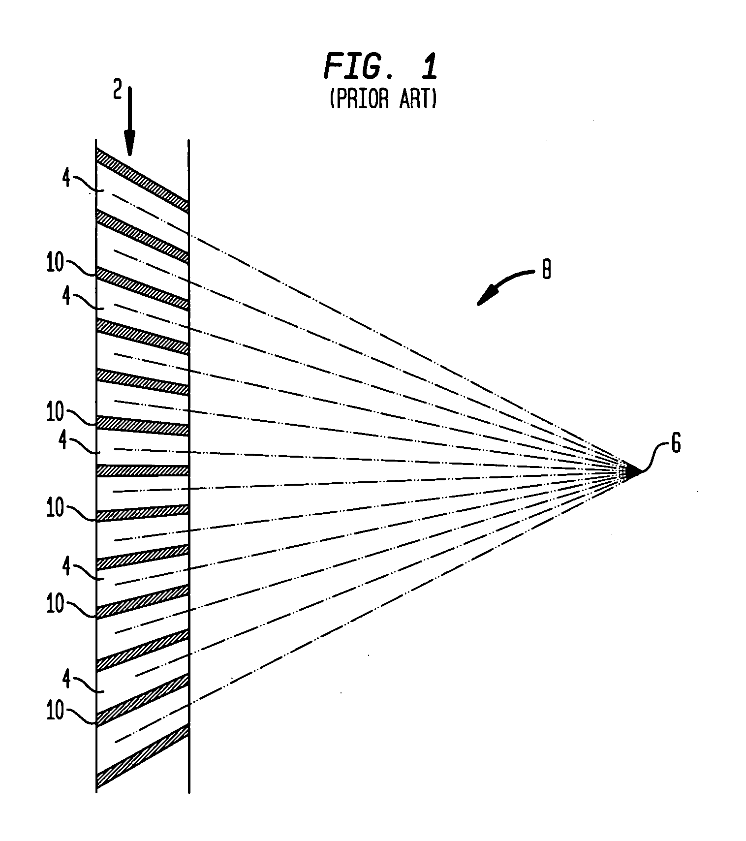
a technology of medical imaging and tracking region, applied in the field of nuclear medicine, can solve the problems of inability to adjust the design of the collimator with respect to length, septa, dimensions and focal or parallel nature of the collimator hole, time-consuming and not necessarily consistent,

Active Publication Date: 2008-11-06
SIEMENS MEDICAL SOLUTIONS USA INC
View PDF2 Cites 22 Cited by
  • Summary
  • Abstract
  • Description
  • Claims
  • Application Information

AI Technical Summary

Problems solved by technology

Collimator design conventionally is non-adaptive, meaning that the design of the collimator with respect to length, septa, dimensions and focal or parallel nature of the collimator holes cannot be adjusted.
In this case, sub-optimal image data will be acquired from view angle N. To avoid this result, it would be necessary for the technician to make manual adjustments at each different view angle, which would be time consuming and not necessarily consistent from view to view.

Method used


Image

  • Tracking region-of-interest in nuclear medical imaging and automatic detector head position adjustment based thereon
  • Tracking region-of-interest in nuclear medical imaging and automatic detector head position adjustment based thereon
  • Tracking region-of-interest in nuclear medical imaging and automatic detector head position adjustment based thereon

Examples


Embodiment Construction

[0013]The present invention will now be described and disclosed in greater detail. It is to be understood, however, that the disclosed embodiments are merely exemplary of the invention and that the invention may be embodied in various and alternative forms. Therefore, specific structural and functional details disclosed herein are not to be interpreted as limiting the scope of the claims, but are merely provided as an example to teach one having ordinary skill in the art to make and use the invention.

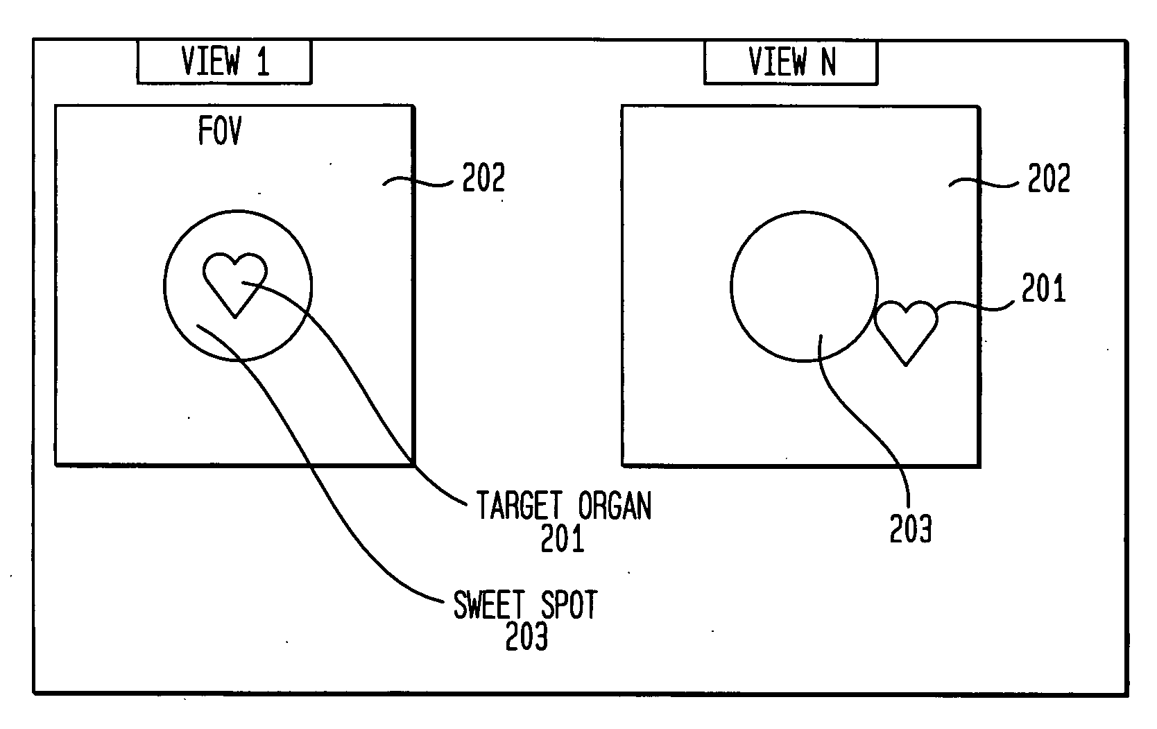
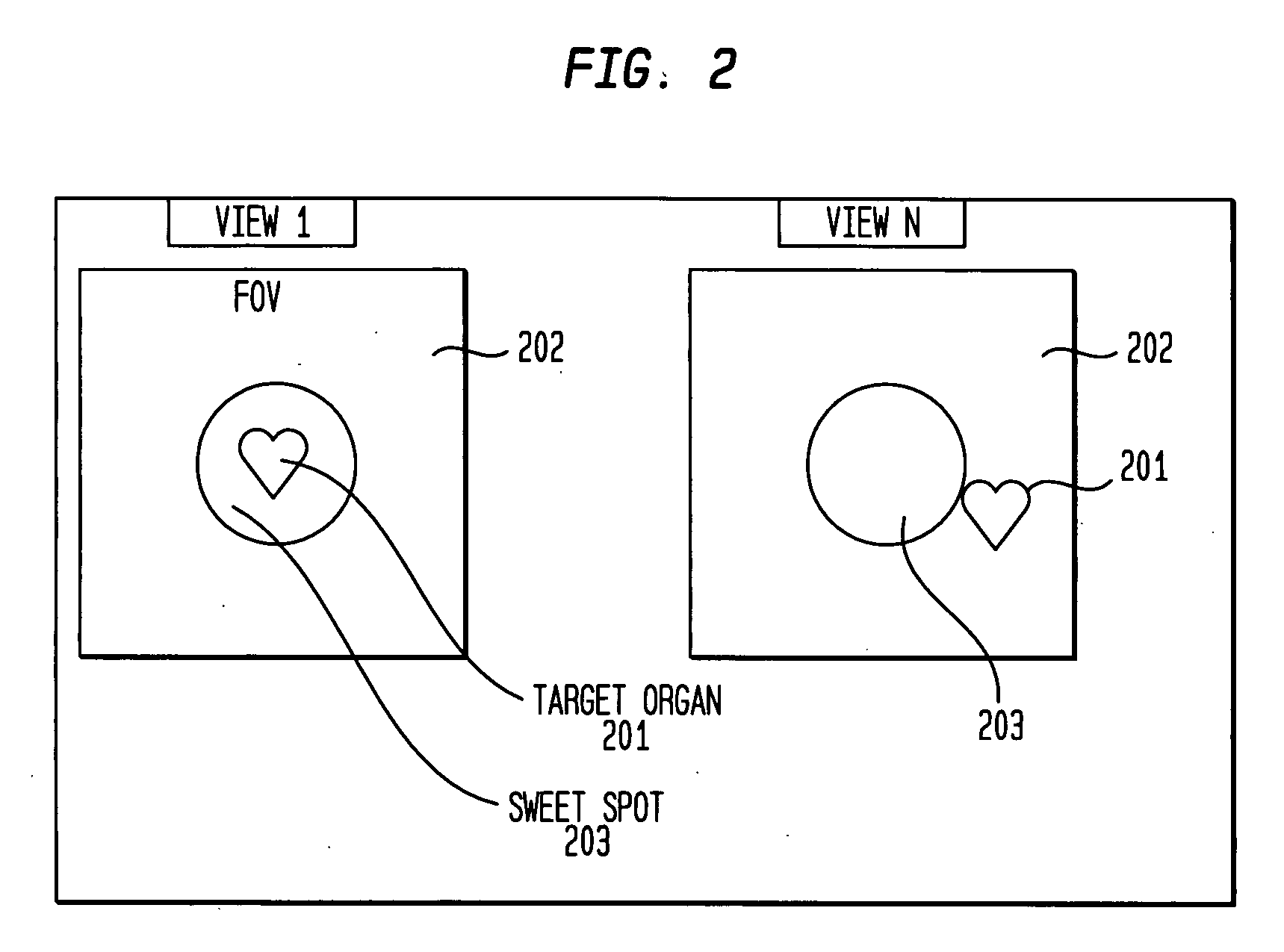
[0014]The invention is based on the fact that the dynamic “wash-in” and “wash-out” of a radiopharmaceutical into tissue, as well as natural organ motion such as the beating of the heart or the movement of the diaphragm during respiration, respectively gives rise to a concentration change in tissue or to a positional change of radioactive concentration c. Thus, in general the radioactive concentration c can be expressed as a function of position and time, or c=c({right arrow over (r)},t)...


PUM


Abstract

A method and system for automatically identifying and tracking a ROI over all planar acquisition view angles of a nuclear imaging system, such as a gamma camera used for SPECT or planar imaging. Temporal intensity variation in emission projection imaging is measured to identify a region of interest (ROI) such as the myocardium. The method automatically tracks the location of the ROI over different planar view angles and adapts detector head orbit and positioning to bring the ROI within a predefined preferred area or so-called “sweet spot” within the FOV of a collimator attached to the front of a scintillation detector surface. After initial positioning of the detector head by the user, the system automatically tracks the ROI location as the detector head(s) rotate about the patient and re-position the detector head(s) appropriately to maintain the ROI within the optimal collimation area of the detector FOV.

Description

FIELD OF THE INVENTION[0001]The present invention generally relates to nuclear medicine, and systems for obtaining nuclear medicine images. In particular, the present invention relates to a method and apparatus for identifying a region-of-interest (ROI) in a nuclear medical image scan, such as an organ or particular region of a patient undergoing imaging, and automatically tracking the identified ROI over different view angles and adjusting detector head positioning so that the identified ROI remains in an optimal area of the field of view (FOV) of the detector head, in particular with respect to a particular collimator being used in the imaging application.BACKGROUND OF THE INVENTION[0002]Nuclear medicine is a unique medical specialty wherein radiation is used to acquire images which show the function and anatomy of organs, bones or tissues of the body. Radiopharmaceuticals are introduced into the body, either by injection or ingestion, and are attracted to specific organs, bones o...

Claims


Application Information

Patent Timeline
Patent Type & Authority Applications(United States)
IPC IPC(8): G01T1/166G06K9/00
CPCA61B6/469G01T1/1648G06T7/0081G06T7/2006G06T2207/10088G06T2207/30048G06T7/11G06T7/215
Inventor VIJA, A. HANS
Owner SIEMENS MEDICAL SOLUTIONS USA INC
Why Eureka
Try Eureka