Caspase-3 Substrate Comprising Imaging Agents

a substrate and imaging agent technology, applied in the direction of tetrapeptide ingredients, enzymes, radiation therapy, etc., can solve the problems of insufficient cell death, inability to regulate apoptosis, unsuitable clinical decision-making in patients with acute coronary syndrome, etc., and achieve good blood clearance and rapid imaging

Inactive Publication Date: 2008-11-20
GUILBERT BENEDICTE +5
View PDF3 Cites 6 Cited by
  • Summary
  • Abstract
  • Description
  • Claims
  • Application Information

AI Technical Summary

Benefits of technology

[0011]There is therefore still a need for an apoptosis imaging agent which permits rapid imaging (eg. within one hour of injection), and with good clearance from blood and background organs.

Problems solved by technology

Failure to regulate apoptosis can give rise to cancers (insufficient cell death) and neuropathologies such as Alzheimer's disease (too much cell death).
There are, however, several problems with this approach.
First, Annexin-5 can also enter necrotic cells to bind PS exposed on the inner leaflet of the cell membrane, which could lead to false-positive results.
This means that the optimal timing of imaging is between 10 and 15 h after injection [Reutelingsperger et al, J. Immunol. Meth., 265 (1-2), 123-32 (2002)], making it unsuitable for clinical decision making in patients with acute coronary syndromes.
This makes imaging of abdominal cell death (eg. in kidney transplants and tumour monitoring) impossible.

Method used


Image

  • Caspase-3 Substrate Comprising Imaging Agents
  • Caspase-3 Substrate Comprising Imaging Agents
  • Caspase-3 Substrate Comprising Imaging Agents

Examples


example 1

Synthesis of Compounds 1-22

a) Peptide Synthesis

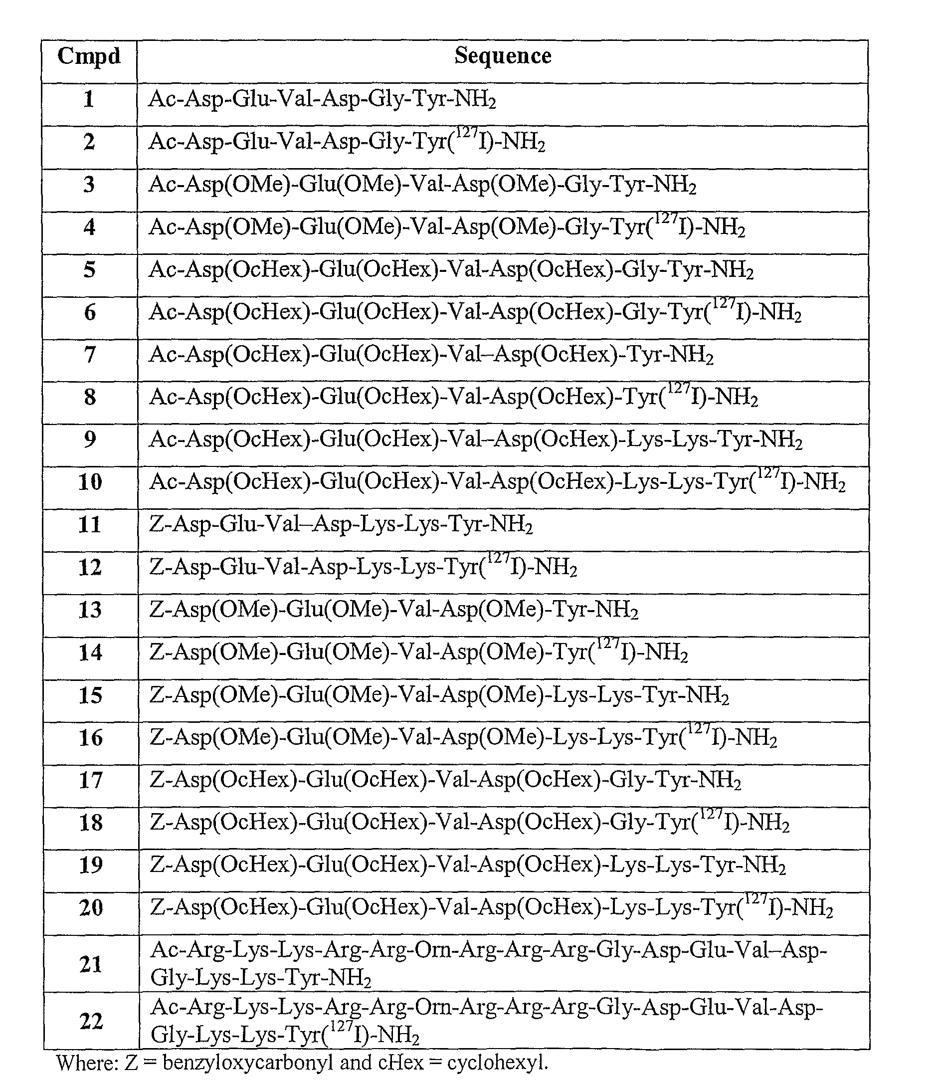
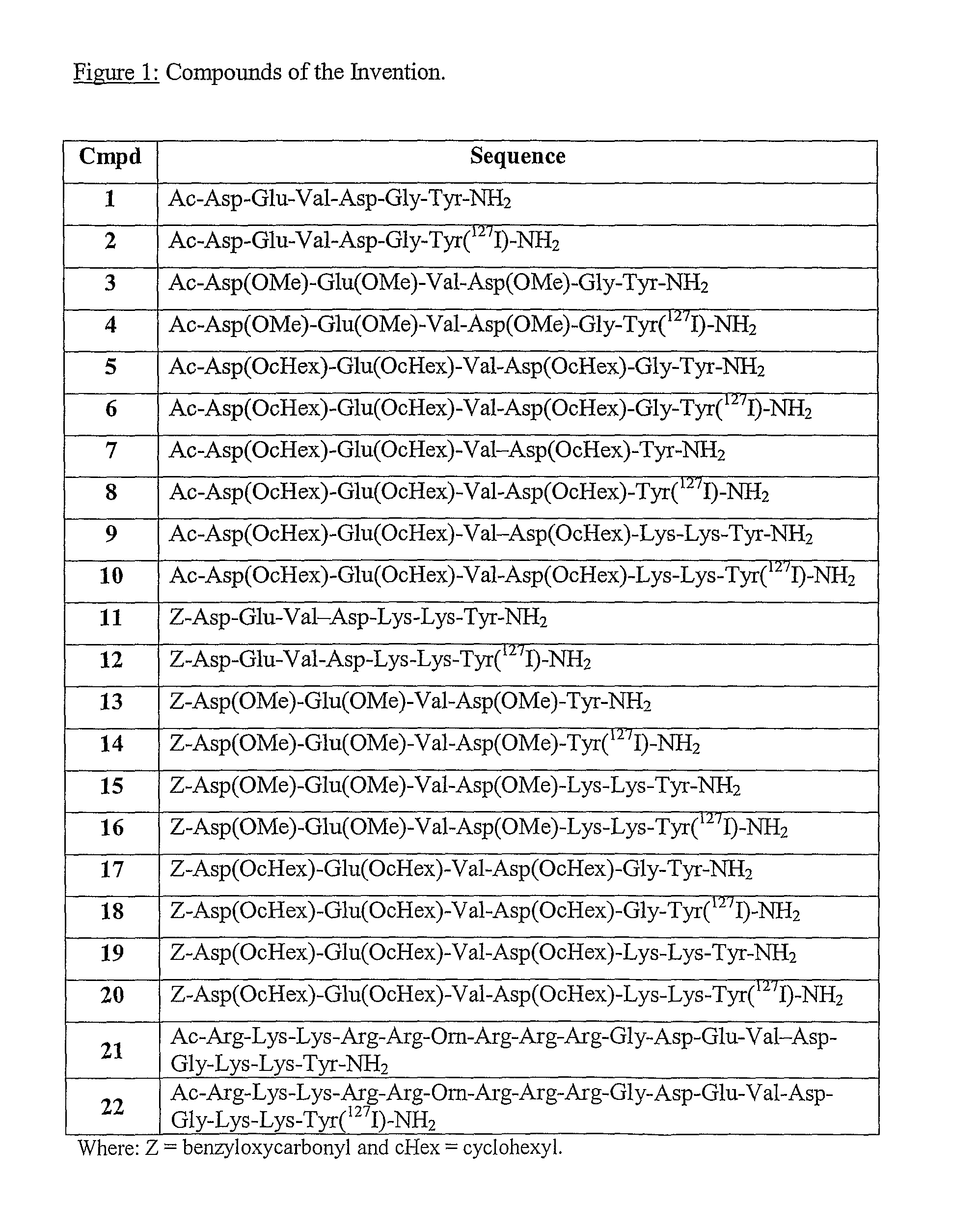
[0121]The peptidyl resin corresponding to the sequences of Compounds 1-22 in FIG. 1 were assembled by standard solid-phase peptide chemistry [Barany et al, Int. J. Peptide Protein Research 30, 705-739 (1987)] on a Rink Amide resin (from NovaBiochem, typical loading 0.73 mmol / g). An Applied Biosystems (Perkin Elmer) model 433A peptide synthesizer was used. The residues (from the carboxyl terminus) were assembled on a 0.25 mmol scale using single couplings (2.5 hours coupling cycles) of a 4-fold molar excess of Fmoc-amino acids (1 mmol cartridges) pre-activated with 2-(1H-benzotriazol-1-yl)-1,1,3,3-tetramethyluronium-hexafluorophosphate (HBTU) / 1-hydroxy-benzotriazole (HOBt) / diisopropylethylamine (DIEA) in NMP. Fmoc-deprotection was achieved with conductivity monitoring using 20% piperidine in N-methylpyrrolidone (NMP). The washing solvent was NMP. The amino acid-side chain protecting groups used were either tert-butyl (tBu) or cyclohexyl ...

example 2

Synthesis of 123I-Labelled Compound 2 (Compound 2A)

Step (a): 127I-Analogue (Compound 2).

[0127]Compound 2 was prepared following the reaction scheme:

[0128]Mass spec analysis of 127I prepared and purified material, confirmed identity.

Step (b): Preparation of Compound 2A.

[0129]To Compound 1 (7411g, 1×10⁻⁷ moles) dissolved in 74 μl water, was added 200 μL pH 4, 0.2M ammonium acetate buffer, 10 μl Na127I in 0.01MNaOH (1×10⁻⁸ moles), ca. 10-30 μl (150-450 MBq) Na123I in 0.05M NaOH and 10 μl 0.001 M PAA solution (1×10⁻³ moles). [123I]-Compound 2 was HPLC purified and diluted in pH 7.4, 50 mM sodium phosphate buffer to 20 and 100 MBq / ml with specific activities of 14 and 45 MBq / nmole respectively. Co-elution with the 1271 standard was observed confirming identity. Good stability (>90%) was observed over 3.5 hours post dilution.

example 3

Synthesis of 123I-Labelled Compound 6 (Compound 6A)

Step (a): 127I-Analogue (Compound 6).

[0130]Compound 6 was prepared following the reaction scheme:

[0131]Mass spec analysis of 127I prepared and purified material, confirmed identity.

Step (b): Preparation of Compound 6A.

[0132]Compound 5 (98 μg, 1×10⁻⁷ moles) dissolved in 98 μl methanol, was added to 100 μl pH4, 0.2M ammonium acetate buffer, 10 μl Na127I in 0.01M NaOH (1×10⁻⁸ moles), ca. 10-30 μl (150-450 MBq) Na123I in 0.05M NaOH and 10 μl 0.001M PAA solution (1×10⁻⁸ moles) post iodonium formation. Compound 6A was HPLC purified and diluted in pH6, 50 mM sodium phosphate buffer to 20 and 100 MBq / ml with specific activities of 13 and 43 MBq / nmole respectively. 10% ethanol was added to aid solubility. Co-elution with the 127I standard was observed confirming identity and Compound 6A. Good stability (>90%) was observed over 3.5 hours post dilution.


PUM

PropertyMeasurementUnit
permeableaaaaaaaaaa
gamma-emitting radioactiveaaaaaaaaaa
positron-emitting radioactiveaaaaaaaaaa

Abstract

The present invention relates to diagnostic imaging agents for in vivo imaging. The imaging agents comprise a synthetic caspase-3 substrate peptide labelled with an imaging moiety suitable for diagnostic imaging in vivo. The invention also provides radiopharmaceutical compositions comprising the imaging agents, together with kits for the preparation of the radiopharmaceuticals. Also described are non-radioactive precursors suitable for the preparation of the imaging agents. The imaging agents are useful for the diagnostic imaging and or therapy monitoring in vivo of various disease states where caspase-3 is involved.

Description

FIELD OF THE INVENTION[0001]The present invention relates to diagnostic imaging agents for in vivo imaging. The imaging agents comprise a synthetic caspase-3 substrate labelled with an imaging moiety suitable for diagnostic imaging in vivo.BACKGROUND TO THE INVENTION[0002]Programmed cell death by apoptosis is a complex process, involving a large number of cellular processes with numerous levels of control. It is initiated by one of two pathways. The first is through an extrinsic pathway initiated via cell surface death receptors and the second is through intrinsic initiators, such as DNA damage by UV radiation. Both of these pathways culminate in the coordinated death of cells which requires energy and, unlike cell death by necrosis, does not involve an inflammatory response. Cells committed to apoptosis present ‘eat me’ signals on their cell surface, which invite other cells to consume them by phagocytosis.[0003]Apoptosis is a critical event in numerous processes within the body. F...

Claims


Application Information

Patent Timeline
Patent Type & Authority Applications(United States)
IPC IPC(8): A61K51/08C12N9/50A61K38/07
CPCA61K38/07A61K51/088
Inventor GUILBERT, BENEDICTECHAMPION, SUEGIBSON, ALEXANDER MARKRICKETTS, SALLY-ANNEAVORY, MICHELLEARBO, BENTE E.
Owner GUILBERT BENEDICTE
Why Eureka
Try Eureka