Methods and Systems for Treating Hiatal Hernias

a technology of hiatal hernia and treatment methods, applied in the field of medical methods and equipment, can solve the problems of insufficient endoesophageal endoscopic visualization, inability to fix stomach and esophagus, etc., and achieve the effect of reducing the hernia

Active Publication Date: 2009-01-15
ADVENT MEDICAL
View PDF10 Cites 31 Cited by
  • Summary
  • Abstract
  • Description
  • Claims
  • Application Information

AI Technical Summary

Benefits of technology

[0012]The present invention provides improved methods and systems for restoring the angle of His and treating hiatal hernias and other conditions of the lower esophagus. While the methods will be suitable for treating patients with GERD, they will be more broadly applicable to patients having or at risk from suffering from hiatal hernias, even if no symptoms of GERD are present. The present invention provides a method for placing and positioning an anchor which connects and secures the esophagus and stomach to the diaphragmatic crus muscle. In order to assure the proper reconfiguration of the angle of His and the reliable capture of the crus muscle, the presence of the crus muscle between the esophagus and stomach is confirmed prior to the placement of the anchor. This restores the native, normal anatomy and thereby treats and reduces the hiatal hernia.
[0015]After the hiatal hernia or other tissue deformity has been reconfigured and the location of the crus muscle has been identified, the lower esophageal wall and the fundus are attached with a fastener that passes through the crus muscle. This is accomplished by deploying a delivery device and advancing the fastener through the delivery device. Fastening can occur in the direction from the esophagus toward the fundus. Alternatively, fastening can occur in the direction from the fundus toward the esophagus. By passing the fastener through the crus muscle, any tendency for the fastener to migrate is limited or eliminated and the resulting attachment is stabilized. The fastener comprises a central portion having two expandable end anchors which expand and engage the esophagus and fundus, respectively.
[0016]Further advantages of this method include a fastener that is stretchable, flexible, removable and adaptable to movement and variations in anatomy. The procedure can be performed by a gastroenterologist rather than surgeons, takes less time than conventional surgery, has fewer complications and side-effects and has lower overall procedural costs than conventional methods. The procedure recreates or augments the natural anatomy.
[0018]The fastener is advanced through a delivery device and passes from the thoracic cavity to the abdominal cavity. Fastening restores the gastrointestinal flap valve, repositions the lower esophageal sphincter against the diaphragmatic crus muscle, enhances the function of the lower esophageal sphincter, and restores the anti-reflux barrier. The angle of His is restored by treating the hiatal hernia.
[0019]A third method according to the present invention comprises treating a hiatal hernia by fastening the lower esophageal wall to the gastric fundus with a fastener that passes through the diaphragmatic crus muscle. The hiatal hernia is first pushed or pulled downwardly to capture the crus muscle at the angle of His. This results in reducing the hernia. Pushing or pushing the hiatal hernia comprises engaging the hernia with the expanded cuff of the endoscope (or other device) and pushing the hernia downward. The hernia can be pushed or pulled down by mechanical pushers, vacuum apparatus, inflatable members, pins, traction devices or the like, either associated with an endoscope or as separate devices. The fastener is advanced through the endoscope and delivered using a catheter to pass the fastener from the thoracic cavity to the abdominal cavity since it passes from the superior to inferior surface of the diaphragmatic crus. Fastening restores the gastro-esophageal flap valve, repositions the lower esophageal sphincter against the crus muscle, enhances the function of the lower esophageal sphincter and restores the anti-reflux barrier. The angle of His is restored by treating the hiatal hernia.

Problems solved by technology

Because the crus muscle cannot be seen visually from the inside of the gastrointestinal lumen, endoesophageal endoscopic visualization is not sufficient and it is usually necessary to ultrasonic image or other imaging or non-imaging techniques to locate the crus muscle prior to deploying the fastener.
Also, the stomach and esophagus are not always fixed, especially in the case of a sliding hiatal hernia, and the structures can move relative to one another and out of alignment as in the normal non-herniated anatomy.

Method used


Image

  • Methods and Systems for Treating Hiatal Hernias
  • Methods and Systems for Treating Hiatal Hernias
  • Methods and Systems for Treating Hiatal Hernias

Examples


Embodiment Construction

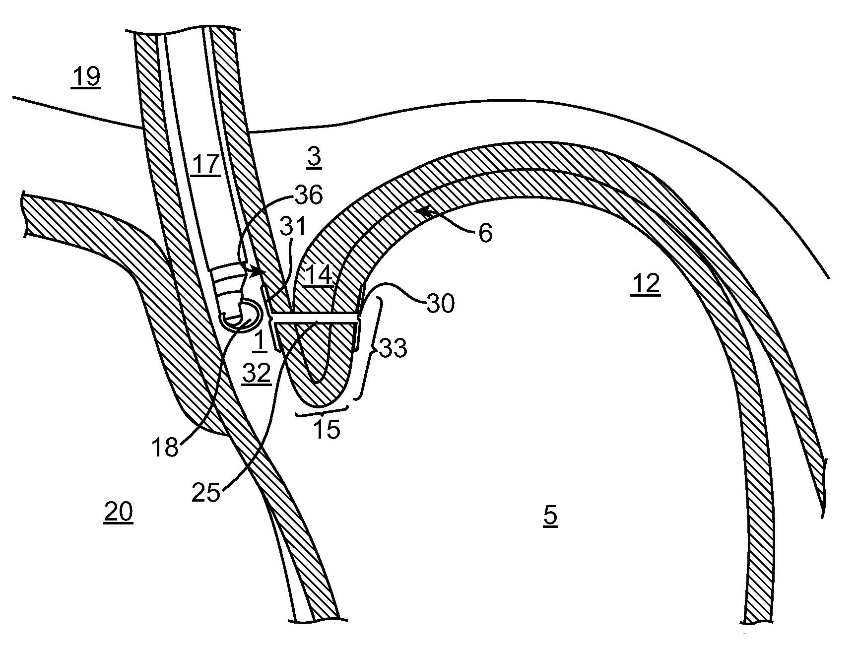
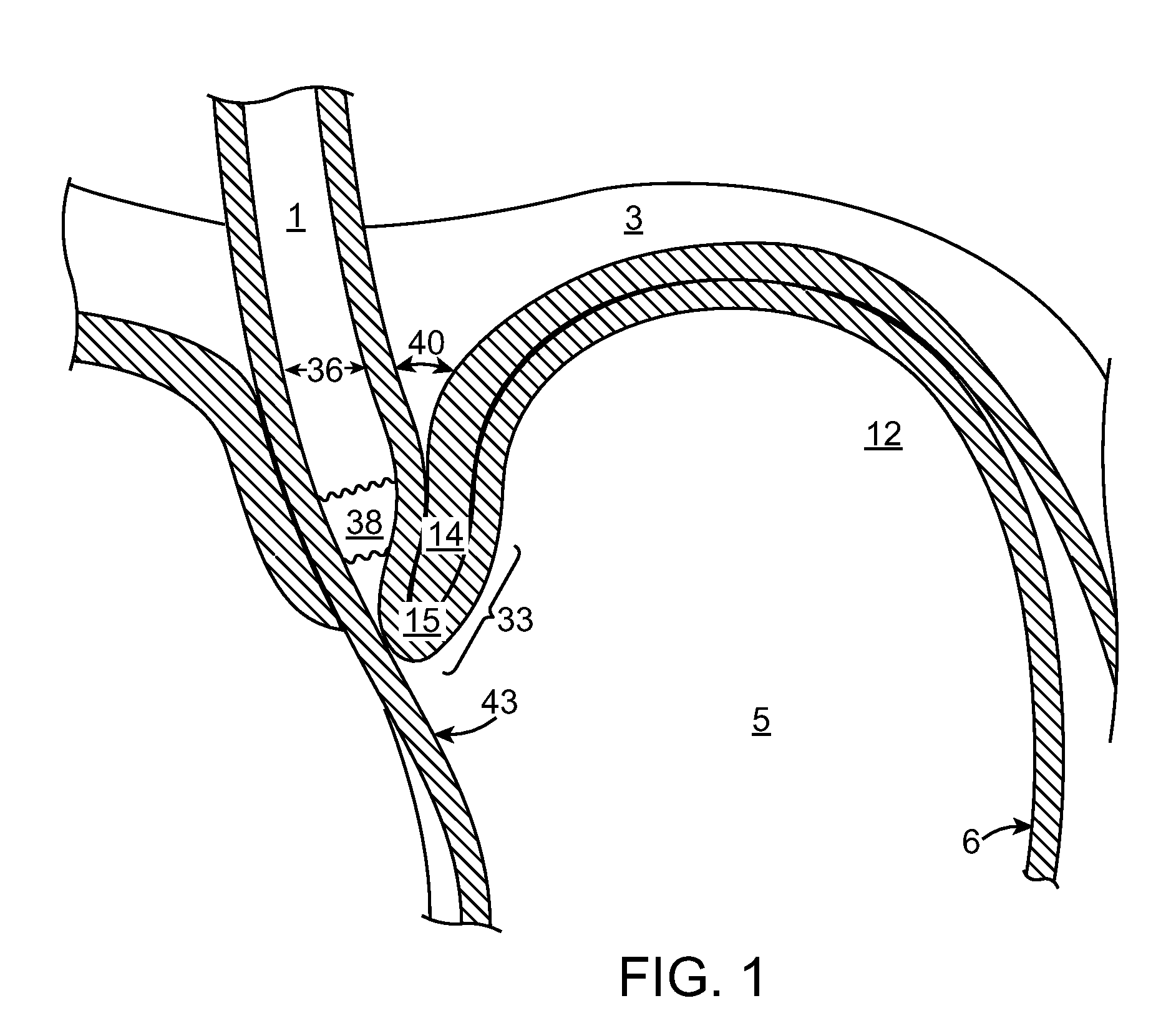
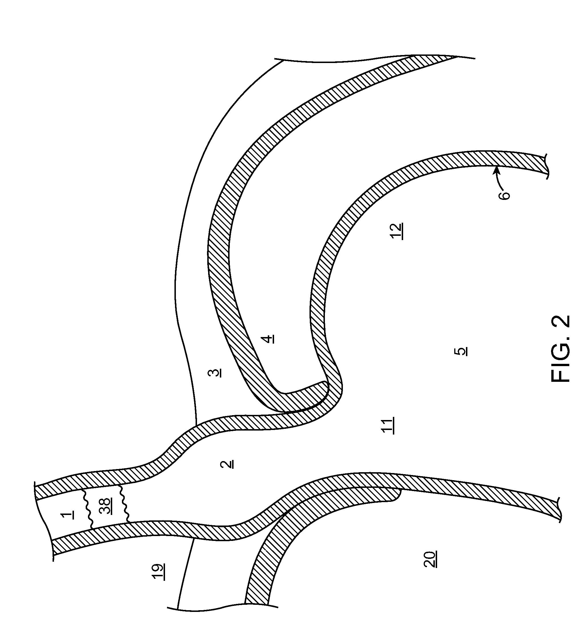
[0040]The medical methods and systems described herein offer improvements over the techniques currently utilized to perform endoscopic procedures to reduce hiatal hernias, restore the angle of His and to treat other conditions of the lower esophagus. The present invention relates to novel methods and systems that fix the distal esophagus and fundus directly to the diaphragmatic crus muscle. The present invention provides several embodiments where the crus is precisely identified followed by the placement of a translumenal anchor which connects, fastens and secures the esophagus and / or stomach to the diaphragmatic crus. This procedure reduces the hiatal hernia and restores the anatomy to its normal configuration.

[0041]FIG. 1 is a cross sectional view of the gastrointestinal tract in the area of the gastroesophageal junction (GEJ), from the esophagus 1 to the stomach 5 depicting a healthy anatomy, including a normal gastro-esophageal valve (GEV) 33 with correct angle of His 15 and cru...


PUM


Abstract

The present invention relates generally to medical methods and systems used to restore the angle of His and treat hiatal hernias and other conditions of the lower esophagus. More particularly, the present invention relates to a method and system that allows fixation of the distal esophagus and fundus of the stomach directly to the diaphragmatic crus muscle. The present invention provides a method where the diaphragmatic crus muscle is identified and precisely located from within and through the gastrointestinal lumen followed by the placement of a translumenal anchor which connects and secures the esophagus and stomach to the diaphragmatic crus muscle. This procedure reduces the hiatal hernia, restores the normal anatomy and treats conditions associated with the lower esophagus.

Description

CROSS-REFERENCE TO RELATED APPLICATIONS[0001]The present application claims the benefit of U.S. Provisional Patent Application No. 60 / 949,746, filed on Jul. 13, 2007 (Attorney Docket No. 026923-000100US), under 37 C.F.R. §1.78, the complete disclosure of which is incorporated herein by reference for all purposes.BACKGROUND OF THE INVENTION[0002]1. Field of Invention[0003]The present invention relates generally to medical methods and apparatus. More particularly, the present invention relates to a method and system for restoring the angle of His and treating and stabilizing hiatal hernias.[0004]Hiatal hernias (also referred to as a hiatus hernia) result when the diaphragm muscle, in particular the diaphragmatic crus, loosens and can no longer firmly hold the esophagus and stomach in their normal position. The esophagus and stomach are able to move in the cranial direction resulting in the stomach being herniated above the diaphragm and into the thoracic cavity. When the hiatal hernia...

Claims


Application Information

Patent Timeline
Patent Type & Authority Applications(United States)
IPC IPC(8): A61B17/08
CPCA61B17/0644A61B2017/00619A61B2017/00827A61B2017/22051A61B2017/0419A61B2017/0445A61B2017/00862
Inventor BINMOELLER, KENNETH F.
Owner ADVENT MEDICAL
Why Eureka
Try Eureka