System and Method of Automatic Prioritization and Analysis of Medical Images

a medical image and automatic prioritization technology, applied in the field of automatic prioritization and analysis of medical images, can solve the problems of delay in diagnosis, delay in diagnosis, and inability to know in advance which patients are at greatest risk,

Active Publication Date: 2009-01-29
MEDIC VISION BRAIN TECH
View PDF14 Cites 187 Cited by
  • Summary
  • Abstract
  • Description
  • Claims
  • Application Information

AI Technical Summary

Problems solved by technology

The radiologist usually does not know in advance which patients are at greatest risk.
Today, in a typical radiology division, a delay in the diagnosis of ambulatory patients can reach several hours or even days.
This increase reflects a delay in the diagnoses.
When dealing with the large volume sets produced by CT and MRI, seconds often add up to minutes.
Unfortunately, the supply of radiologists has not keep up with the demand and there is a growing shortage of several thousands in the number of well-trained, qualified radiologists only in the US.
Though the CAD system aids radiologists during the diagnosis process, the aforementioned throughput problems and the delays remain unchanged.
Moreover, none of the known CAD systems is designed to aid in a general diagnosis of the brain.
Though such a system can improve the workflow, the delays in the diagnosis process are not substantially decreased, inter alia, because the order of the received 3D medical images remains constant.
Though such CAD systems improve the quality of each analysis, they fail to improve the throughput of the diagnosis.
Though aiding in the detection process, the currently used CAD systems do not overcome the difficulties in the current diagnosis workflow of medical images.
Moreover, the currently used CAD systems do not aid in the detection of indications in some market segments such as brain MRI scans, brain CT scans etc.

Method used


Image

  • System and Method of Automatic Prioritization and Analysis of Medical Images
  • System and Method of Automatic Prioritization and Analysis of Medical Images
  • System and Method of Automatic Prioritization and Analysis of Medical Images

Examples


Embodiment Construction

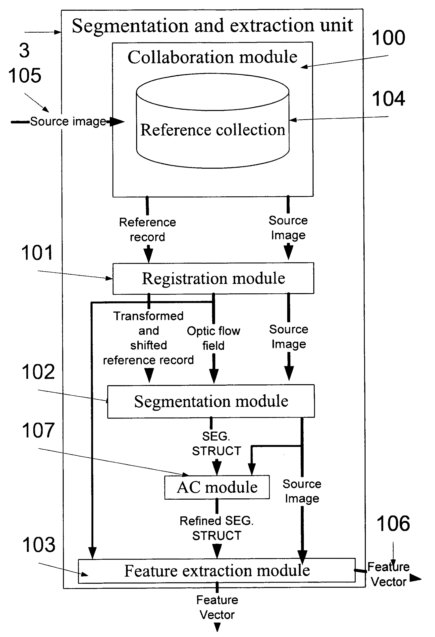
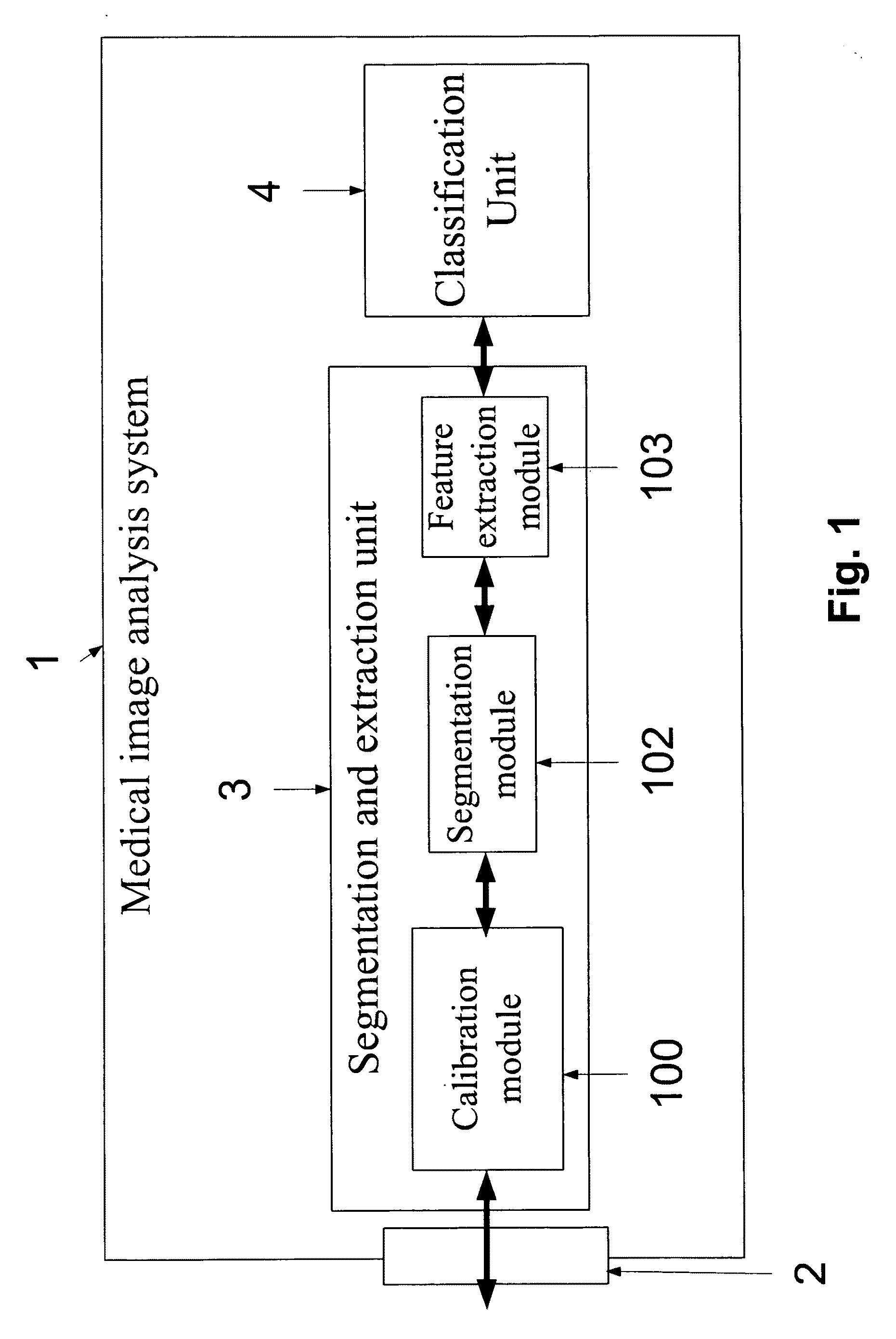
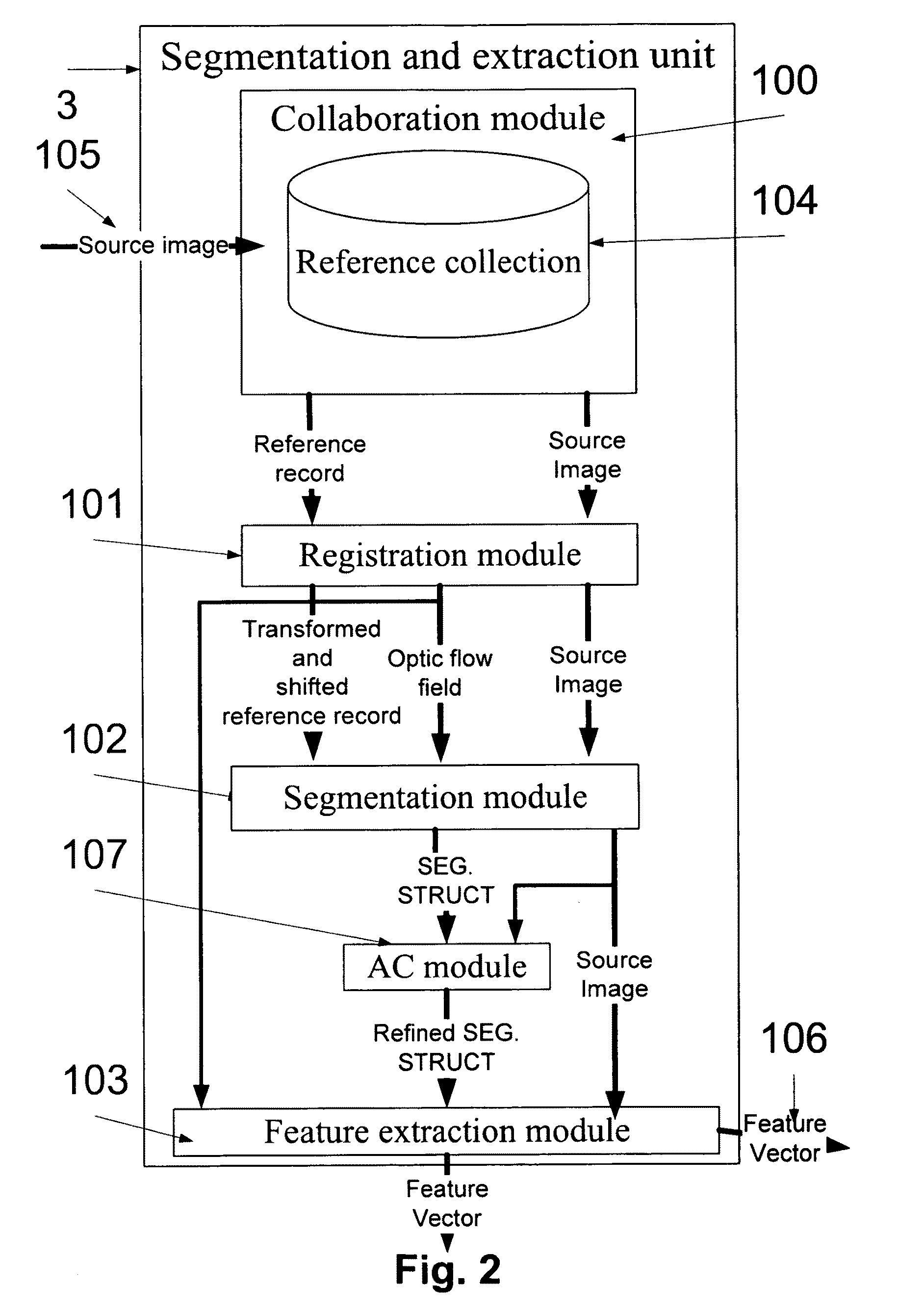
[0143]The present embodiments comprise a real-time autonomous prioritization and alerting system for 3D medical images, such as CT scans. The system provides a solution for a number of problems, inter alia, the shortage in radiologists and the critical period between the carrying out of a CT scan and the diagnosis thereof. The system receives a 3D medical image, analyzes it, and preferably alerts when pathology is found. Preferably, the system gives the 3D medical image a priority-level that indicates the urgency of the case. Preferably, the system is used for analyzing and prioritizing a number of 3D medical images. The priority level of each one of the 3D medical images allows, inter alia, prioritization of the scans which are pending on the working station, therefore allowing the radiologist to diagnose critical cases first. In addition, the system enhances the radiologists' throughput in two or more levels. First, the system extracts high-probability normal cases from the queue ...


PUM


Abstract

A system for analyzing a source medical image of a body organ. The system comprises an input unit for obtaining the source medical image having three dimensions or more, a feature extraction unit that is designed for obtaining a number of features of the body organ from the source medical image, and a classification unit that is designed for estimating a priority level according to the features.

Description

FIELD AND BACKGROUND OF THE INVENTION[0001]The present invention relates to analysis of three-dimensional (3D) medical images and, more particularly, but not exclusively to automatic analysis of 3D medical images that depicts an organ or a human body system, especially the brain, or a section thereof by identified suspected indications of different diseases or disorders.[0002]Systems and devices for visualizing the inside of living organisms are among the most important medical developments in the last thirty years. Systems like computerized tomography (CT) scanners and magnetic resonance imaging (MRI) scanners allow physicians to examine internal organs or areas of the body that require a thorough examination. In use, the visualizing scanner outputs a 3D medical image, such as a sequence of computerized cross-sectional images of a certain body organ, which is then interpreted by specialized radiologists.[0003]Commonly, a patient is referred for a visual scan by a general practition...

Claims


Application Information

Patent Timeline
Patent Type & Authority Applications(United States)
IPC IPC(8): G06K9/00G06V10/24G06V10/40G16H30/40G16H50/20G16H70/60
CPCG06F19/321G06F19/345G06F19/3487G06K9/32G06K9/46G06K9/6206G06T2207/30016G06T7/0012G06T7/0046G06T7/0089G06T2207/20128G06T2207/20161G06K2209/053G06T7/75G06T7/149G16H50/20G16H15/00G16H70/60G16H30/40G06V10/24G06V10/40G06V10/754G06V2201/032
Inventor BAR-AVIV, EZERDAHAN, ELLRANDEVIR, ZVI
Owner MEDIC VISION BRAIN TECH
Why Eureka
Try Eureka