Coating stents with cyclic rgd peptides or mimetics
- Summary
- Abstract
- Description
- Claims
- Application Information
AI Technical Summary
Benefits of technology
Problems solved by technology
Method used
Image
Examples
example 1
RGD Peptide Binds to Titanium-Oxide Containing Nitinol Coupons
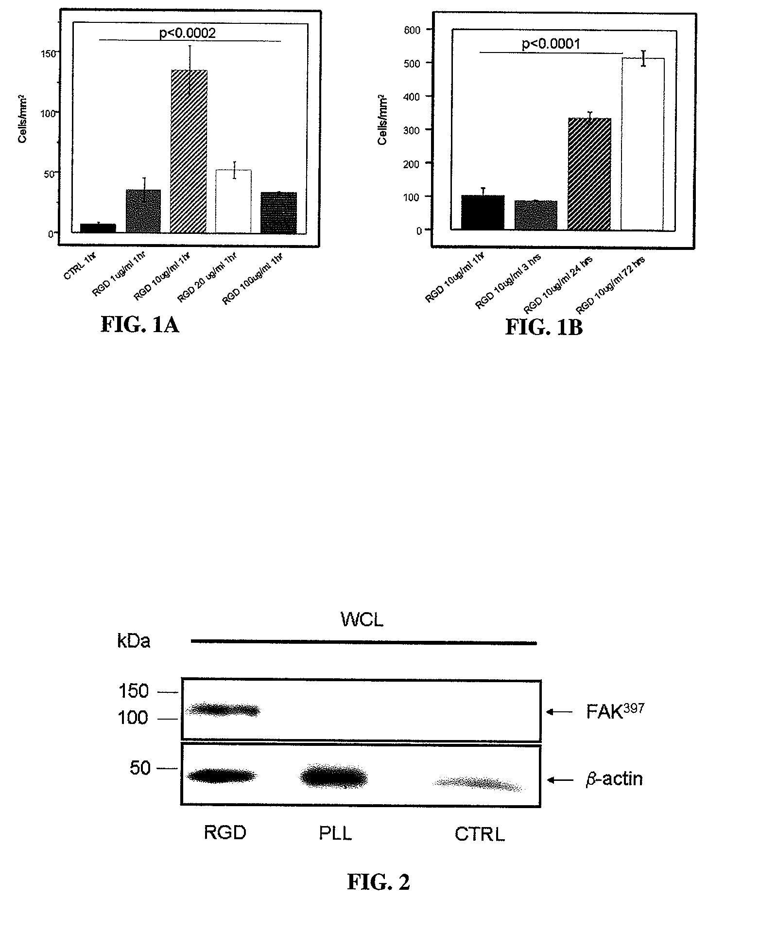
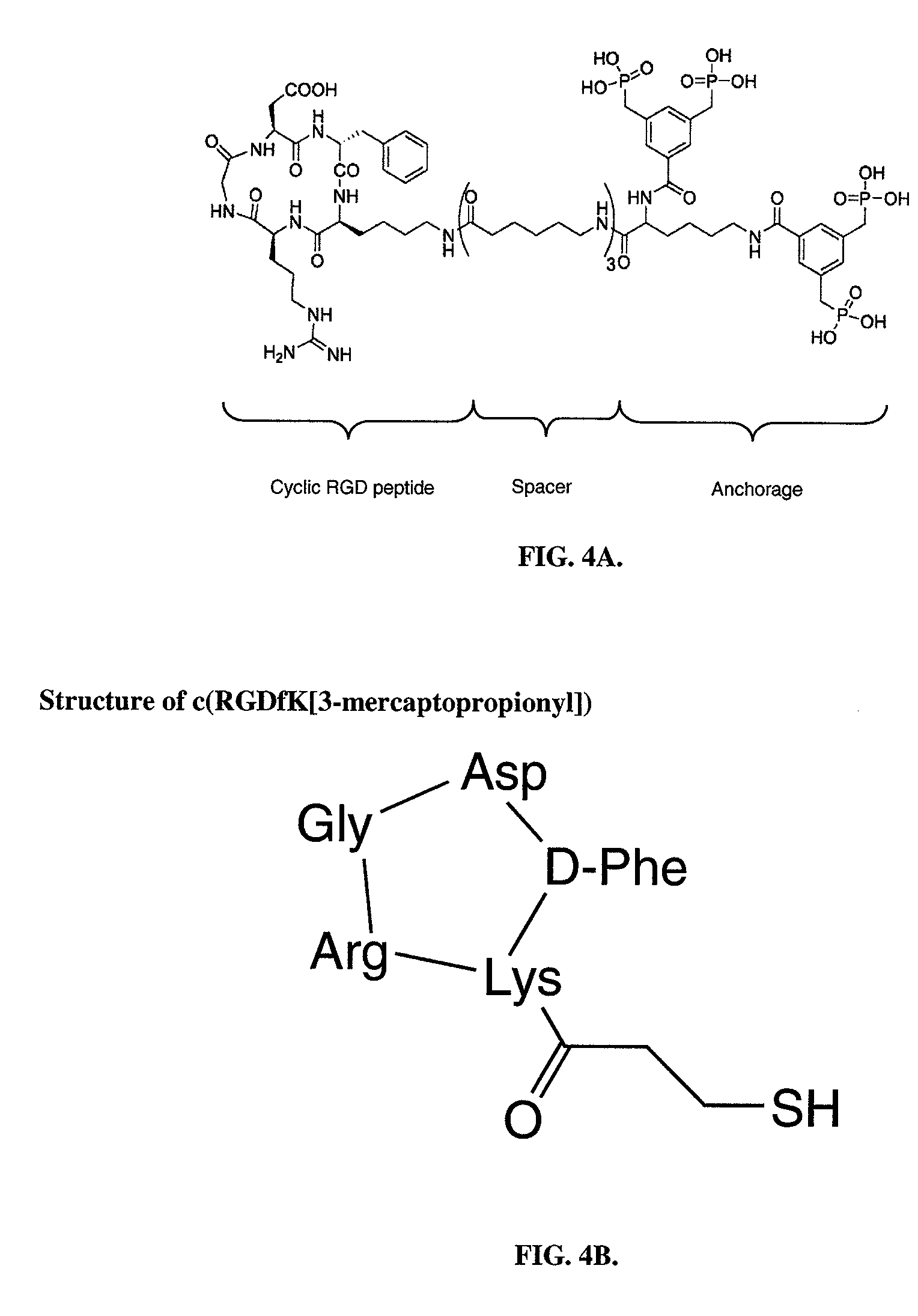
[0101]In preliminary experiments, the phosphonate anchored cyclic RGD peptide was shown to specifically bind to Titanium-oxide containing Nitinol coupons. For these experiments, small Nitinol coupons (˜2×2 cm) were coated over night with cyclic RGD peptide solubilized at concentrations of 1, 10, 20 and 100 μg / mL. Applicants found specific binding of RGD peptide over a wide range of doses, with a maximum at 10 μg / mL. Binding of RGD peptide was indirectly proven in a cell adhesion experiment utilizing human umbilical endothelial vein cells (HUVECs). Cells were seeded on RGD-coated and control (BSA coated) coupons at a concentration of approximately 1×10⁵ / mL for 1 hour, 3 hours, 24 hours and 72 hours and attachment of the cells was quantified following immunofluorescent labeling and counting the number of cells per area in 6 randomly selected regions. See FIGS. 1A and 1B.
example 2
Cyclic RGD Peptide Works through Specific Activation of Integrins
[0102]In a separate analysis, applicants focused on the signal pathways that are involved in the process of cellular attachment and anchorage following coating with RGD peptide. It has been reported that the currently used RGD peptide is highly specific for alpha v beta 3 integrin (Meyer, J., 2006) which is abundantly expressed on various cell types. Importantly, endothelial cells express large amounts of alpha v beta 3 integrin, necessary for cellular adhesion, proliferation and migration (Cheresh, D. A., 1987). Upon binding to RGD peptide, integrins form cluster and allow the binding of focal adhesion kinase (FAK), which gets activated via phosphorylation of distinct tyrosine groups. In the current experiment applicants have proven the hypothesis that specific binding of alpha v beta 3 integrin to immobilized RGD peptide results in intracellular activation of focal adhesion kinase (FAK), thus demonstrating an approac...
example 3
Advanced Attachment of Endothelial Cells on RGD Coated Stents In Vitro
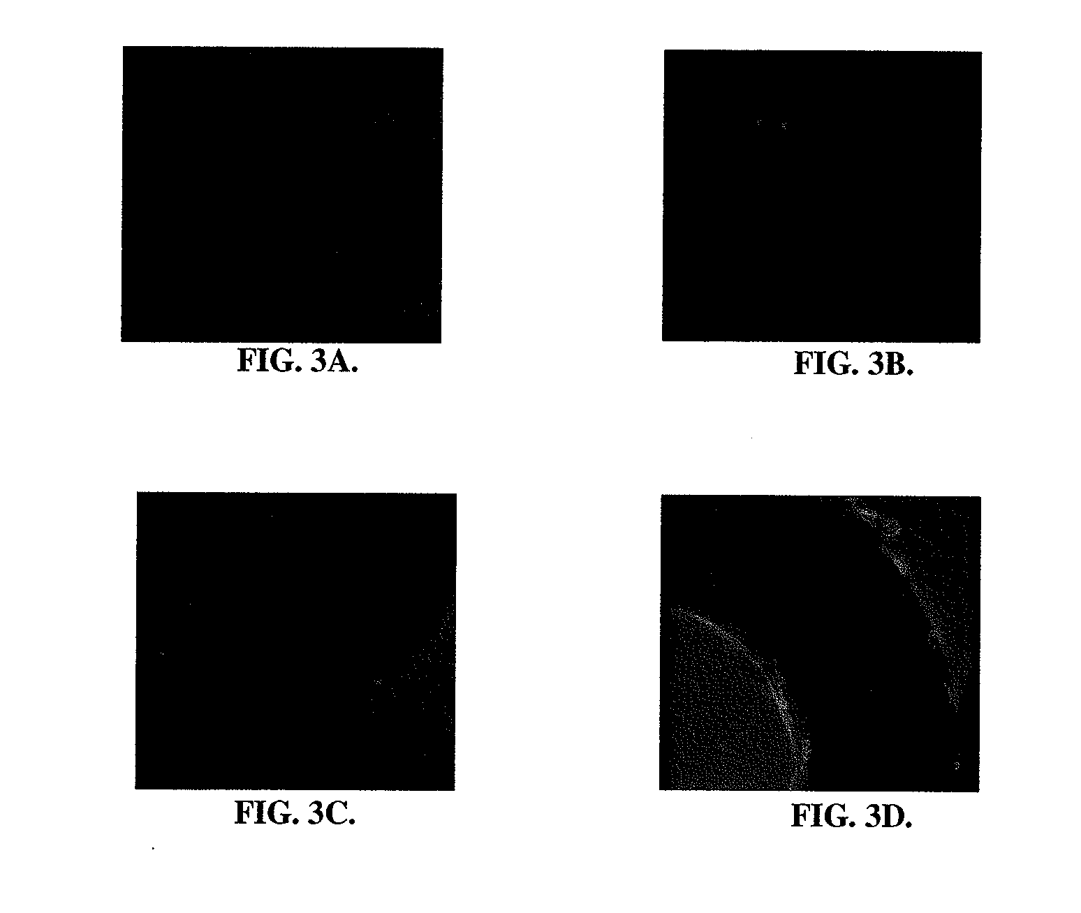
[0103]To prove the concept of advanced binding of endothelial cells to RGD coated stents, in vitro experiments were conducted both under static and dynamic conditions. Cells were seeded on Nitinol stents coated with RGD peptide or BSA (control) under static conditions for 3 hours. Following fixation of cells and immunofluorescent staining with Alexa-Fluor phalloidin, the number of adherent endothelial cells were quantified in 3 randomly selected regions. For dynamic adhesion assays, Nitinol stents were cut into small stripes and inserted into customized Ibidi μ-Slides™ (Ibidi, Germany). Flow-through was accomplished for 24 hours and the number of adherent cells quantified. There was significantly greater adherence of endothelial cells to RGD coated stents, as compared to control stents. See FIGS. 3A-3D.
PUM
| Property | Measurement | Unit |
|---|---|---|
| Fraction | aaaaa | aaaaa |
| Fraction | aaaaa | aaaaa |
| Therapeutic | aaaaa | aaaaa |
Abstract
Description
Claims
Application Information
 - R&D
- Intellectual Property
- Life Sciences
- Materials
- Tech Scout
- Unparalleled Data Quality
- Higher Quality Content
- 60% Fewer Hallucinations
Browse by: Latest US Patents, China's latest patents, Technical Efficacy Thesaurus, Application Domain, Technology Topic, Popular Technical Reports.
© 2025 PatSnap. All rights reserved.Legal|Privacy policy|Modern Slavery Act Transparency Statement|Sitemap|About US| Contact US: help@patsnap.com


