Device and method for the performance of ophthalmological operations

a technology for ophthalmological operations and devices, applied in the field of devices and methods for treating glaucoma, can solve the problems of not ensuring that only the separating tissue between the anterior aqueous chamber and the schlemm's canal is destroyed, and the destruction of the tissue behind the schlemm's canal, so as to avoid accidental tissue injury

Inactive Publication Date: 2009-04-30
HAEFLIGER EDUARD ANTON
View PDF16 Cites 23 Cited by
  • Summary
  • Abstract
  • Description
  • Claims
  • Application Information

AI Technical Summary

Benefits of technology

[0011]Preferably the suction channel is provided with a cutting tool having a cutting edge extending maximally to the orifice of the suction channel. This has the advantage that the cutting edge exerts its effect only on tissue which is sucked into the suction channel. Thus, accidental tissue injury can be avoided. In particular, tissue located behind Schlemm's canal is not injured.

Problems solved by technology

Such a device can not guarantee that only the separating tissue between the anterior aqueous chamber and Schlemm's canal is destroyed.
Even the most careful handling can e.g. lead to the destruction of tissue which is behind Schlemm's canal i.e. tissue located on the side of Schlemm's canal facing away from the anterior aqueous side.

Method used


Image

  • Device and method for the performance of ophthalmological operations
  • Device and method for the performance of ophthalmological operations
  • Device and method for the performance of ophthalmological operations

Examples


second embodiment

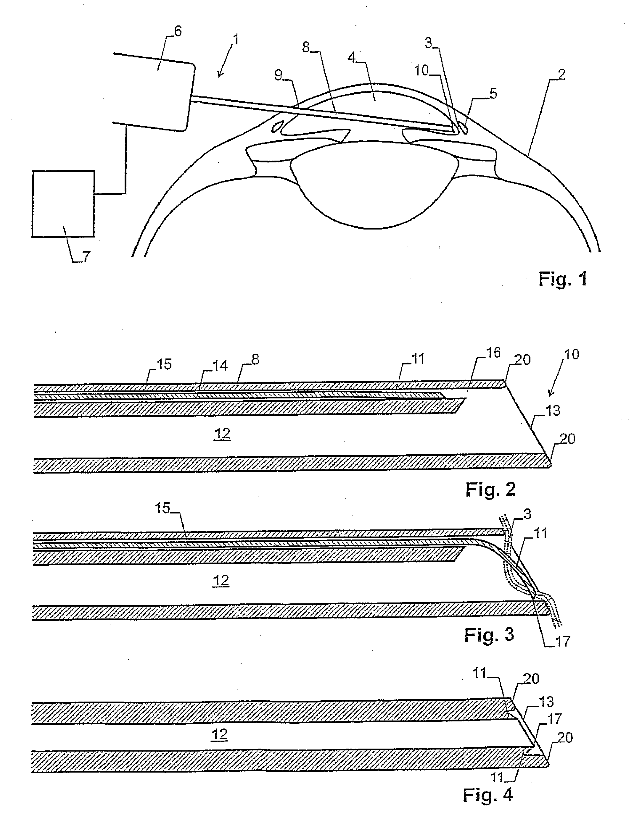
[0030]the device according to the invention is depicted in FIG. 4. In this embodiment the separation means or the cutting tool 11, respectively, are formed by a circular or oval cutting edge 17, respectively, which extends along the inner wall of suction channel 12. The cutting edge 17 is arranged closely to the orifice 13 or at the orifice 13. When separating tissue 3 is sucked into the orifice 13 it meets the cutting edge 17 and is cut off.

third embodiment

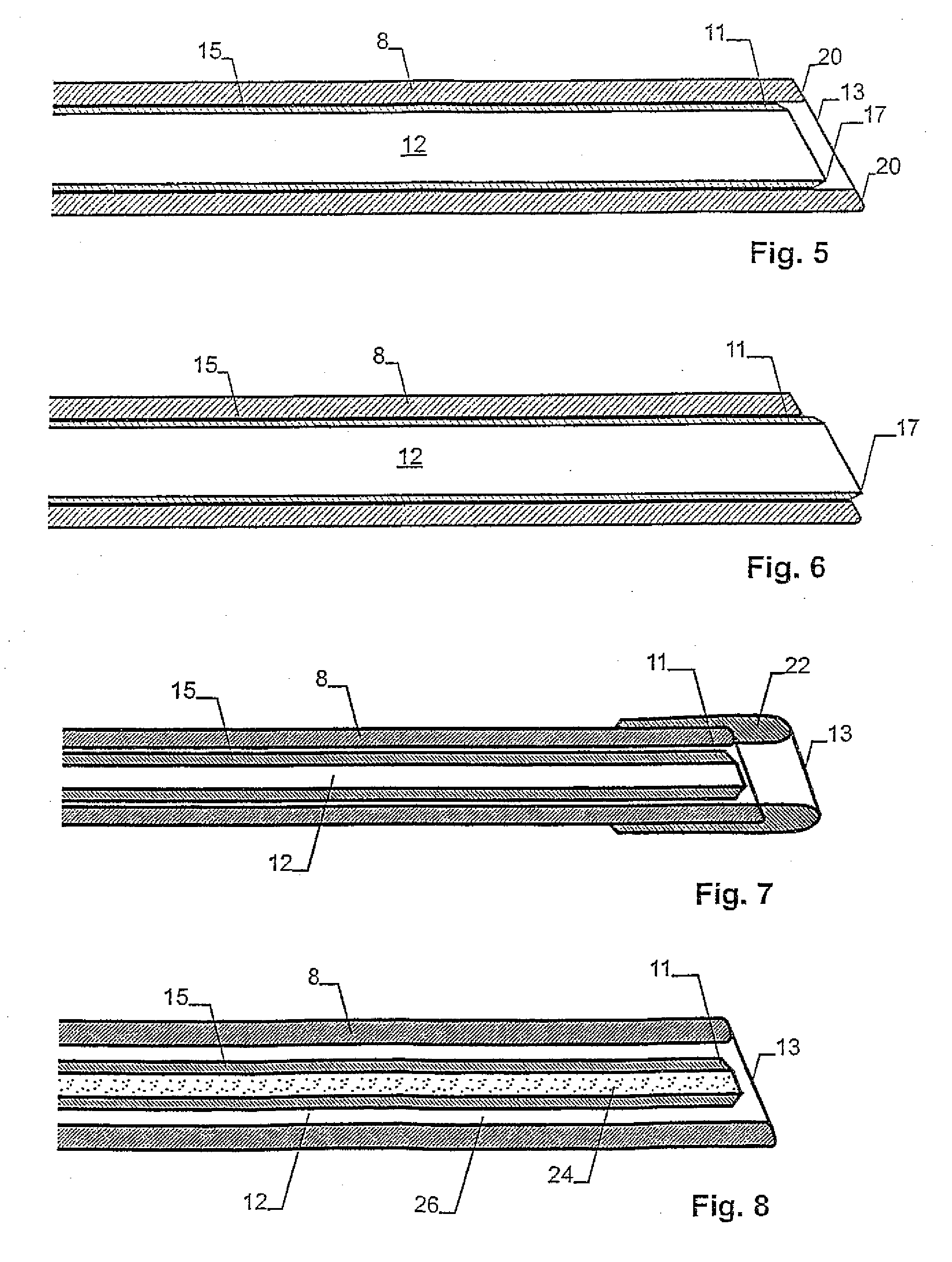
[0031]the device according to the invention is depicted in FIGS. 5 and 6. In this embodiment the cutting tool 15 is formed by a tube section which is arranged in the suction channel 12. The distal end of the tube forms the cutting tool 11 with cutting edge 17.

[0032]Preferably the cutting device 15 or the cutting tool 11 is longitudinally displaceable and can be moved from the neutral position shown in FIG. 5 to the separation position shown in FIG. 6. In the neutral position the cutting edge 17 is behind the orifice 13, in the separation position the cutting edge 17 protrudes beyond the orifice 13 up to a given distance. The distance corresponds approximately to the typical width of the separating tissue 3. In this manner, separating tissue which is sucked in by the orifice 13 can be cut off.

[0033]It is conceivable that the cutting device 15 is rotatable about its longitudinal axis so that it can be turned at least over a limited angular range compared to the probe 8. This allows to...

first embodiment

[0036]The suction channel 12 can have a round or square crossection. In particular in the first embodiment according to FIG. 2 and FIG. 3 the crossection is at least in the area of the distal end 10 rather square so that the cutting tool can glide over the complete crossection while in the embodiments according to FIGS. 4 to 6 the crossection is preferably rather round or elliptic.

[0037]In a further embodiment as shown in FIG. 7, a sleeve 22 is arranged as support device at the distal end of the probe 8. It is made of a material which is more deformable than the probe and allows a more tight seal of the suction channel 12 towards the tissue.

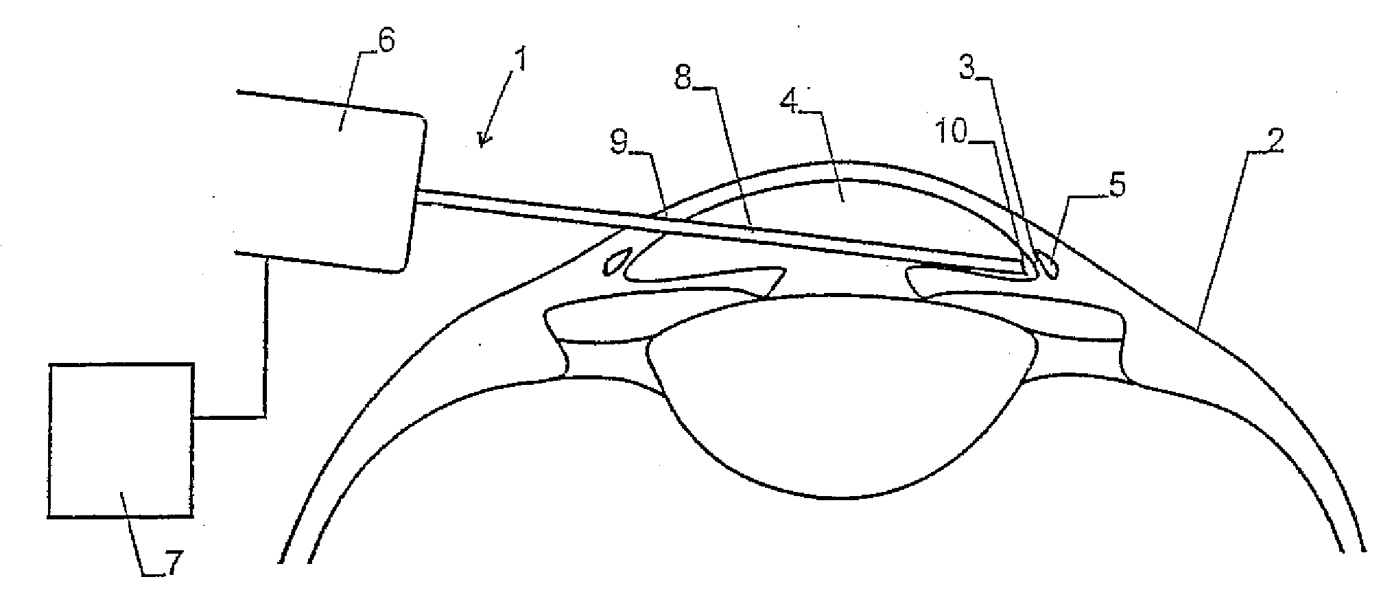
[0038]In the application of the device described herein, the distal end 10 of probe 8 is guided through the anterior aqueous chamber 4 to separating tissue 3 whereby the cutting tool 11 of the embodiments according to FIG. 2 and FIG. 3 or FIGS. 5 and 6, respectively, is at this stage in neutral position. Then, a piece of separating tissue is suck...


PUM


Abstract

The invention relates to a device for performing ophtalmological operations, especially for treating glaucoma. Said device comprises a probe in which a suction channel is arranged. A cutting tool is arranged in the suction channel, by which means separating tissue extending to Schlemm's canal and being sucked into the suction channel can be removed. The device enables openings to be created between the front chamber and Schlemm's canal in a targeted and careful manner.

Description

PRIORITY CLAIM[0001]This Application is a divisional of and claims the benefit if U.S. patent application Ser. No. 10 / 850,722 filed on May 21, 2004.CROSS REFERENCES TO RELATED APPLICATIONS[0002]This application claims the priority of international patent application PCT / IB01 / 02224, filed Nov. 22, 2001, the disclosure of which is incorporated herein by reference in its entirety.TECHNICAL FIELD[0003]The invention relates to a device and a method for treating glaucoma by generating openings between the anterior aqueous chamber and Schlemm's canal according to the preamble of the independent claims.[0004]WO 01 / 37767 describes several devices for treating of glaucoma. These devices are provided with a probe which can be introduced into the anterior aqueous chamber of the eye in order to generate openings in the tissue separating the anterior aqueous chamber and Schlemm's canal. In an embodiment this probe has a channel which e.g. can be connected to a suction unit or an irrigation unit. ...

Claims


Application Information

Patent Timeline
Patent Type & Authority Applications(United States)
IPC IPC(8): A61F9/007
CPCA61F9/00781
Inventor HAEFLIGER, EDUARD ANTON
Owner HAEFLIGER EDUARD ANTON
Why Eureka
Try Eureka