
Expanded nk cells

a technology of expanded and activated nk cells and cytotoxicity, which is applied in the direction of biocide, plant growth regulators, blood/immune system cells, etc., can solve the problems of inadapting the dose of cells to clinical trials, affecting the clinical effect of autologous nk cells, and affecting the treatment effect of mm patients, etc., to achieve the effect of increasing the effect of administered cells and increasing the cytotoxicity of expanded and activated nk

Inactive Publication Date: 2009-05-14
CELLPROTECT NORDIC PHARMA
View PDF1 Cites 24 Cited by
  • Summary
  • Abstract
  • Description
  • Claims
  • Application Information

AI Technical Summary

Benefits of technology

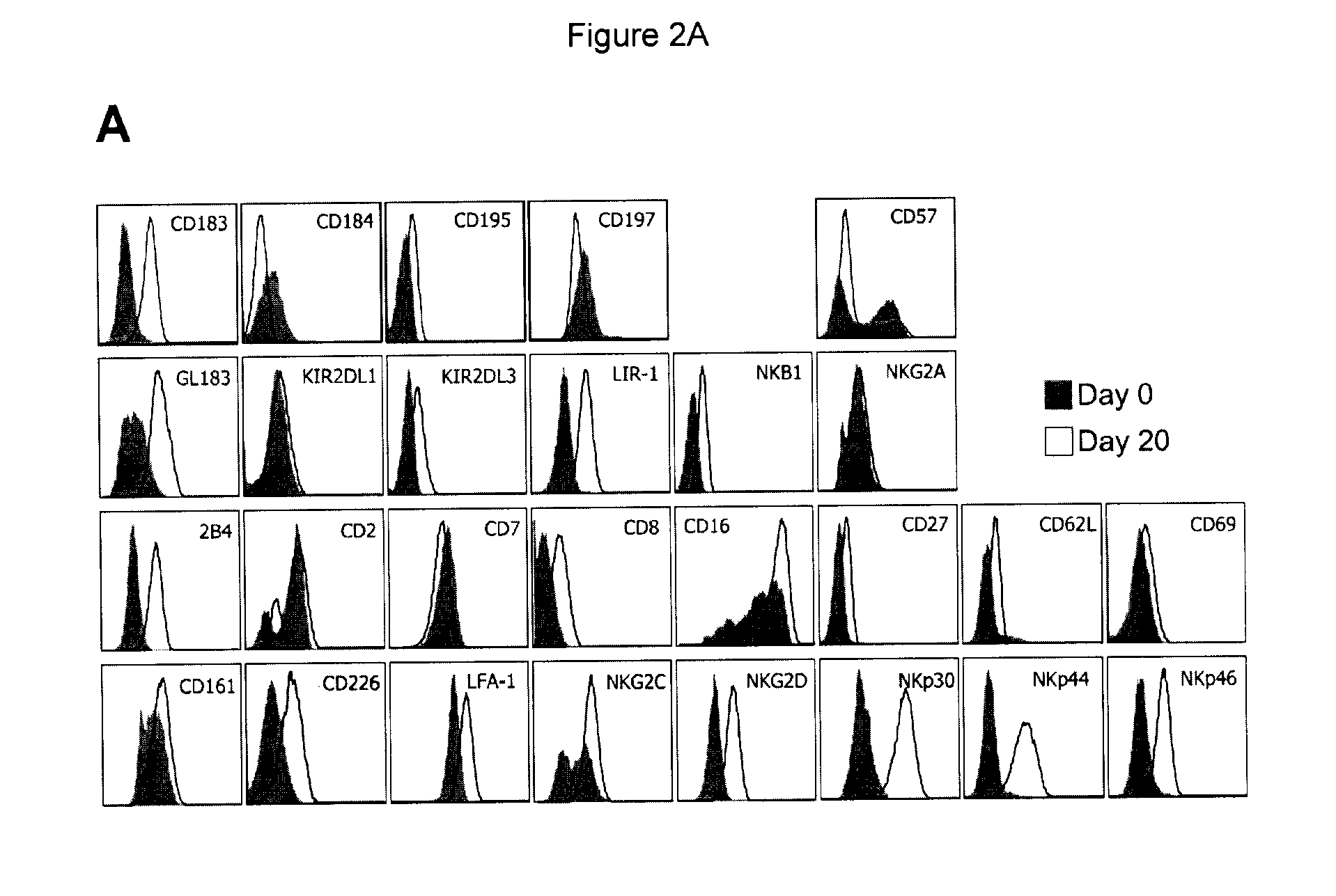
[0031]The increased cytotoxicity of the expanded and activated NK cells can also be an effect mediated by an upregulation of a combination of receptors. Thus, in yet another embodiment the present invention relates to expanded and activated NK cells having an upregulated expression of more than one receptor. Thus, recognition of the target cell by NK cells can involve a combination of receptors that synergistically deliver activating signals.
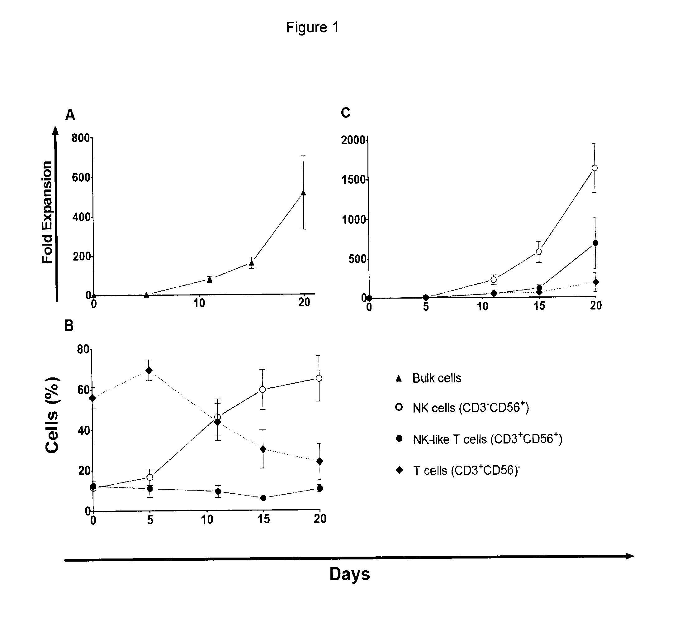
[0032]Another embodiment of the present invention relates to a composition comprising the expanded and active NK cells as described above. In addition to the NK cells, a composition according to the invention may also contain any suitable physiological buffer, such as, but not limited to, phosphate buffered saline. Suitable physiological buffers are well known to the skilled person. Other substances, such as, but not limited to cytokines such as IL-2 and its derivatives, immunomodulatory drugs and proteosome inhibitors, that increase the effect of the administered cells can also be added to the composition or administered separately. In one embodiment the composition also contains lower levels of other cells. Non-expanded PBMCs usually contain a mixture of different cells, for example, NK cells (CD56+CD3−), NK-like T cells (CD56+CD3+) and T cells (CD56−CD3+). After expansion NK cells are the dominating cell type in the culture but also other cell types can be present in the expanded culture.
[0044]Administration, alone or in combination with the NK cells of the invention, of subcutaneous IL-2 or its derivatives as well as immunomodulatory drugs such as, but not limited to, thalidomide or proteosome inhibitors such as, but not limited to, bortezomib, may further increase the effect of administered cells.

Problems solved by technology

However the great majority of patients with MM are incurable due to the persistence of minimal residual disease.
Clinical trials that have been performed using autologous NK cells were hampered by the fact that the cell dose was inadaptable to the demands of clinical trials.

Method used


Image

  • Expanded nk cells
  • Expanded nk cells
  • Expanded nk cells

Examples


example 1

Ex Vivo Expansion of NK Cells from PBMCs

[0055]Material & Methods: The culture conditions for the expansion of cytotoxic cells have previously been optimized on PBMCs from healthy individuals (Carlens et al., Hum. Immunol. 2001; 62:1092-1098). Briefly, PBMCs were initially thawed and cultured in T25 flasks (TPP, Trasadingen, Switzerland) at a concentration of 0.5×10⁶ cells / ml in CellGro SCGM serum-free medium (CellGenix, Freiburg, Germany) with the addition of 5% human serum (Biowhittaker-Cambrex, Md., USA) and 500 U / ml rhlL-2 (Proleukin®, Chiron Corporation, Emeryville, Calif., USA). For the first 5 days, the medium was further supplemented with anti-CD3 antibody (Orthoclone OKT-3, Ortho Biotech Inc., Raritan, N.J., USA) to a final concentration of 10 ng / ml. On day 5 of culture, the OKT-3-containing medium was washed out, and fresh medium with IL-2 (500 U / ml) and 5% human serum was added. The cultures were then replenished with fresh medium every other day throughout the culture per...

example 2

Flow Cytometry Based Phenotyping of NK Cells and NK Ligands on Multiple Myeloma (MM) Cells

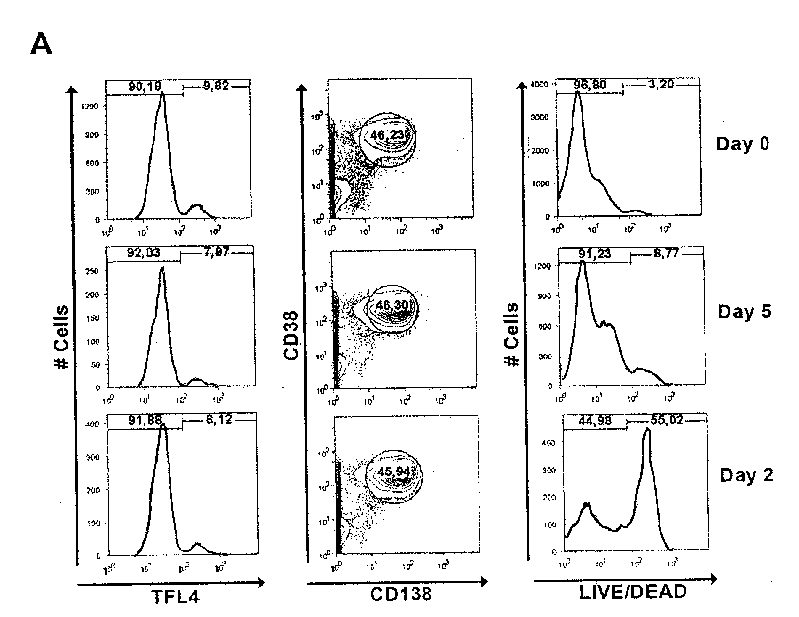
[0057]Material & Methods: The cell phenotype and expansion dynamics of subpopulations were analyzed by flow cytometry on days 0, 5-6, 9-10, 14-15 and 20 of culture using standard procedures with fluorochrome conjugated mAbs against the following surface antigens CD3, CD14, CD38, CD56 and CD138.

[0058]Day 0 and day 20 cells from all patients were subjected to a more detailed immunophenotypic analysis. To avoid inter-acquisition variability, all frozen samples were simultaneously thawed for a detailed phenotypic characterization of CD56+CD3− (NK) cell subset by flow cytometry. This panel included fluorochrome conjugated mAbs against the following surface antigens: CD2 (RPA-2.10), CD3 (UCHT-1), CD4 (SK3), CD7 (M-T701), CD8 (HIT8a), CD14 (MOP9), CD16 (3G8), CD19 (HIB19), CD25 (M-A251), CD27 (M-T271), CD38 (HIT2), CD56 (B159), CD57 (NK-1), CD161 (DX12), CD183 (3D12), CD184 (12G5), CD195 (2D7 / CCR5), C...

example 3

Evaluation of Cell-Mediated Cytotoxicity

[0062]Material & Methods: The cytotoxic capacity of NK cells before and after expansion was evaluated in vitro with a standard 4 hour 51 Cr-release assay against NK-sensitive K562 cells. Because the 51 Crrelease assay is not suitable for primary MM cells 16-18, a flow cytometry based cell mediated cytotoxicity assay was used.

[0063]For the 51 Cr-release assay, K562 target cells were labelled with 100 μCi of 51Cr for one hour at 37° C., washed twice with PBS, and resuspended in 1 ml of RPMI medium. A total of 3×10⁴ target cells in 100 μl RPMI medium was placed in triplicates into V-bottom 96-well plates and incubated for 4 hours with 100 μl of effector cells at appropriate concentrations to obtain effector:target ratios from 1:3 to 10:1. Aliquots of supernatants were then counted using a Packard Cobra Auto-Gamma 5000 Series Counting System (Meriden, Conn., USA). The percentage of specific 51Cr release was calculated according to the formula: per...


PUM

PropertyMeasurementUnit
densityaaaaaaaaaa
fluorescent cell counting assayaaaaaaaaaa
fluorescence intensityaaaaaaaaaa

Abstract

The present invention relates to expanded NK cells. The NK cells have been expanded ex vivo, are activated and have a cytotoxic phenotype. The cytotoxicity against malignant cells is markedly increased compared to non-expanded NK cells. The invention also relates to a method of treatment.

Description

TECHNICAL FIELD[0001]The present invention relates to expanded natural killer (NK) cells being cytotoxic against malignant cells and a method of treatment of malignant disease.BACKGROUND ART[0002]NK cells are cytotoxic lymphocytes that lyse certain tumor and virus infected cells without any prior stimulation or immunization. NK cells are also potent producers of various cytokines, e.g. IFN-γ, TNF-α, GM-CSF and IL-3. Therefore, NK cells are also believed to function as regulatory cells in the immune system, influencing other cells and responses.[0003]In humans, NK cells are broadly defined as CD56+CD3− lymphocytes. The cytotoxic activity of NK cells is tightly controlled by a balance between the activating and inhibitory signals from receptors on the cell surface. A main group of receptors that inhibits NK cell activation are the inhibitory killer immunoglobulin-like receptors (KIRs). Upon recognition of self MHC class I molecules on the target cells, these receptors deliver an inhib...

Claims


Application Information

Patent Timeline
Patent Type & Authority Applications(United States)
IPC IPC(8): A61K35/12C12N5/08A61P35/00A61K39/00C12N5/0783
CPCA61K35/12A61K2039/5158C12N2501/515C12N2501/23C12N5/0646A61P35/00A61K2039/804A61K2239/46A61K39/4613A61K39/4644A61K35/17A61K2035/124C12N2500/90A61K2039/572A61K2039/585C12N2500/32C12N2500/44C12N2500/84C12N2501/2302C12N2501/33
Inventor DILBER, SIRACALICI, EVREN
Owner CELLPROTECT NORDIC PHARMA
Learn More