Automated Detection of Cell Colonies and Coverslip Detection Using Hough Transforms

a technology of hough transform and cell colony, applied in the field of automatic microscopy, can solve the problems of slow system throughput, inability to use morphological methods that enhance the image of colonies, and inability to achieve the effect of enhancing the image of colonies

Active Publication Date: 2009-05-21
LEICA BIOSYST IMAGING
View PDF5 Cites 22 Cited by
  • Summary
  • Abstract
  • Description
  • Claims
  • Application Information

AI Technical Summary

Benefits of technology

[0012]In one embodiment, a method for an automatic detection of cell colonies, includes: acquiring digital images of sub-areas of a slide; generating background intensity images; forming background-subtracted sub-area images by subtracting the minimum of a morphologically processed image and the original digital image from the original image; generating shrunk sub-area images by shrinking the background-subtracted sub-area images by a predefined scale factor; stitching the shrunk sub-area images into a composite image representing the entire slide; converting the composite image into a binary edge image by the application of an automatic thresholding technique; passing the binary edge image through a Hough transform to produce a Hough space transformed image; analyzing the Hough space transformed image to identify peaks representing a coverslip edge location; subjecting the Hough space transformed image to an inverse Hough transform to convert the peaks back to the edges of the coverslip in the binary edge image; setting to zero all pixels of the comp...

Problems solved by technology

Operators are required to manually identify colonies (by drawing around them) resulting in slow system throughput.
Furthermore, focus mapping can be slow or inaccurate due to sparse cell populations on colony slides.
However, those methods do not use morphological methods that enhance the image of the colonies of interest, neither do they associate the metaphases with the colonies.
They also do not disclose a coverslip detection.
They do not disclose background subtraction, morphological methods for colony detection, thresholding, association of metaphases with the colonies, or the coverslip detection.
An accurate identification of the edges of a coverslip on a...

Method used


Image

  • Automated Detection of Cell Colonies and Coverslip Detection Using Hough Transforms
  • Automated Detection of Cell Colonies and Coverslip Detection Using Hough Transforms
  • Automated Detection of Cell Colonies and Coverslip Detection Using Hough Transforms

Examples


Embodiment Construction

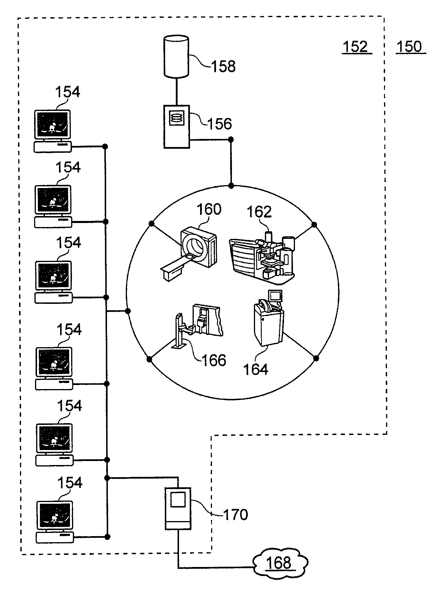
[0029]The embodiments of the present invention can be used to automatically detect the location of cell colonies on a specimen slide, as a precursor to automatically finding metaphase cells and associating them with each colony. The location of the colonies is determined by image analysis. The image can be generated by scanning a slide on an automated microscope with motorized x, y and z axes, capturing images at multiple positions with a CCD camera and stitching these images into a mosaic representing the entire scanned area. The embodiments of the present invention may also use a Hough transform to identify the position of coverslips over the specimen slides, whereby the search for the colonies can be limited to the area under the coverslip.

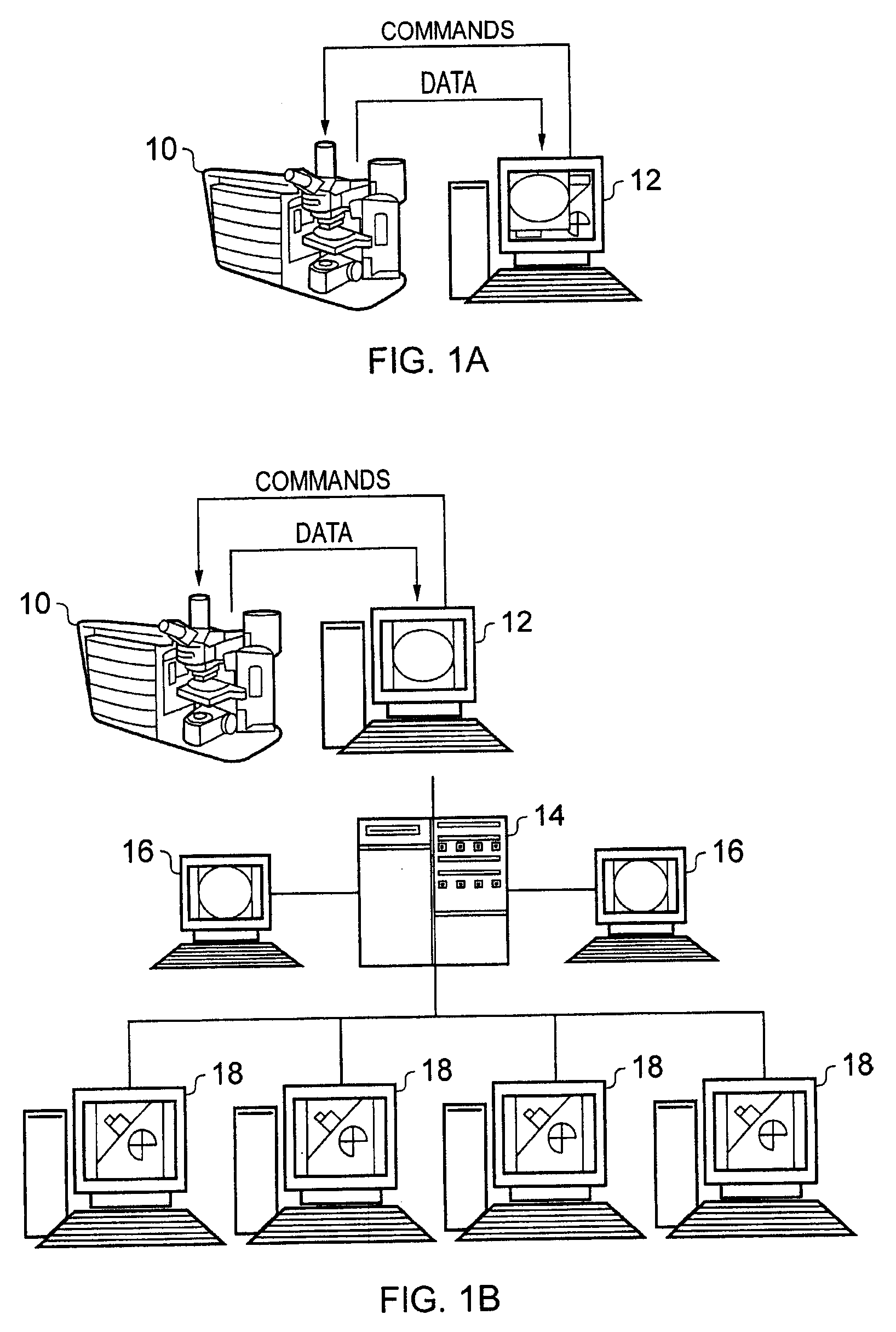
[0030]FIG. 1A schematically illustrates a microscope system for capturing images of a sample. The microscope unit 10 captures digital images of a sample under investigation and the digital images are transferred to computer 12 where they are st...


PUM


Abstract

The present invention provides methods and systems for automatic detection of the location of cell colonies on a specimen slide, in particular under the coverslip of a specimen slide. Slide scanning can be performed using an automated microscope with motorized axes. The location of the colonies can be determined by image analysis, which is followed by automatically finding metaphase cells and associating them with each colony. The invention also provides an automated, Hough-transform-based method for identifying the location of the slide coverslip and, if desired, analyzing only the image area contained within the coverslip.

Description

CROSS-REFERENCES TO RELATED APPLICATIONS[0001]The present application claims benefit of two provisional applications: 60 / 981,712 (Automated Detection of Cell Colonies) and 60 / 981,694 (Automated Coverslip Detection by Image Analysis Using Hough Transforms), both filed on Oct. 22, 2007, the entire contents of which are herein incorporated by reference.BACKGROUND OF THE INVENTION[0002]The present invention relates generally to automated microscopy, and more specifically to improvements in the automatic detection of cell colonies' location on a glass sample slide. Additionally, the glass sample slide may be covered with a coverslip that protects cell colonies from contamination and damage. The coverslip area is that area of a specimen slide where most of focus mapping, image capture and image analysis needs to take place, because cell colonies reside underneath the coverslip. Thus the edges of the coverslip, which denote the area of interest for automated microscopy, need to be reliably...

Claims


Application Information

Patent Timeline
IPC IPC(8): G06K9/00H04N5/335
CPCG01N33/5005G02B21/34G06K9/00127G06T7/0012G06T7/0083G06T2207/30024G06T2207/20036G06T2207/20061G06T2207/20144G06T2207/20224G06T2207/10056G06T7/12G06T7/194G06V20/69
Inventor GREGSON, MARK
Owner LEICA BIOSYST IMAGING
Why Eureka
Try Eureka