Accurate Pelvic Fracture Detection for X-Ray and CT Images

Active Publication Date: 2012-06-07
VIRGINIA COMMONWEALTH UNIV
View PDF2 Cites 47 Cited by
  • Summary
  • Abstract
  • Description
  • Claims
  • Application Information

AI Technical Summary

Benefits of technology

[0013]According to another aspect of the invention, the system and method employs advanced image processing techniques to analyze pelvic CT images to correctly segment bone tissue in a fully automated manner. The invention implements wavelet processing, Laplacian filtering, morphology operations, a series of region growing techniques and gradient based segmentation methods to create an automated segmentation system and provides an automated decision making system that provides physicians with reliable recommendations for the treatment of traumatic pelvic injuries.
[0014]According to a further aspect of the inventi

Problems solved by technology

These maintain the natural curvature of the pelvic structure and help prevent the model from converging to false edges; a common problem in low resolution X-ray images.
Although splines have been used in medica

Method used


Image

  • Accurate Pelvic Fracture Detection for X-Ray and CT Images
  • Accurate Pelvic Fracture Detection for X-Ray and CT Images
  • Accurate Pelvic Fracture Detection for X-Ray and CT Images

Examples


Embodiment Construction

[0029]The embodiment of the invention is described in terms of a system on which the methods of the invention may be implemented. The system is composed of various imaging components, databases and computational interfaces that one of ordinary skill in the computational arts will be familiar with. The methods of the invention are described with reference to flowcharts which illustrate the logic of the processes implemented. The flowcharts and the accompanying descriptions are sufficient for one of ordinary skill in the computer programming and image processing arts to prepare the necessary code to implement the embodiment of the invention.

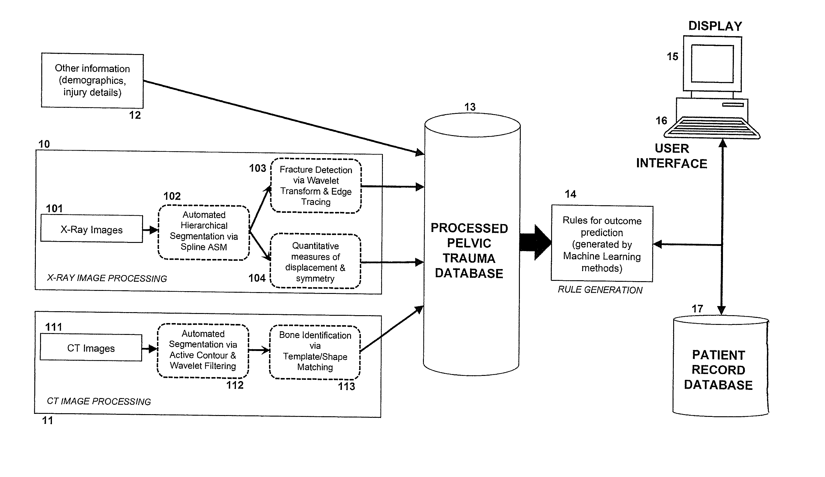
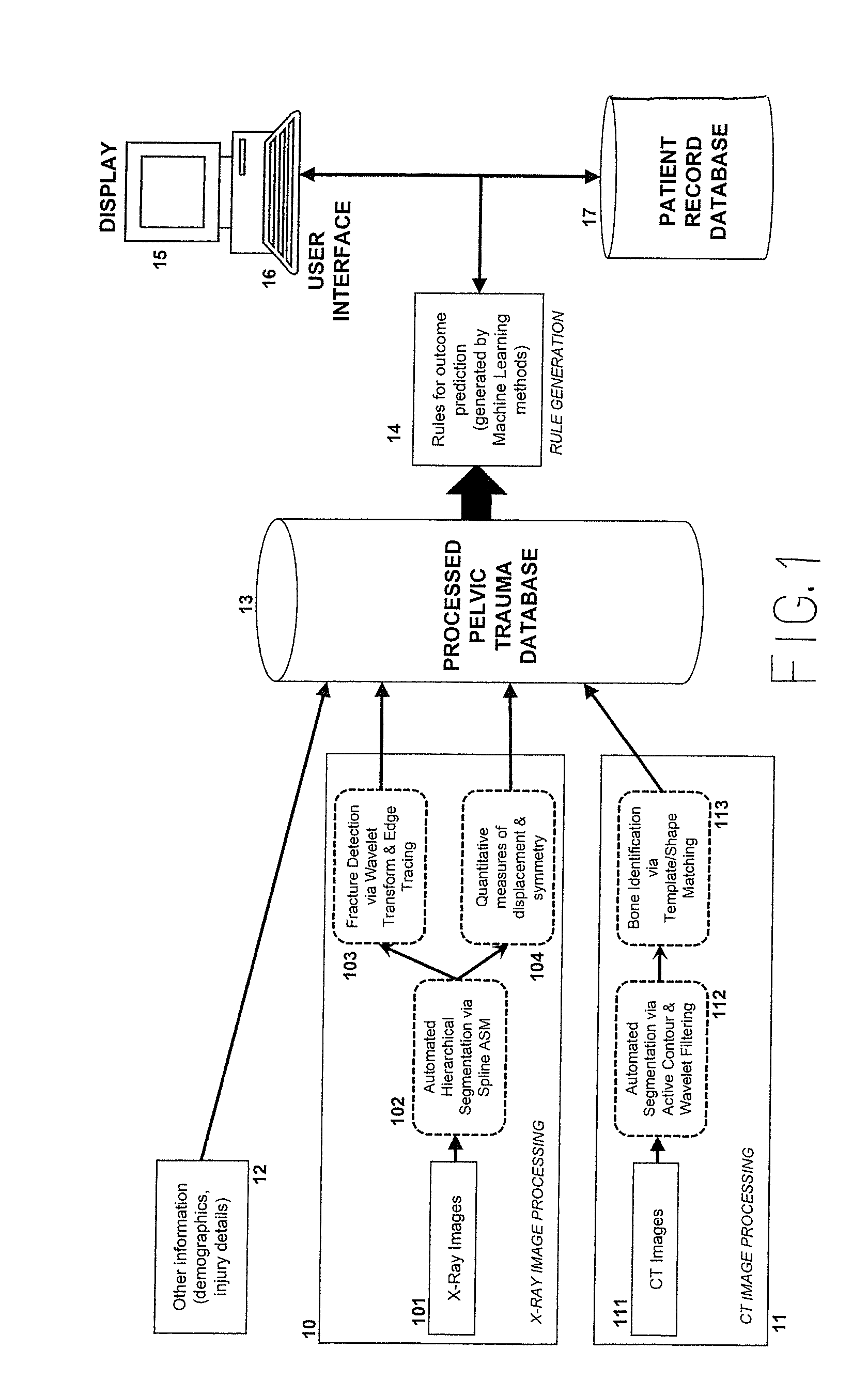
[0030]Referring now to the drawings, and more particularly to FIG. 1, there is presented in block diagram form an overview of the decision support system framework, and the function that the X-ray and CT components serve. X-ray image processing is performed at 10, the X-ray images being input at 101. CT image processing is performed at 11, the CT i...


PUM


Abstract

Accurate pelvic fracture detection is accomplished with automated X-ray and Computed Tomography (CT) images for diagnosis and recommended therapy. The system combines computational methods to process images from two different modalities, using Active Shape Model (ASM), spline interpolation, active contours, and wavelet transform. By processing both X-ray and CT images, features which may be visible under one modality and not under the other are extracted and validates and confirms information visible in both. The X-ray component uses hierarchical approach based on directed Hough Transform to detect pelvic structures, removing the need for manual initialization. The X-ray component uses cubic spline interpolation to regulate ASM deformation during X-ray image segmentation. Key regions of the pelvis are first segmented and identified, allowing detection methods to be specialized to each structure using anatomical knowledge. The CT processing component is able to distinguish bone from other non-bone objects with similar visual characteristics, such a blood and contrast fluid, permitting detection and quantification of soft tissue hemorrhage. The CT processing component draws attention to slices where irregularities are detected, reducing the time to fully examine a pelvic CT scan. The quantitative measurement of bone displacement and hemorrhage area are used as input for a trauma decision-support system, along with physiological signals, injury details and demographic information.

Description

STATEMENT OF GOVERNMENT INTEREST[0001]The U.S. Government has a paid-up license in this invention and the right in limited circumstances to require the patent owner to license on reasonable terms as provided for by the terms of Grant No. 05-0033-02 awarded by U.S. Medical Research and Material Command Combat Casualty Care Research Program and by the terms of NSF Grant No. IISO758410.DESCRIPTIONBackground of the Invention[0002]1. Field of the Invention[0003]The present invention generally relates to a system and method for the application of advanced image processing techniques to detect key pelvic structures and, more particularly, to the automated analysis of X-ray radiographs and Computed Tomography (CT) images to provide diagnostic recommendations for physicians and radiologists. As used herein, “X-ray images” refer to plain film radiographs.[0004]2. Background Description[0005]Pelvic fractures are among the most life-threatening injuries that can be suffered by a major trauma pa...

Claims


Application Information

Patent Timeline
IPC IPC(8): A61B6/00G06F15/18
CPCA61B6/032A61B6/505A61B6/5217A61B6/5235G06T2207/30008G06T7/0081G06T2207/10081G06T2207/10116G06T2207/20212G06T7/0012G06T7/11G16H50/30
Inventor NAJARIAN, KAYVANVASILACHE, SIMINASMITH, REBECCAWARD, KEVIN R.
Owner VIRGINIA COMMONWEALTH UNIV
Why Eureka
Try Eureka