
Biopsy Needle Assembly and a Device for Taking a Tissue Sample

Inactive Publication Date: 2009-06-04
JANSSENS JAAK PH
View PDF12 Cites 29 Cited by
  • Summary
  • Abstract
  • Description
  • Claims
  • Application Information

AI Technical Summary

Benefits of technology

[0010]It is an advantage of the present invention that consecutive tissue samples may be taken without the need of removing the biopsy needle assembly after each sample been taken. It is as well an advantage of the present invention that EMI interferences, e.g. from the driving motors may be avoided.
[0011]An advantage of the present invention is the combined action of a helix and suction or a vacuum to localize the specimen into the receiving room during and before cutting.
[0012]Another advantage is the lack of noise during the biopsy procedure. Normal vacuum assisted biopsies make a suction noise in the tubings and vacuum generator.
[0013]A biopsy needle assembly for taking tissue sample as subject of the present invention comprises
[0014]an outer cannula having a front end and a back end, the front end being for insertion in a tissue and having a cutting edge for cutting a tissue sample. The cannula serves also as coaxial guide in order to prevent seeding by repeated punctures;
[0015]a hollow biopsy needle located inside and coaxially with this cannula; the hollow biopsy needle has an inner cavity, a front end and a back end, the front end being provided with a tissue receiving means; this tissue receiving means cooperating with the cutting edge of the cannula for receiving a cut tissue sample;

Problems solved by technology

The use of a vacuum unit coupled to the assembly by means of tubing, has the disadvantage that the tubing may hinder the physician in his or her work.
This requires significant size increase of the vacuum unit.
It is also a disadvantage of the presently known vacuum assisted biopsy needle assemblies that an additional manipulation or action of the physician is needed in order to activate the vacuum at the end of the biopsy needle.
As medical interventions become more and more complex, such additional action to activate the vacuum may easily be overlooked.
It is as well a disadvantage that most often, when a physician is to take several consecutive tissue samples, the biopsy needle assembly is to be brought in as many times as tissue samples are needed.
This obviously causes discomfort for both patient and physician, may result in more damage caused to the patient by the consecutive penetration of the body and may cause seeding of diseased, e.g. tumour cells along the extraction pathway.
It is also a disadvantage of presently known assemblies, that driving means such as motors are located in the hand hold device, which may cause interference, such as EMI interference, with the other apparatuses used during the medical manipulation, e.g. scanning equipment such as MRI scanners which are used in conjunction with the biopsy needle to ensure accurate placing of the needle tip.

Method used


Image

  • Biopsy Needle Assembly and a Device for Taking a Tissue Sample
  • Biopsy Needle Assembly and a Device for Taking a Tissue Sample
  • Biopsy Needle Assembly and a Device for Taking a Tissue Sample

Examples


Embodiment Construction

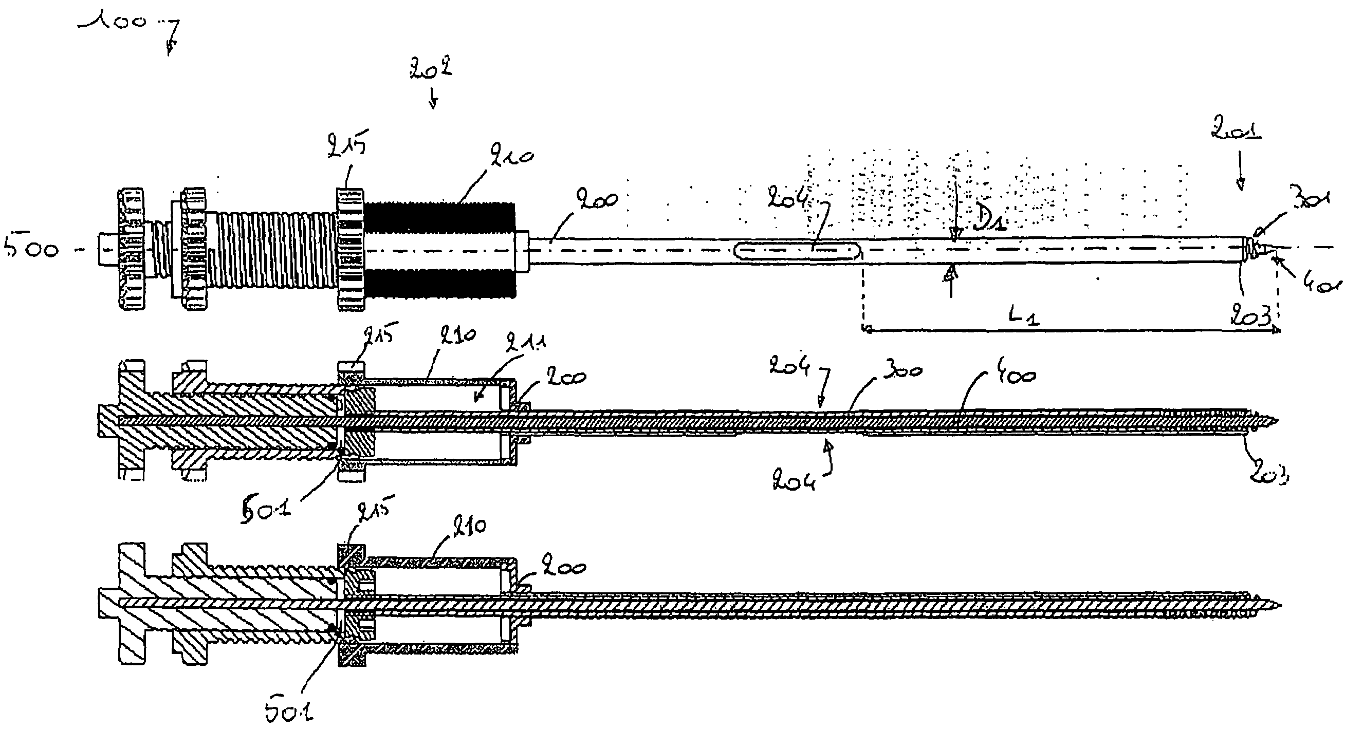
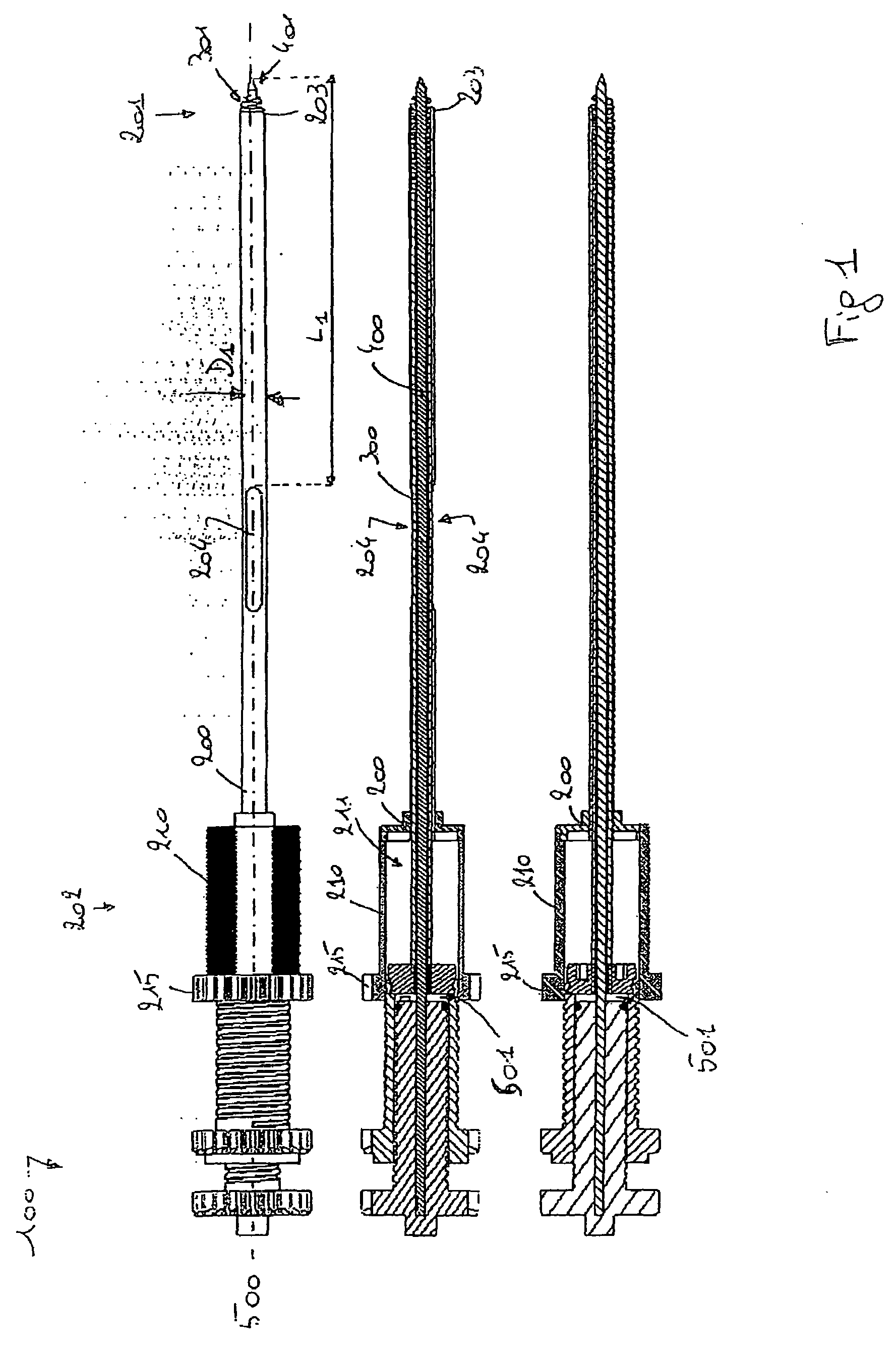
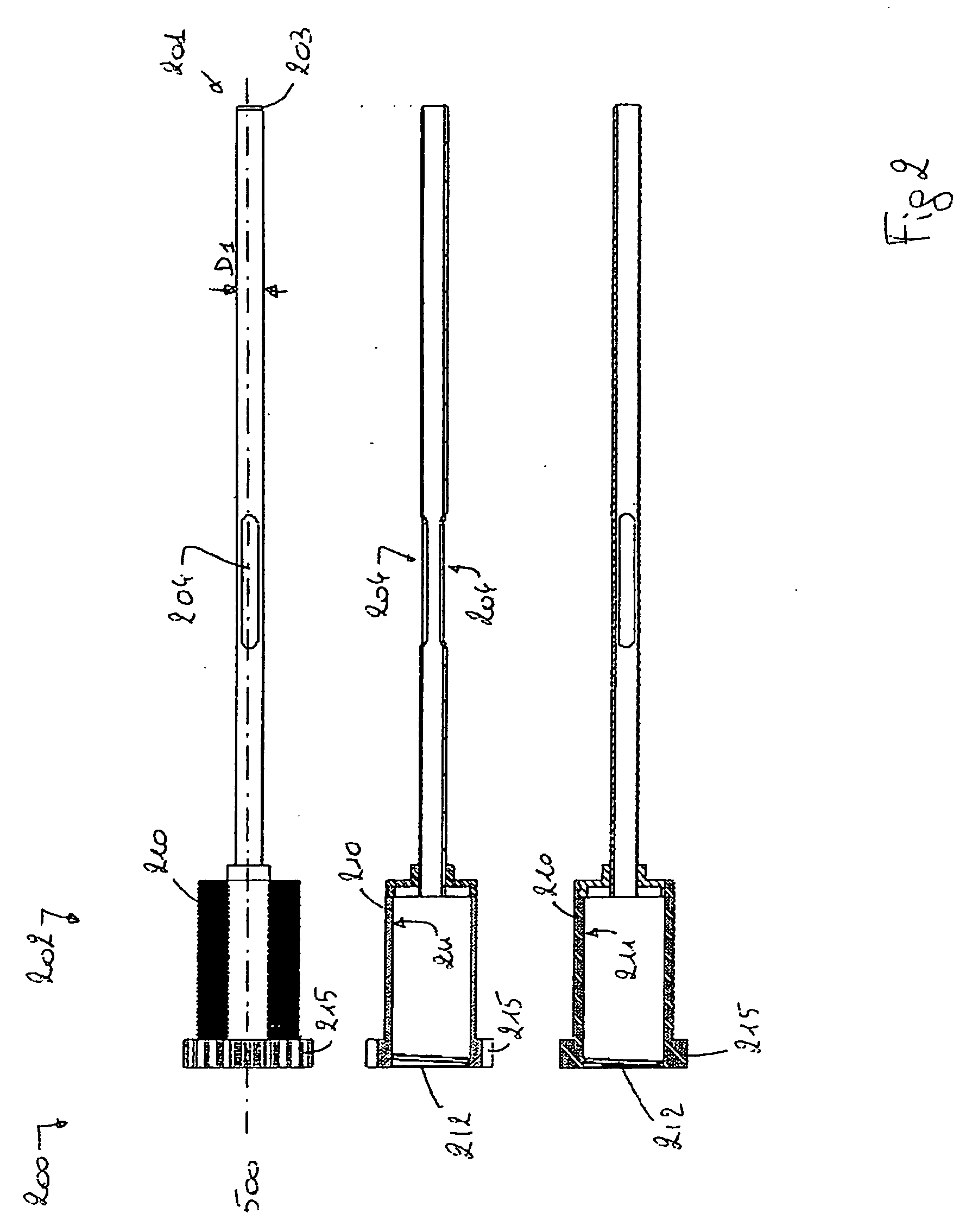
[0049]The present invention will be described with respect to particular embodiments and with reference to certain drawings but the invention is not limited thereto but only by the claims. The drawings described are only schematic and are non-limiting. In the drawings, the size of some of the elements may be exaggerated and not drawn on scale for illustrative purposes.

[0050]The dimensions and the relative dimensions do not correspond to actual reductions to practice of the invention.

[0051]Furthermore, the terms first, second, third and the like in the description and in the claims, are used for distinguishing between similar elements and not necessarily for describing a sequential or chronological order. It is to be understood that the terms so used are interchangeable under appropriate circumstances and that the embodiments of the invention described herein are capable of operation in other sequences than described or illustrated herein.

[0052]Moreover, the terms top, bottom, over, ...


PUM


Abstract

A biopsy needle assembly as subject of the present invention for taking tissue sample comprises an outer cannula having a front with a cutting edge end and a back end; a hollow biopsy needle located inside and coaxially with the cannula, having an inner cavity. The front end if the needle is provided with a tissue receiving means cooperating with the cutting edge of cannula; an inner needle having located inside and coaxially with the hollow biopsy needle; a vacuum unit coupled to the hollow biopsy needle; the cannula and hollow biopsy needle being mutually axially moveable. The vacuum unit is adapted to create a lowered air pressure in the inner cavity when the front end of the hollow biopsy needle is axially moved away from the front end of the inner needle. The vacuum unit is located uniaxially with the cannula and hollow biopsy needle. The invention further relates to a device for taking a tissue sample by means of a biopsy needle assembly as subject of the present invention.

Description

TECHNICAL FIELD OF THE INVENTION[0001]The present invention relates to biopsy needles and biopsy needle assemblies and devices for sampling tissue from human or animal body, using such biopsy needles or needle assemblies.BACKGROUND OF THE INVENTION[0002]Biopsy needle assemblies are already well known in different forms. As an example, US2003 / 0114773A1 describes a biopsy needle assembly comprising an outer cannula for insertion in a tissue of which the front end has a cutting edge for cutting the tissue sample. The biopsy needle assembly comprises further a hollow biopsy needle located inside and coaxially with the cannula, at the front end being provided with a tissue receiving means, such as a helical screw. The tissue receiving means cooperates with the cutting edge of cannula for receiving cut tissue sample. The biopsy needle assembly further can comprise an inner needle or localisation needle, having a front end and a back end, this inner needle being located inside and coaxiall...

Claims


Application Information

Patent Timeline
IPC IPC(8): A61B10/02
CPCA61B10/0266A61B2010/0208A61B10/0283
Inventor JANSSENS, JAAK PH.
Owner JANSSENS JAAK PH
Features
  • Generate Ideas
  • Intellectual Property
  • Life Sciences
  • Materials
  • Tech Scout
  • Unparalleled Data Quality
  • Higher Quality Content
  • 60% Fewer Hallucinations
Learn More