Method and System for Treating Acute Heart Failure by Neuromodulation

a neuromodulation and heart failure technology, applied in the field of acute heart failure treatment methods and systems, can solve the problems of increased heart contractility, increased myocardium consumption, and excess mortality caused by cardiac arrhythmias

Active Publication Date: 2009-07-02
THE CLEVELAND CLINIC FOUND
View PDF56 Cites 12 Cited by
  • Summary
  • Abstract
  • Description
  • Claims
  • Application Information

AI Technical Summary

Benefits of technology

[0005]In an embodiment, the present invention provides a method of treating acute heart failure in a patient in need thereof comprising inserting a delivery device into a pulmonary artery and positioning the delivery device at a pulmonary trunk of the pulmonary artery. The method also comprises applying a therapy signal to at least one sympathetic cardiopulmonary fiber surrounding the pulmonary trunk to treat the acute heart failure. The at least one sympathetic cardiopulmonary fiber affects heart contractility more than heart rate.
[0...

Problems solved by technology

However, inotropic agents have both chronotropic and inotropic effects and characteristically increase heart contractility at the expense of significant increments in oxygen consumption secondary to elevations in heart rate.
As a r...

Method used


Image

  • Method and System for Treating Acute Heart Failure by Neuromodulation
  • Method and System for Treating Acute Heart Failure by Neuromodulation
  • Method and System for Treating Acute Heart Failure by Neuromodulation

Examples


example 1

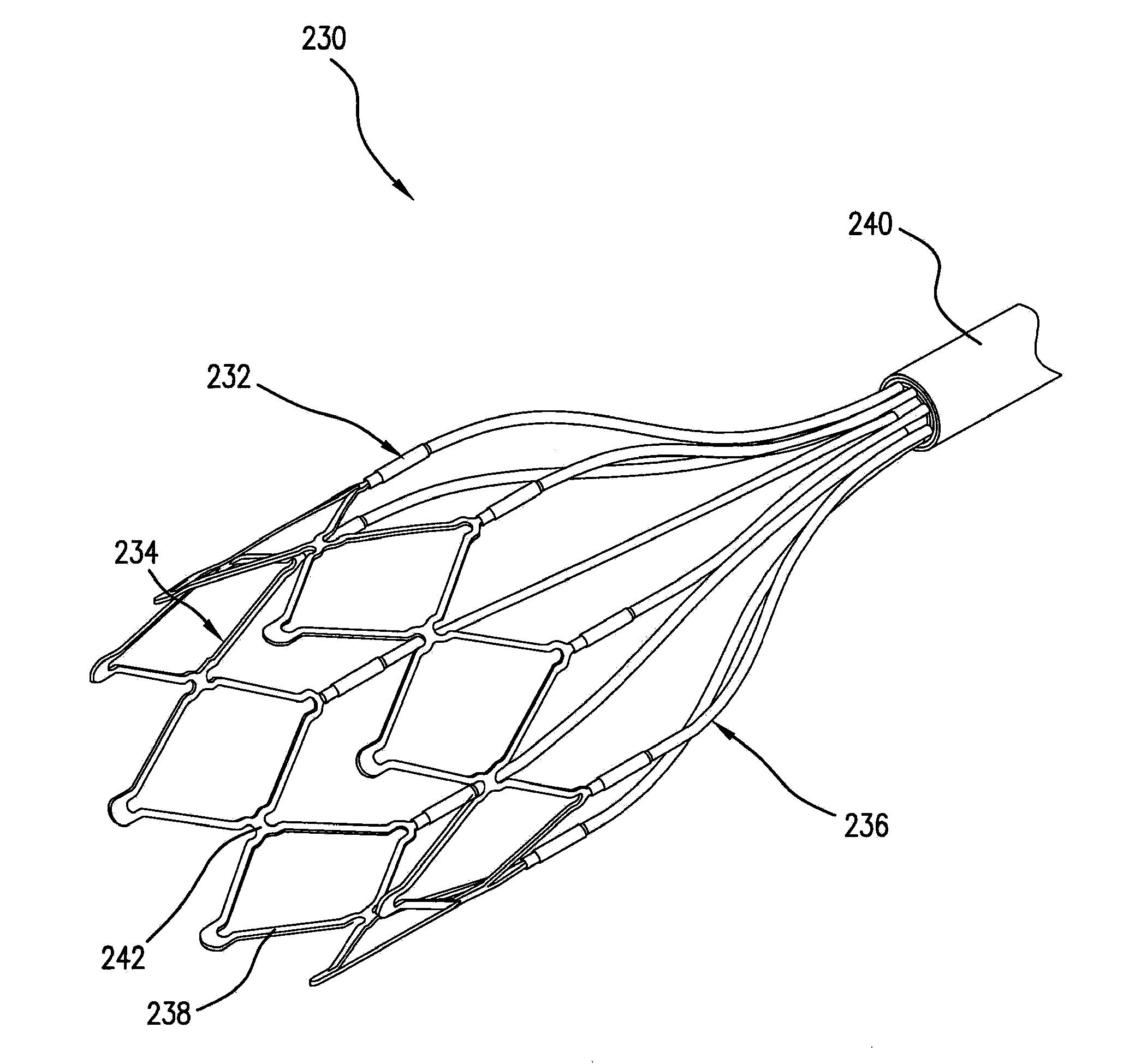
[0031]Six open-chest dogs were instrumented with a left ventricle conductance catheter and an aortic flow probe. Modified electrode-catheters were placed inside the pulmonary artery under echocardiographic and fluoroscopic guidance in five dogs. In the last dog, a stent-based electrode, as illustrated in FIG. 3, was used. Stimulation was applied at 20 Hz, 0.4 ms, and 15-25 mA. The corresponding hemodynamic effects are reported as averages of 30 second periods of continuous recording.

[0032]Pressure variation in the left ventricle over time increased in all dogs. The average increment was 25.7% (+ / −11.8) and the average of maximum increase variation was 28.3 (+ / −8.9). Emax was measured in the last animal, showing a 45% increase. The average reduction of RR interval during stimulation was 3.3% (+ / −10.4).

[0033]Therefore, electrical modulation via a pulmonary artery catheter can produce positive inotropic effects with minimal changes in heart rate.

example 2

[0034]Eight open-chest dogs are instrumented with a left ventricle conductance catheter and an aortic flow probe. Modified electrode-catheters are placed inside the pulmonary artery under echocardiographic and fluoroscopic guidance in five dogs. In three dogs, a stent-based electrode, as illustrated in FIG. 3, is used. Stimulation is applied at 20 Hz, 0.4 ms, and 15-25 mA. The corresponding hemodynamic effects are reported as averages of 30 second periods of continuous recording.

[0035]FIG. 6 shows the percentage changes in dP / dt max for each dog when compared to the baseline. Specifically, FIG. 6 shows the peak dP / dt max % change achieved during modulation and the average % change in dP / dt max in a period of 30 seconds. There is significant increase in dP / dt max during modulation ranging from 21-22 to 44-45%. However, there is no significant change between the average and the peak value.

[0036]FIG. 7 compares the average dP / dt max to the percentage increase in heart rate in the eight...


PUM


Abstract

Methods and systems of treating acute heart failure by applying a therapy signal at least one sympathetic cardiopulmonary fiber surrounding the pulmonary trunk that affects heart contractility more than heart rate. Methods and systems also include adjusting the signal to effectuate treatment.

Description

CROSS-REFERENCE TO RELATED APPLICATIONS[0001]This application claims priority to U.S. Provisional Application No. 60 / 873,021 (filed 6 Dec. 2006), which is incorporated by reference in its entirety herein. The present application is also related to U.S. application Ser. No. 11 / 222,766 filed on Sep. 12, 2005, which is incorporated by reference herein.FIELD OF THE INVENTION[0002]The present invention relates to methods and systems for treating acute heart failure by electrically modulating at least one sympathetic cardiopulmonary fiber.BACKGROUND OF THE INVENTION[0003]Diseases or injuries causing or resulting in acute heart failure are widespread. The goals of therapy in acute heart failure are often to correct the hemodynamic instability and address decompensation in order to increase patient mortality. One treatment option for acute heart failure is the administration of inotropic agents, such as dopamine and dobutamine. However, inotropic agents have both chronotropic and inotropic ...

Claims


Application Information

Patent Timeline
IPC IPC(8): A61N1/365A61B18/18A61N1/362
CPCA61N1/36114A61N1/3627A61M5/14212A61N1/36585A61N1/0565A61B18/1492A61B2018/00267A61B2018/00351A61B2018/00577A61B2018/00648A61B2018/00827A61B2018/00892A61N1/0568
Inventor MACHADO, SANDRAPENN, MARCREZAI, ALI R.SMITH, ALEX
Owner THE CLEVELAND CLINIC FOUND
Why Eureka
Try Eureka