Image normalization for computer-aided detection, review and diagnosis

a computer-aided detection and image normalization technology, applied in image enhancement, image analysis, instruments, etc., can solve the problems of losing important information in digital images, and achieve the effect of robust handling of detection and diagnosis tasks

Inactive Publication Date: 2009-09-24
THREE PALM SOFTWARE
View PDF4 Cites 12 Cited by
  • Summary
  • Abstract
  • Description
  • Claims
  • Application Information

AI Technical Summary

Benefits of technology

[0011]The conventional normalization methods attempt to normalize images from one modality to another modality. For example, in mammography CAD, normalization is usually applied to the digital FFDM or CR images to make their appearance look like digitized film images. The disadvantage of this normalization direction is that it may lose important information in the digital images, such as, skin line at low film OD (optical density) range. This

Problems solved by technology

The disadvantage of this normalization direction is that it may lose important informa

Method used


Image

  • Image normalization for computer-aided detection, review and diagnosis
  • Image normalization for computer-aided detection, review and diagnosis
  • Image normalization for computer-aided detection, review and diagnosis

Examples


Embodiment Construction

[0016]The method of the present invention analyzes individual image characteristics, generates transformation parameters dynamically in an optimized manner, and applies the generated transform to normalize images as they were acquired from a consistent pseudo-modality.

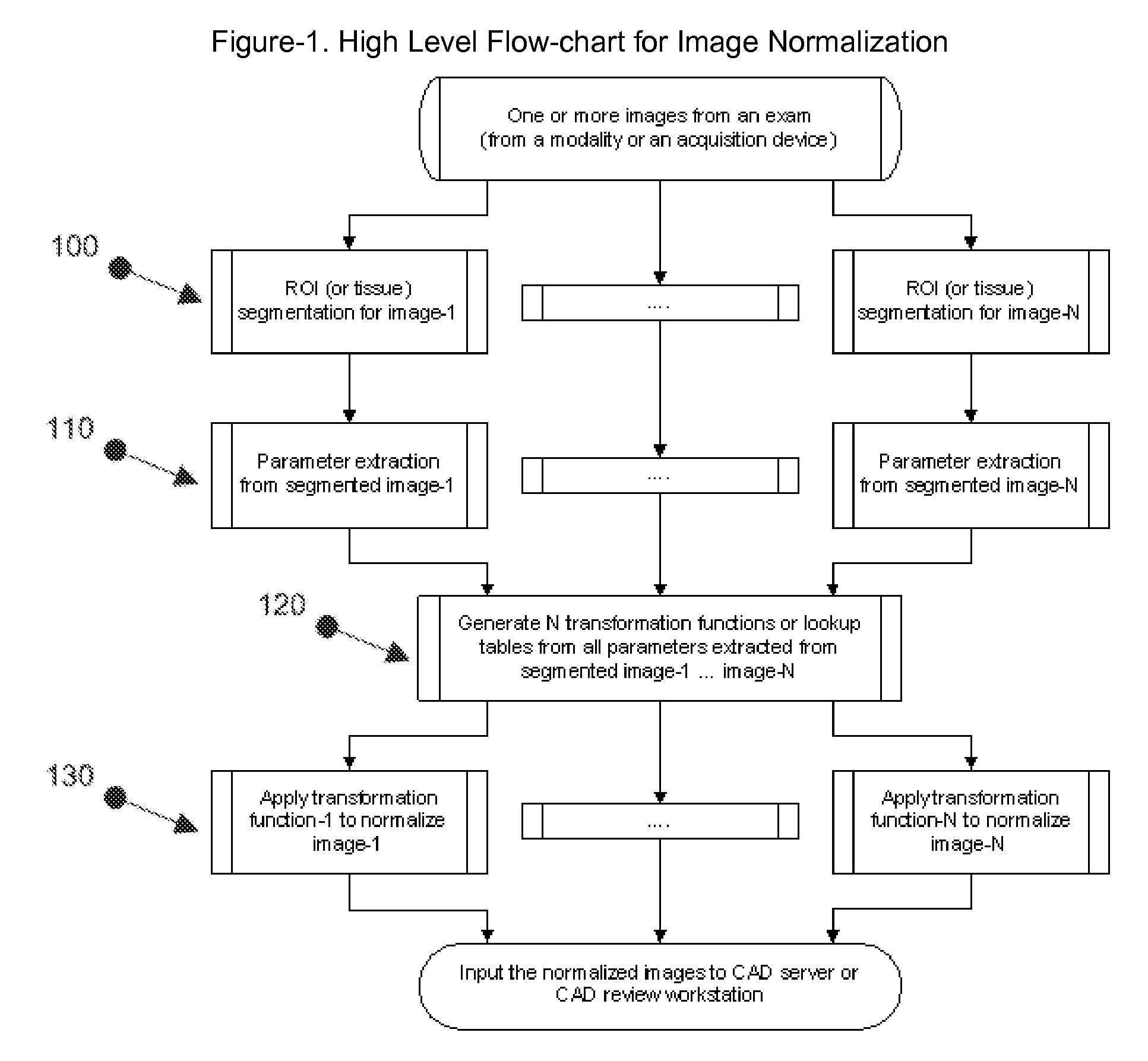
[0017]Referring to FIG. 1, a block diagram of the high level flow-chart for image normalization starts from inputting one or more images from an exam and sometime plus from another exam of the same patient that was taken previously.

[0018]In step 100, segmentation to obtain a region of interest, which usually is a body part, such as breast tissue inside breast border.

[0019]In step 110, a set of image characteristic parameters, such as, mean and standard deviation, for each image are extracted from the region of interest in each image.

[0020]In step 120, all sets of parameters from all images are processed to obtain a set of optimal parameters for each image transformation function.

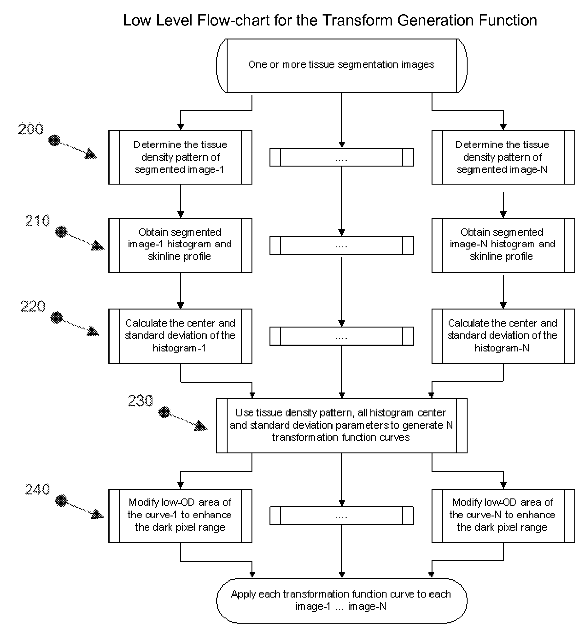
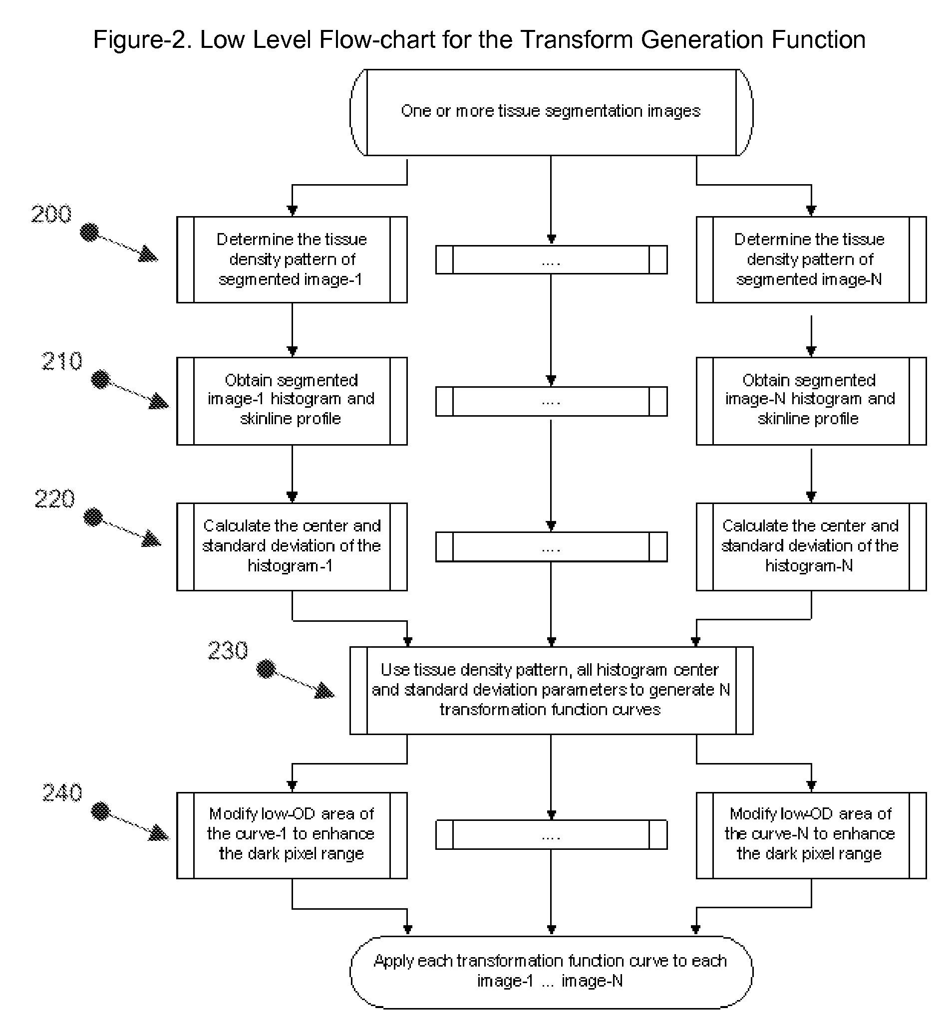
[0021]In step 130, the transformation fu...


PUM


Abstract

A method and apparatus for processing medical images from one of a plurality of digital acquisition modalities or manufacturers with different imaging condition is proposed for creating consistent appearance of the images. The method comprises (1) tissue segmentation to isolate the region of interest; (2) dynamic extraction of the optimal parameters for image transformation from the segmented region; (3) generation of a transformation function from the individual image optimized parameters; and (4) use of the transformation function to produce images that have consistent image characteristics. This method also applies to multiple images from a single study or multiple studies. The transformed images can be used for computer-aided lesion detection, review and diagnosis.

Description

CROSS-REFERENCE TO RELATED APPLICATIONSU.S. Patent Documents[0001]U.S. Pat. No. 7,072,498 B1 July 2006 Roehrig et al. “Method and apparatus for expanding the use of existing computer-aided detection code”[0002]U.S. Pat. No. 6,584,216 B1 June 2003 Nyl et al. “Method for standardizing the MR image intensity scale”[0003]U.S. Pat. No. 6,483,933 B1 November 2002 Shi et al. “Digital-to-film radiographic image conversion including a network”[0004]U.S. 60 / 906,304 March 2007 “Image normalization for computer-aided detection, review and diagnosis”STATEMENT REGARDING FEDERALLY SPONSORED RESEARCH OR DEVELOPMENT[0005]Not Applicable.REFERENCE TO SEQUENCE LISTING, A TABLE, OR A COMPUTER PROGRAM LISTING COMPACT DISC APPENDIX[0006]Not Applicable.BACKGROUND OF THE INVENTION[0007]The present invention relates generally to the field of medical imaging analysis. Particularly, the present invention relates to a method and system for utilizing image normalization algorithms in conjunction with computer-ai...

Claims


Application Information

Patent Timeline
IPC IPC(8): G06K9/00
CPCG06T5/008G06T5/40G06T2207/30068G06T2207/20144G06T2207/20008G06T7/194
Inventor ZHANG, HEIDI DAOXIANHEFFERNAN, PATRICK BERNARD
Owner THREE PALM SOFTWARE
Why Eureka
Try Eureka