Method and composition for hyperthermally treating cells in the eye with simultaneous imaging

a technology of simultaneous imaging and hyperthermia, which is applied in the field of composition for hyperthermia treatment of cells at a site in the body, can solve the problems of insufficient use of chemotherapy drugs alone to treat a tumor or cancerous site, adversely affecting the patient's health, and immediate cell death

Inactive Publication Date: 2009-09-24
GHOLAM A PEYMAN
View PDF7 Cites 25 Cited by
  • Summary
  • Abstract
  • Description
  • Claims
  • Application Information

AI Technical Summary

Benefits of technology

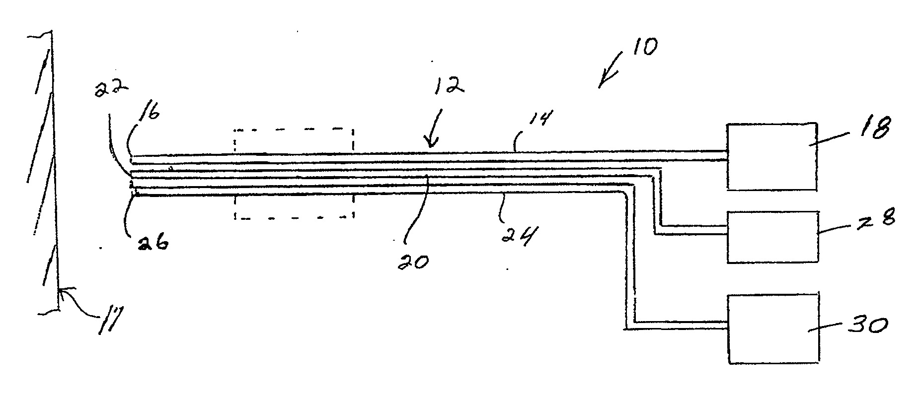
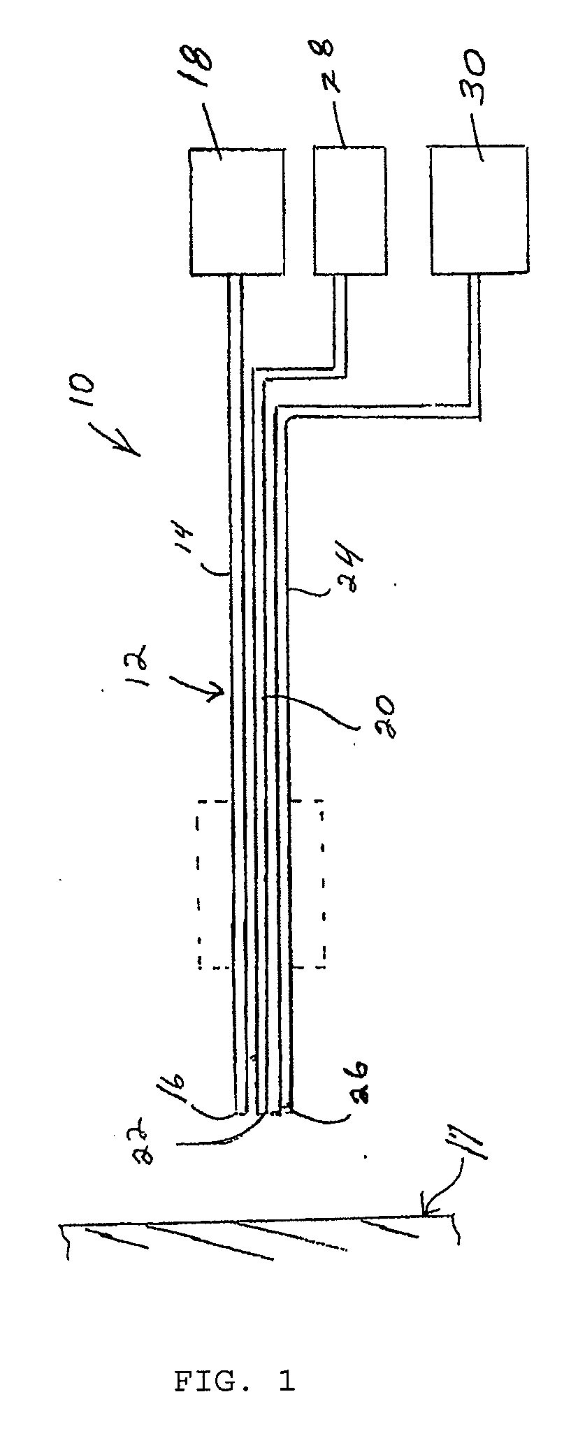
[0024]The aspects of the invention are further attained by providing a method of hyperthermally treating tissue of an animal. The method comprises the step of introducing a temperature indicating substance into the bloodstream of the animal to flow through a target site. The temperature indicating substance includes a first fluorescent dye encapsulated in a first temperature sensitive liposome. The first fluorescent dye is releasable from the first liposome by heating to a temperature of at least 42° C. A second fluorescent dye encapsulated in a second temperature sensitive liposome is also included. The second fluorescent dye is releasable from the second liposome by heating to a temperature of at least 50° C. The target site is heated to a temperature of at least 42° C. The first fluorescent dye is fluoresced to indicate an effective temperature for hyperthermally treating the tissue without releasing the second fluorescent dye from the second liposomes.

Problems solved by technology

Cell death begins to occur when the cells are heated to a temperature of about 5° C. or more above the normal body temperature of 37° C. Applying radiation to a localized site in the body, such as a tumor or other area containing undesired cells, can heat the cells at the site to temperatures in excess of 60° C. Such high temperatures causes a phenomenon known as protein denaturation to occur in the cells, which results in immediate cell death.
Although the techniques mentioned above can be suitable for preventing certain types of cell proliferation and certain sites in the body, several drawbacks with these techniques exist.
For example, often the use of chemotherapy drugs alone to treat a tumor or cancerous site is insufficient to kill the undesired cells.
Moreover, the chemotherapy drugs also kill many normal healthy cells along with the cancerous cells, which can adversely affect the patient's health.
The use of radiation in conjunction with chemotherapy can have a more detrimental effect on the cancerous cells.
However, as with chemotherapy, radiation often kills normal healthy cells, such as those in front of or behind the site of interest, along with the cancerous cells.
Moreover, the intense heating of the cells can cause the cells to coagulate and thus block the capillaries at the site of interest.
In addition, the above techniques have not been used to prevent unwanted cell proliferation at certain locations in the eye, such as at the retina or at the lens capsule.
Because the retina is very sensitive, conventional radiation techniques can be too severe to treat cancerous cells on, in or under the retina.
This growth can result in the cells covering all or a substantial portion of the front and rear surfaces of the lens capsule, which can cause the lens capsule to become cloudy and thus adversely affect the patient's vision.
However, it is often difficult to remove all of the unwanted cells.
Hence, after time, the unwanted cells typically will grow back, thus requiring further surgery.

Method used


Image

  • Method and composition for hyperthermally treating cells in the eye with simultaneous imaging
  • Method and composition for hyperthermally treating cells in the eye with simultaneous imaging

Examples


Embodiment Construction

[0027]The present invention is directed to a method and composition for hyperthermally treating tissue. In particular, the invention is directed to as method for heating tissue above a temperature effective to kill tissue cells or inhibit multiplication of cells below the protein denaturization temperature of the tissue.

[0028]The method of the invention introduces a composition into the bloodstream of the body in a location to flow into or through a target site to be treated. A heat source is applied to the target site to heat the tissue in the target site for a time sufficient to hyperthermally treat the tissue and activate the composition. As used herein, the term “hyperthermal” refers to a temperature of the cell or tissue that kills or damages the cells without protein denaturization.

[0029]The composition contains a temperature indicator that is able to provide a visual indication when a minimum or threshold temperature is attained that is sufficient to hyperthermally treat the ...


PUM


Abstract

A method and composition for hyperthermally treating cells in the eye with simultaneous imaging. The heat (temperature) production inside the eye ( target) tissue is imaged. The desired temperature is achieved using a laser and photoacoustic imaging technique. Hyperthermia treatment of tissue in a target site applies a heat source to kill cells without protein denaturation. The method introduces an encapsulated dye that is released at a selected temperature in the target site to indicate that a threshold temperature has been reached to hyperthermally treat the tissue. In one embodiment, the composition releases the dye at a temperature of 42° C. to 50° C., and preferably about 45° C. to 49° C. The composition which can be a liposome composition encapsulating the dye can be introduced to the bloodstream of the patient to flow through the target site.

Description

[0001]This application is a Continuation-in-Part of U.S. patent application Ser. No. 11 / 485,352, filed Jul. 13, 2006, the entirety of which is hereby expressly incorporated by reference herein.FIELD OF THE INVENTION[0002]The present invention relates to a method and composition for hyperthermally treating cells at a site in the body. More particularly, the present invention relates to a method for treating cells at a target site in the body, such as at a lens capsule of an eye, tumors, and exudative ARM (age related macular degeneration) by applying thermal energy to the target site to heat the cells to a temperature which will kill the cells or impede cell multiplication without exceeding the protein denaturation temperature of the tissue.BACKGROUND OF THE INVENTION[0003]Several techniques currently exist for treating cells at a selected site in the body with heat or chemicals to kill or impede multiplication of those cells to prevent undesired cell proliferation. For example, nume...

Claims


Application Information

Patent Timeline
Patent Type & Authority Applications(United States)
IPC IPC(8): A61F9/007A61B18/04A61B8/00A61M37/00
CPCA61K49/222A61K41/0052
Inventor PEYMAN, GHOLAM A.
Owner GHOLAM A PEYMAN
Why Eureka
Try Eureka