Medical Imaging Processing and Care Planning System

a medical imaging and care planning technology, applied in the field of medical radiation therapy system, can solve the problems of ineffective or impaired radiotherapy treatment, mis-identifying images of patients, and known systems for patient radiotherapy treatment, and achieve the effect of detection and more accurate treatmen

Active Publication Date: 2009-10-22
SIEMENS MEDICAL SOLUTIONS USA INC
View PDF9 Cites 55 Cited by
  • Summary
  • Abstract
  • Description
  • Claims
  • Application Information

AI Technical Summary

Benefits of technology

[0004]A system enables detection and more accurate treatment of secondary occurrences of cancerous lesions in radiotherapy patients, for example, by automatic comparison of radiotherapy 3D X-Ray images taken on different treatment occasions and stages for update and re-planning of treatment and for verification of correct patient and image association. A medical radiation therapy system and workflow includes a task processor for providing task management data for initiating image comparison tasks prior to performing a session of radiotherapy. An image comparator, coupled to the task processor, in response to the task management data, compares a first image of an anatomical portion of a particular patient used for planning radiotherapy for the particular patient, with a second image of the anatomical portion of the particular patient obtained on a subsequent date, by image alignment and comparison of image element representative data of aligned first and second images to determine an image difference representative value and determines whether the image difference representative value exceeds a first predetermined threshold. An output processor, coupled to the image comparator, initiates generation of an alert message indicating a need to review planned radiotherapy treatment for communication to a user in response to a determination that the image difference representative value exceeds a predetermined threshold.

Problems solved by technology

Known systems for patient radiotherapy treatment are also vulnerable to mis-identifying images or a patient associated with an image.
This results in potentially ineffective or impaired radiotherapy treatment.

Method used


Image

  • Medical Imaging Processing and Care Planning System
  • Medical Imaging Processing and Care Planning System
  • Medical Imaging Processing and Care Planning System

Examples


Embodiment Construction

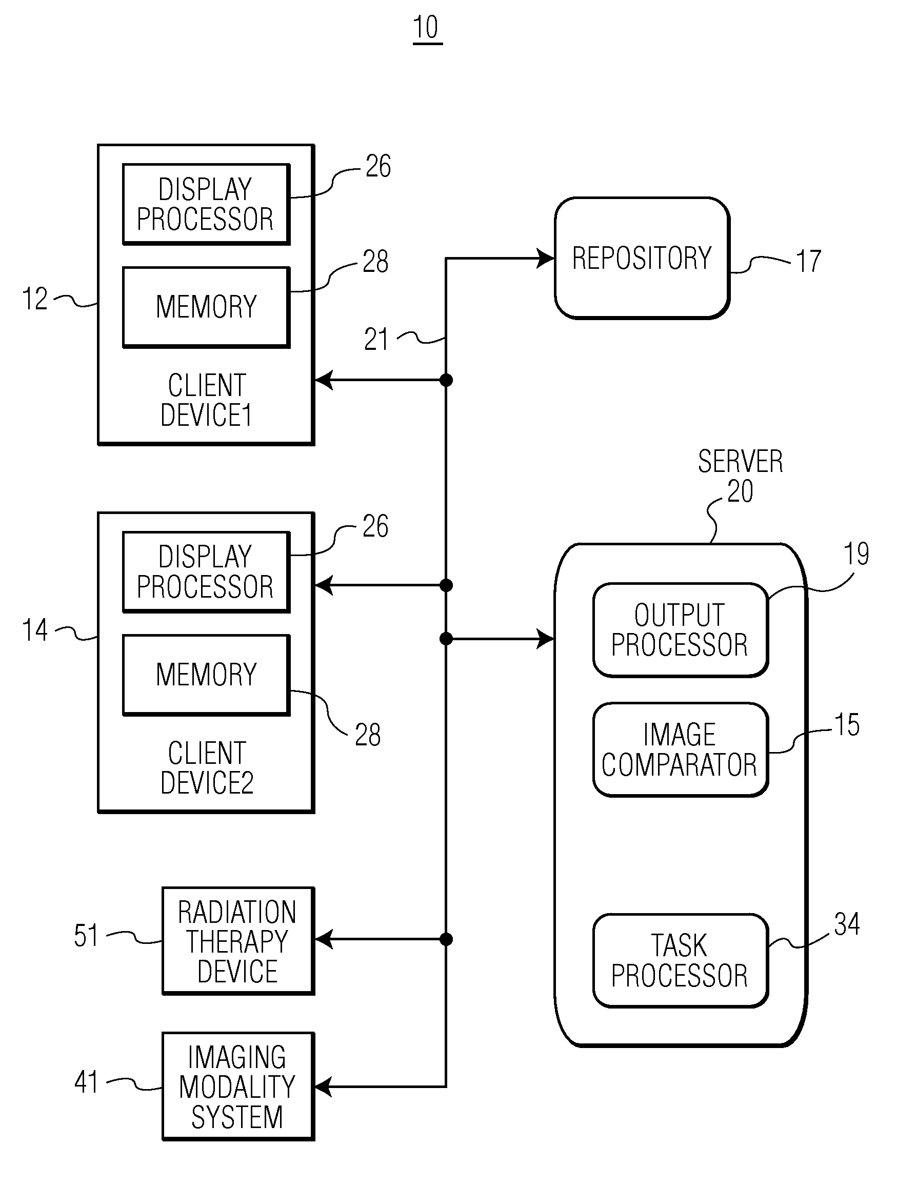
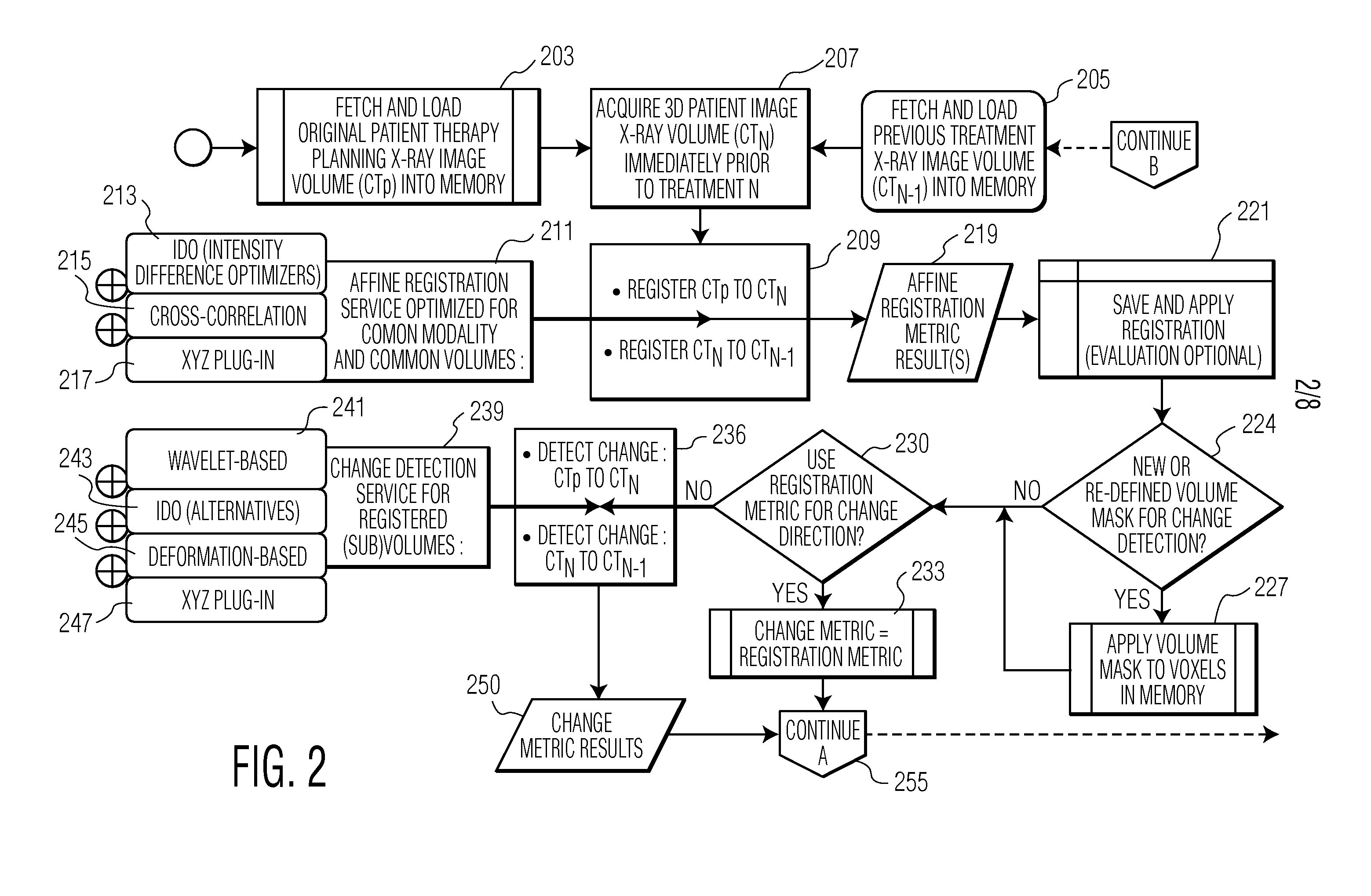
[0012]A system enables detection and more accurate treatment of secondary occurrences of cancerous lesions in radiotherapy patients, for example. In radiotherapy, 3D X-Ray initial images are acquired to plan a treatment process. Subsequent images (of which many dozen may be taken) may be used to position a patient, but are typically not used to re-plan and refine the treatment process, nor are subsequent images used to incrementally or in ensemble, determine anatomical changes (substantive or otherwise) with respect to an initial planning image. The system provides updated and re-planned treatment based on a more accurate assessment of patient condition and targeting of affected cancerous regions. Furthermore, the system provides an indication of incorrect patient selection based on a probability measure indicating a likelihood of correct image-to-patient association derived from a measure of a degree of similarity of images. The system advantageously mutually compares subsequent pa...


PUM


Abstract

A system automatically compares radiotherapy 3D X-Ray images and subsequent images for update and re-planning of treatment and for verification of correct patient and image association. A medical radiation therapy system and workflow includes a task processor for providing task management data for initiating image comparison tasks prior to performing a session of radiotherapy. An image comparator, coupled to the task processor, in response to the task management data, compares a first image of an anatomical portion of a particular patient used for planning radiotherapy for the particular patient, with a second image of the anatomical portion of the particular patient obtained on a subsequent date, by image alignment and comparison of image element representative data of aligned first and second images to determine an image difference representative remainder value and determines whether the image difference representative remainder value exceeds a first predetermined threshold. An output processor, coupled to the image comparator, initiates generation of an alert message indicating a need to review planned radiotherapy treatment for communication to a user in response to a determination the image difference representative remainder value exceeds a predetermined threshold.

Description

[0001]This is a non-provisional application of provisional application Ser. No. 61 / 046,918 filed Apr. 22, 2008, by H. P. Shukla et al.FIELD OF THE INVENTION[0002]This invention concerns a medical radiation therapy system and workflow involving comparing images of patient anatomy used for planning radiotherapy to trigger review of planned radiotherapy treatment, patient identity verification and associated alert message generation.BACKGROUND OF THE INVENTION[0003]In known systems for patient radiotherapy treatment, 3D X-Ray images are taken to plan a treatment process. Subsequent images (of which many dozens may be taken) may be used to position the patient, but are typically not used to re-plan and refine the treatment process, nor are subsequent images used to incrementally or in ensemble, determine anatomical changes (substantive or otherwise) with respect to an initial planning image. Known systems for patient radiotherapy treatment are also vulnerable to mis-identifying images o...

Claims


Application Information

Patent Timeline
Patent Type & Authority Applications(United States)
IPC IPC(8): A61N5/10G06K9/00
CPCA61N5/1049A61N5/1065G06T2207/30004G06T7/2053G06T2207/10072A61N2005/1061G06T7/0016G06T7/254
Inventor SHUKLA, HIMANSHU P.ZALESKI, JOHN R.
Owner SIEMENS MEDICAL SOLUTIONS USA INC
Why Eureka
Try Eureka