Radiation imaging apparatus and method for driving the same

a technology of radiation imaging apparatus and imaging apparatus, which is applied in the field of radiation, can solve the problems of small x-ray dose that can be received by the x-ray sensor unit, inability to observe the acquired x-ray image, and inability to penetrate through the subject, so as to reduce the radiation dose

Active Publication Date: 2009-11-12
CANON KK
View PDF2 Cites 9 Cited by
  • Summary
  • Abstract
  • Description
  • Claims
  • Application Information

AI Technical Summary

Benefits of technology

[0022]Exemplary embodiments of the present invention are directed to a radiation imaging apparatus and its driving method capable of acquiring a radiographic image that includes a sufficient amount of internal information of a subject and is suitable for observation. Additionally, exemplary embodiments of the present invention are directed to a radiation imaging apparatus and its driving method capable of reducing a radiation dose that may be absorbed by the subject.
[0023]According to an aspect of the present invention, a radiation imaging apparatus irradiates a subject with a radioactive ray emitted from a radiation generation unit to capture a radiographic image based on a radioactive ray that has penetrated through the subject. The radiation imaging apparatus includes a histogram calculation unit configured to calculate a histogram resulting from pixel values of the radiographic image, and a radiation irradiation condition determination unit configured to determine radiation irradiation conditions for a radioactive ray to be emitted from the radiation generation unit in such a way as to flatten a distribution of the histogram.

Problems solved by technology

However, if the amount of the irradiation X-dose that may be absorbed by the subject is small, the amount of the X-ray that can penetrate through the subject is small.
As a result, the X-ray dose that can be received by the X-ray sensor unit is small.
The acquired X-ray image may be inappropriate to observe the subject.
If the X-ray dose that may enter the image intensifier exceeds the dynamic range, it may be difficult to “acquire an X-ray image that is suitable for observation.” More specifically, in this case, halation (i.e., overexposure) may occur on the acquired X-ray image.
If the X-ray dose that may enter the image intensifier is below the dynamic range, it may be difficult to “acquire an X-ray image that is suitable for observation” because of underexposure on the X-ray image.
As described above, when the X-ray sensor unit is an image intensifier, it is generally very difficult to adequately satisfy both the purpose of “reducing the amount of X-ray that may be absorbed by the subject” and another purpose of “acquiring an X-ray image that is suitable for observation.”
Therefore, the X-ray image “A” may not be an X-ray image that includes a sufficient amount of internal information of the subject 1000 and is suitable for observation.
Therefore, reducing the X-ray dose that may be absorbed by the subject may not be considered sufficiently.

Method used


Image

  • Radiation imaging apparatus and method for driving the same
  • Radiation imaging apparatus and method for driving the same
  • Radiation imaging apparatus and method for driving the same

Examples


Embodiment Construction

[0046]The following description of exemplary embodiments is illustrative in nature and is in no way intended to limit the invention, its application, or uses. It is noted that throughout the specification, similar reference numerals and letters refer to similar items in the following figures, and thus once an item is described in one figure, it may not be discussed for following figures. Various exemplary embodiments, features, and aspects of the invention will be described in detail below with reference to the drawings.

[0047]The following exemplary embodiments of the present invention are directed to an X-ray imaging apparatus that uses an X-ray as a radioactive ray. The present invention is also applicable to a radiation imaging apparatus that captures a radiographic image based on another radioactive ray, such as α-ray, β-ray, and γ-ray.

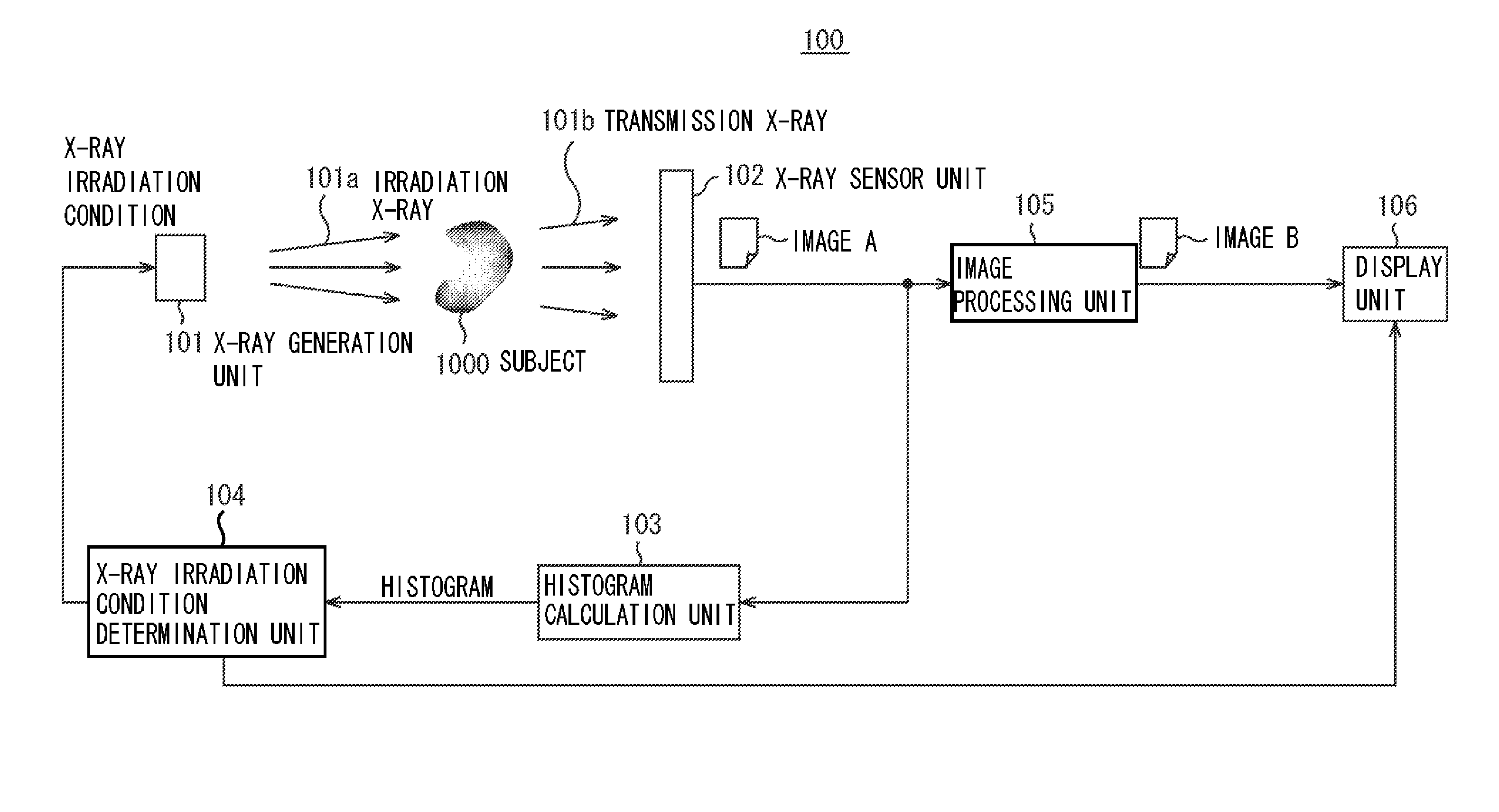
[0048]FIG. 1 illustrates an example of the configuration of an X-ray imaging apparatus (i.e., a radiation imaging apparatus) according to a first...


PUM

PropertyMeasurementUnit
thicknessaaaaaaaaaa
thicknessaaaaaaaaaa
thicknessaaaaaaaaaa

Abstract

A radiation imaging apparatus irradiates a subject with a radioactive ray emitted from a radiation generation unit to capture a radiographic image based on a radioactive ray that has penetrated through the subject. The radiation imaging apparatus includes a histogram calculation unit configured to calculate a histogram resulting from pixel values of the radiographic image, and a radiation irradiation condition determination unit configured to determine radiation irradiation conditions for a radioactive ray to be emitted from the radiation generation unit in such a way as to flatten a distribution of the histogram.

Description

BACKGROUND OF THE INVENTION[0001]1. Field of the Invention[0002]The present invention relates to a radiation imaging apparatus that irradiates a subject with a radioactive ray emitted from a radiation generation unit to capture a radiographic image based on the radioactive ray that has penetrated through the subject, and also relates to a method for driving the radiation imaging apparatus.[0003]2. Description of the Related Art[0004]There is a conventional X-ray imaging apparatus that can capture an image of a subject with an X-ray (i.e., one of various kinds of radioactive rays), so that the acquired X-ray image can be used for a medical care. In this case, the X-ray imaging apparatus includes an X-ray generation unit that can emit an X-ray toward the subject. The X-ray imaging apparatus acquires an X-ray image based on the X-ray having penetrated through the subject. Users can observe an internal state of the subject based on the acquired X-ray image. The X-ray imaging apparatus m...

Claims


Application Information

Patent Timeline
Patent Type & Authority Applications(United States)
IPC IPC(8): G01N23/04H05G1/10
CPCA61B6/542A61B6/00
Inventor TAKAHASHI, KAZUHIKO
Owner CANON KK
Why Eureka
Try Eureka