Method for Detecting Disease-Related Marker Using Gastric Mucosal Lavage Fluid

a gastric mucosal and marker technology, applied in the field of disease-related marker detection, can solve the problems of insufficient total resection ratio, physical limitation of extending the spot into the “area”, and difficult macroscopic evaluation in most cases, and achieves the effects of prolonging the time of examination, improving quality, and facilitating sample collection

Inactive Publication Date: 2010-03-18
SAPPORO MEDICAL UNIVERSITY +1
View PDF1 Cites 2 Cited by
  • Summary
  • Abstract
  • Description
  • Claims
  • Application Information

AI Technical Summary

Benefits of technology

[0020]Regarding the samples and methods of the present invention, a gastric mucosal washes obtained by washing the gastric mucosal surface, after removal of the mucus attached to the gastric mucosa of a subject, is used, and this is essentially different from the method described in non-patent document 7, wherein a mere gastric washing resulting from endoscopy which contains a large quantity of gastric juice is used. When a mere gastric washing was used as a sample, sufficient DNA for PCR could not be obtained in approximately 20% of the cases, and execution of Southern blotting was impossible in approximately 40% of the cases, as described in non-patent document 7; because certain degrees of physical and economical burdens are required for subjects in the execution of endoscopy, the use of the conventional method, wherein gastric washings that frequently require sample re-collection are used, could never be used in clinical sites. However, when the present method using a gastric mucosal washes is used, a sample containing markers of superior quality can easily be obtained; because a waste fluid derived from endoscopy can be utilized as a sample material, following various advantages are obtained.
[0021]Namely, because washing liquids conventionally disposed of are used as materials in the samples and methods of the present invention, various possible risks occurring in other test methods utilizing an endoscope (such as risk of bleeding (biopsy), allergy, development of kidney damages (pigment dispersion method), prolongation of the time of examinations (magnification endoscopy), expensive apparatus (magnification endoscopy, NBI: Narrow Band Imaging) do not newly occur, and in addition, because it is easy to collect samples, almost no burden is applied to physicians operating endoscopy and assisting nurses. Moreover, in the present method, since a sample collection tube is fixed at the middle of the closed circuit of an endoscopic apparatus, samples can be protected from contamination and no additional investment in the fac...

Problems solved by technology

However, different from total gastrectomy that had been the major treatment method until the increase of early gastric-cancer cases, a part of the stomach remains in the above treatment methods, so that care must be taken to the recurrence of cancer in the remaining stomach.
Furthermore, since preoperative evaluation of the above early cancers by macroscopic diagnosis does not have a 100% diagnostic performance at present, the total resection ratio does not reach 100%; thus, the judgment and evaluation of remnant cancer has become very important.
However, with conventional gastric endoscopy, it is naturally impossible to pick up minute cancer that cannot be macroscopically selec...

Method used


Image

  • Method for Detecting Disease-Related Marker Using Gastric Mucosal Lavage Fluid
  • Method for Detecting Disease-Related Marker Using Gastric Mucosal Lavage Fluid
  • Method for Detecting Disease-Related Marker Using Gastric Mucosal Lavage Fluid

Examples


example 1

Sample Collection Container

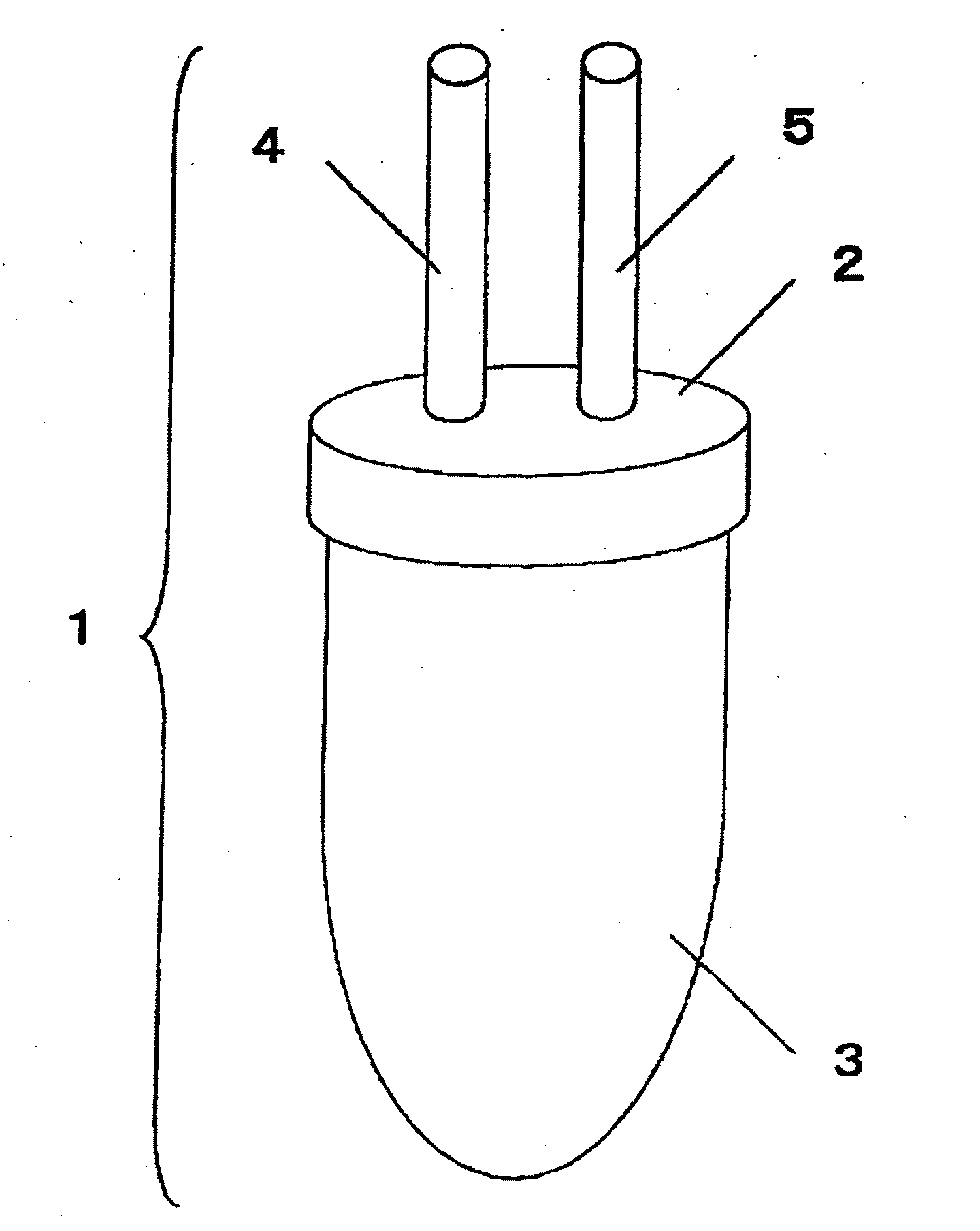
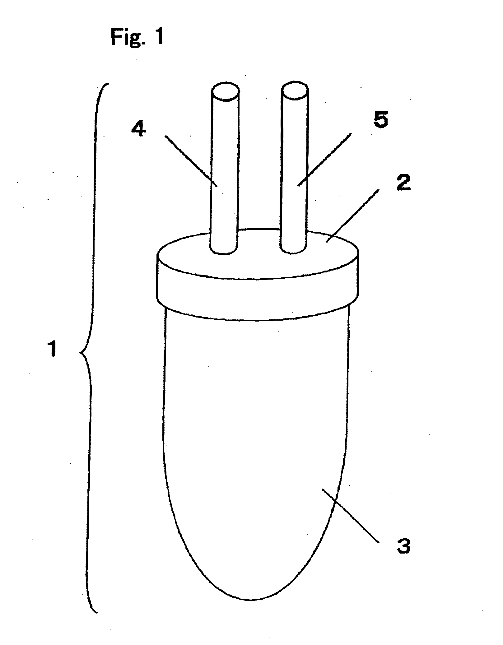
[0079]FIG. 1 shows an example of the sample collection container of the present invention. The sample collection container (1) consists of the cap-shaped upper part of the sample collection container (2) and the lower part of the sample collection container (3) with a volume of 50 ml, and the two parts are air-tightly joined by screwing. The upper part of the sample collection container (2) is equipped with two connecting portions, i.e., the Endoscope-side connecting portion (4) and the aspirator-side connecting portion (5), enabling airtight connection with a suction line of an endoscopic apparatus. In addition, 0.5 ml of 0.5-M EDTA (special grade, Wako Pure Chemical Industries, Ltd.) is sealed into the sample collection container (1).

example 2

Preparation of Samples

[0080]From the suction bottle of the endoscopic apparatus (EVIS LUCERA, Olympus Corporation), a suction tube that connects the connector was dismantled and connected to the Endoscope-side connecting portion of said sample collection container, and at the same time, the aspirator-side connecting portion of said sample collection container was connected to the connecting portion of the scope side of the suction bottle, thus forming a closed circuit.

[0081]To human subjects with various gastric disorders, the pretreatment agent prepared by dissolving 20,000 units of Pronase® (Pronase® MS, Kaken Pharmaceutical Co., Ltd.) and 1 g of sodium hydrogen carbonate into a solution of diluting 4 ml of dimethicone (Gascon® drop, Kissei Pharmaceutical Co., Ltd., this contains 80 mg of dimethylpolysiloxane) in 50-100 ml of tap water was administered, then 10-15 min later, the subjects were rotated around their body axis for 2 to 3 times while they were lying, and subjected to n...

example 3

Evaluation of Quality and Quantity of DNA

i) Evaluation of Quality of DNA

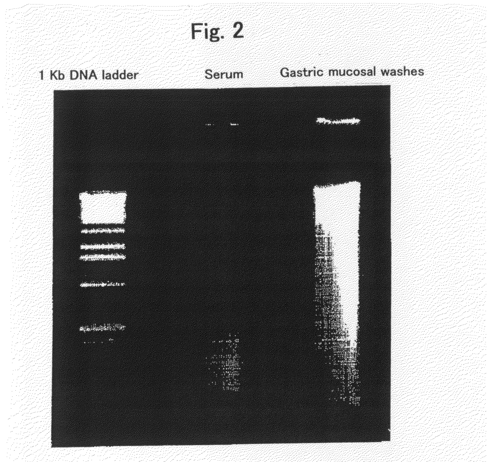
[0084]The quality of DNA contained in the above samples derived from human subjects with gastric cancer was evaluated by electrophoresis. Electrophoresis was performed in 1% agarose gel at 100 V for 30 min, by diluting 1-2 μg of a DNA sample to 10 μl of H2O. FIG. 2 shows the evaluation results by electrophoresis. The left lane indicates a positive control (1 Kb DNA extension ladder, Cat. No. 10511-012, Invitrogen), and the middle lane indicates a serum sample, and the right lane indicates a gastric mucosal washes. Any of the results demonstrates that the gastric mucosal washes comprises DNA with a quality better than that in the serum samples and comparable to that in the biopsy samples.

ii) Evaluation of Quantity of DNA

[0085]The amounts of DNA collected from the samples derived from human subjects with gastric cancer were evaluated by spectrometry. The following samples were used: from subjects with gastric canc...


PUM

PropertyMeasurementUnit
Massaaaaaaaaaa
Temperatureaaaaaaaaaa

Abstract

The invention relates to a sample containing a gastric mucosa lavage fluid collected from a subject who has received a gastric mucus removal treatment and a method for detecting a disease-related marker using the same. By using the sample of the invention, the disease-related marker can be detected conveniently, less invasively, highly sensitively and highly accurately.

Description

TECHNICAL FIELD[0001]The present invention relates to a method for detecting a disease-related marker, more specifically, the method using gastric mucosal washes, a sample containing gastric mucosal washes, a pretreatment agent used for the method, and an apparatus used for the method.BACKGROUND ART[0002]Cancer has been the leading cause of death in Japan for the past 20 years. When evaluating the cancer as the cause of death by cancer site, gastric cancer has continuously been among the high-ranked cancers; however, details of gastric-cancer cases have apparently been changing in recent years. While the number of patients with gastric cancer stages III and IV represented by advanced gastric cancer accounted for the majority of patients in the past, currently more than 50% of the patients are those of stage I. Stage I is classified into stage Ia and stage Ib, and both are characterized by the fact that the cancer is limited to the submucosal layer; that is, they are classified as so...

Claims


Application Information

Patent Timeline
IPC IPC(8): C12Q1/68A61B5/00
CPCC12Q1/6806C12N15/1003G01N33/15
Inventor WATANABE, YOSHIYUKITOYOTA, MINORUIMAI, KOHZOHSHINOMURA, YASUHISAITOH, FUMIOTOKINO, TAKASHI
Owner SAPPORO MEDICAL UNIVERSITY
Why Eureka
Try Eureka