Diagnostic imaging apparatus

a diagnostic imaging and x-ray technology, applied in the field of electromechanical endoscopy equipment, can solve the problems of inability to precisely diagnose at the m level, difficult to detect a large intestine pit lesion inside the tissue, and inability to observe up to the inside of the tissue, so as to achieve high precision and easy visual determination of the possibility of a large intestine pit lesion

Inactive Publication Date: 2010-03-18
FUJIFILM CORP
View PDF8 Cites 11 Cited by
  • Summary
  • Abstract
  • Description
  • Claims
  • Application Information

AI Technical Summary

Benefits of technology

[0024]Thus, diagnostic support can be performed so that a suspicion of a large intestine lesion is easily visually determined.
[0025]According to the present invention, the shape of large intestine pits can be observed up to the deep part of the tissue throughout the large intestine mucosa layer. Particularly, it is possible to cut out a plurality

Problems solved by technology

However, for the observed image of an endoscope by normal light, mainly, a living body tissue surface can be observed, but observation is not possible up to the inside of the tissue, and it is difficult to detect a large inte

Method used


Image

  • Diagnostic imaging apparatus
  • Diagnostic imaging apparatus
  • Diagnostic imaging apparatus

Examples


Embodiment Construction

[0043]Best modes for carrying out the present invention will be described below.

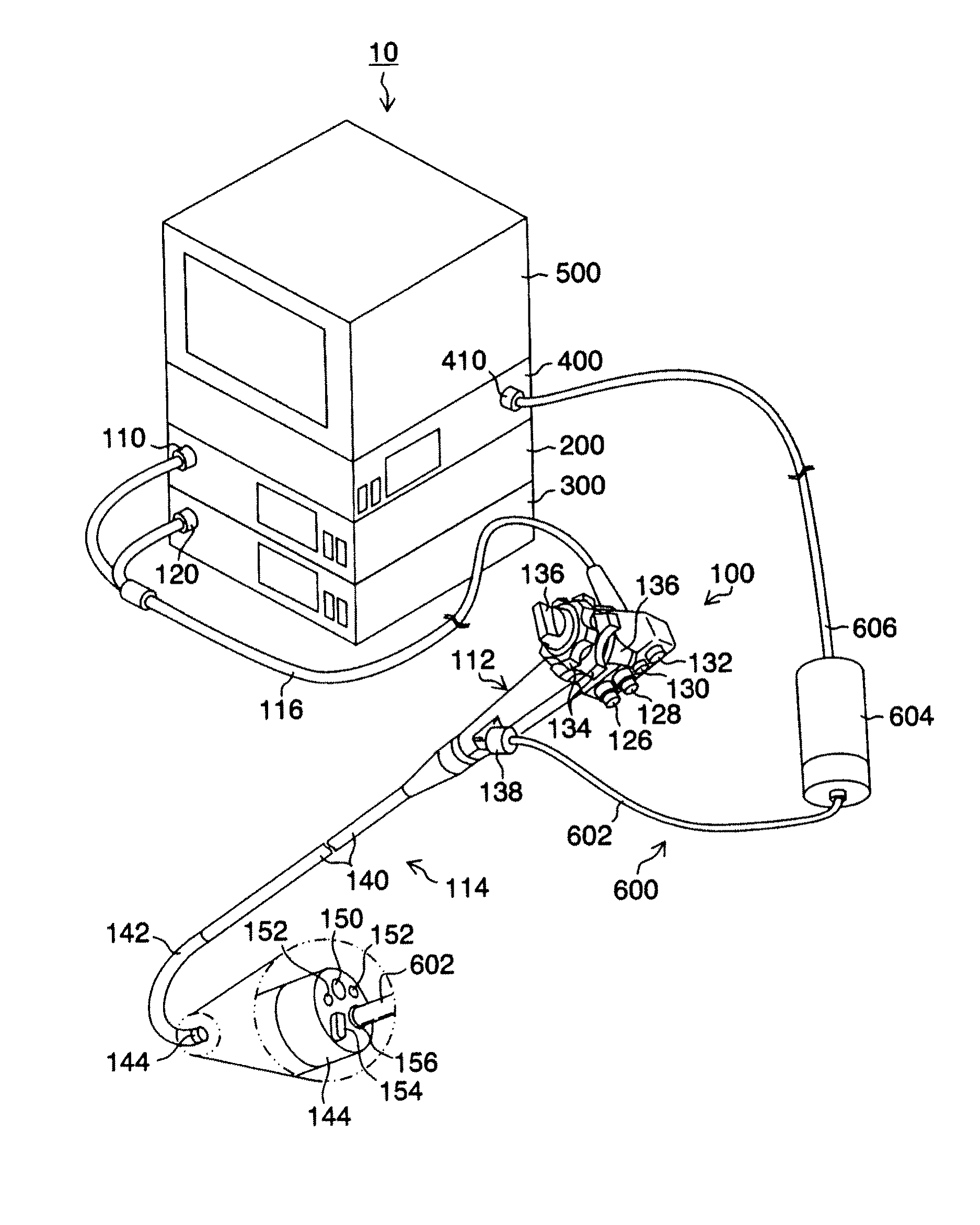
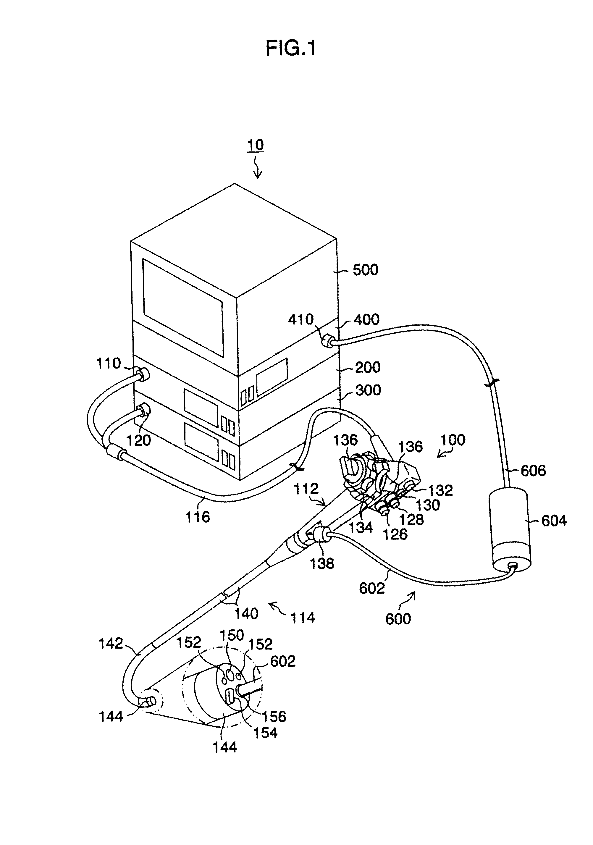
[0044]FIG. 1 is an appearance view showing a diagnostic imaging apparatus 10 according to the present invention.

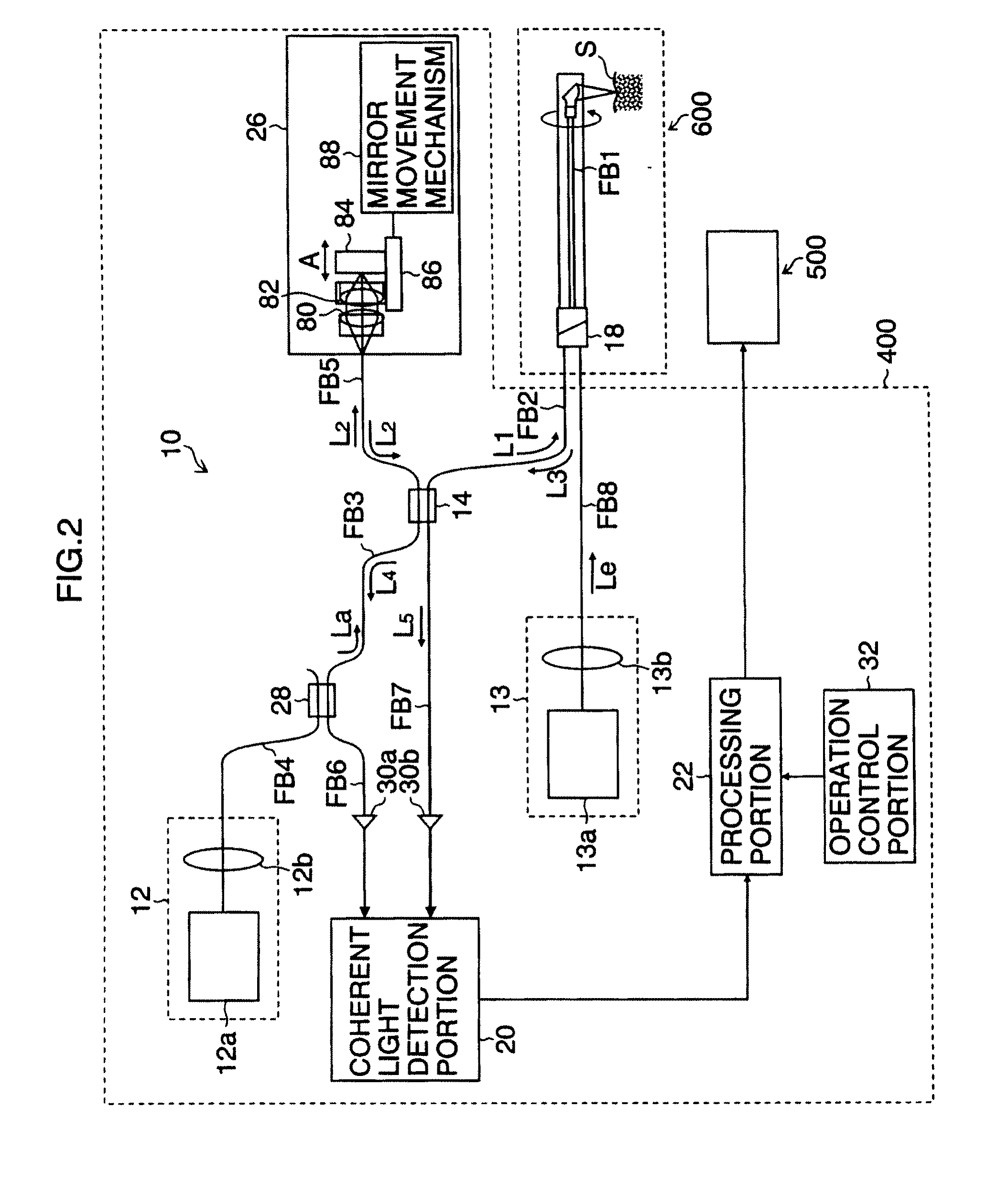
[0045]As shown in the figure, this diagnostic imaging apparatus 10 is mainly composed of an endoscope 100, an endoscope processor 200, a light source apparatus 300, an OCT processor 400, and a monitor apparatus 500. The endoscope processor 200 may be configured to contain the light source apparatus 300.

[0046]The endoscope 100 comprises a hand operation portion 112 and an insertion portion 114 provided continuously to this hand operation portion 112. An operator grasps and operates the hand operation portion 112, and inserts the insertion portion 114 into the body of a test subject to perform observation.

[0047]A forceps insertion portion 138 is provided in the hand operation portion 112, and this forceps insertion portion 138 is in communication with the forceps port 156 of a tip portion 144. In...


PUM


Abstract

A diagnosis apparatus in which a lesion in the large intestine can be diagnosed with high precision by detecting pits is provided. A three-dimensional optical tomographic image is obtained by the configuration of an apparatus using an endoscope and an optical probe, images in X-Y planes perpendicular to the depth direction of a living body tissue are cut out at a plurality of depth positions, based on the three-dimensional tomographic image data, and a pit pattern shape highlighted image is generated from their average image to perform diagnostic support.

Description

BACKGROUND OF THE INVENTION[0001]1. Field of the Invention[0002]The present invention relates to an electronic endoscope apparatus and an OCT apparatus and to a diagnostic imaging apparatus in which three-dimensional volume data is obtained to perform the diagnosis of a lesion in the large intestine.[0003]2. Description of the Related Art[0004]Conventionally, as an endoscope apparatus for observing the inside of the body cavity of a living body, electronic endoscope apparatuses, in which illumination light illuminates in the body cavity of a living body, and an image formed by reflected light is picked up and displayed on a monitor or the like, have been widely spread and used in various fields.[0005]Also, as in Japanese Patent Application Laid-Open No. 2002-148185, many endoscope apparatuses comprise a forceps port, and by a probe introduced into the body cavity through this forceps port, the biopsy and treatment of a tissue in the body cavity can be performed.[0006]In recent years...

Claims


Application Information

Patent Timeline
IPC IPC(8): A61B5/05G06K9/00
CPCA61B5/0066A61B5/4255A61B5/0084
Inventor WATANABE, DAISUKEKINJO, NAOTO
Owner FUJIFILM CORP
Why Eureka
Try Eureka