Radiation imaging and therapy apparatus for breast

a radiation imaging and therapy apparatus technology, applied in the direction of diagnostics, therapy, instruments, etc., can solve the problems of unavoidable upsizing of the entire apparatus, difficult to reproduce the location of the affected part in the radiation application therapy apparatus for accurate treatment of the affected part in the breast, and needing contrast agents

Inactive Publication Date: 2010-03-25
FUJIFILM CORP
View PDF14 Cites 45 Cited by
  • Summary
  • Abstract
  • Description
  • Claims
  • Application Information

AI Technical Summary

Benefits of technology

[0013]According to the one aspect of the present invention, the location of a lesion within the breast can be detected based on the radiation image obtained by application of low-energy radiation from the first radiation generating unit, and high-energy

Problems solved by technology

However, the image-guided radiation therapy apparatus can be used for all body organs as targets of examinations, and there is a problem that a contrast agent is necessary for an examination of breast cancer because an X-ray having high penetrating power generated by an X-ray tube voltage equal to or more than 100 kV for image formation is used.
Further, since image-guided radiation therapy apparatus is used for a

Method used


Image

  • Radiation imaging and therapy apparatus for breast
  • Radiation imaging and therapy apparatus for breast
  • Radiation imaging and therapy apparatus for breast

Examples


Embodiment Construction

[0026]Hereinafter, preferred embodiments of the present invention will be explained in detail with reference to the drawings. The same reference numbers are assigned to the same component elements and the description thereof will be omitted. In the following embodiments, the case where an X-ray is used as radiation will be explained, however, the present invention can be applied to cases of using α-ray, β-ray, γ-ray, electron ray, ultraviolet ray, or the like.

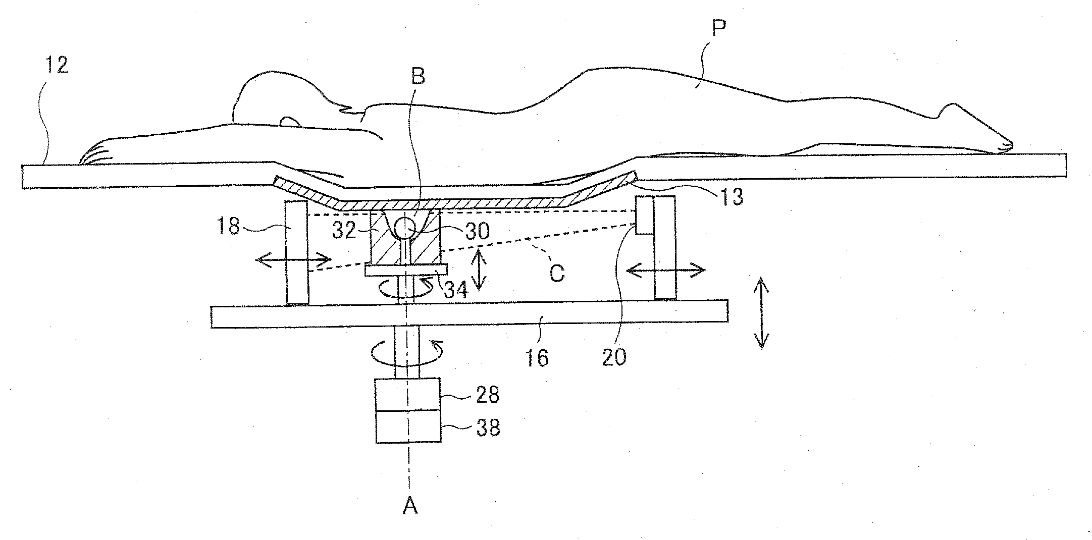
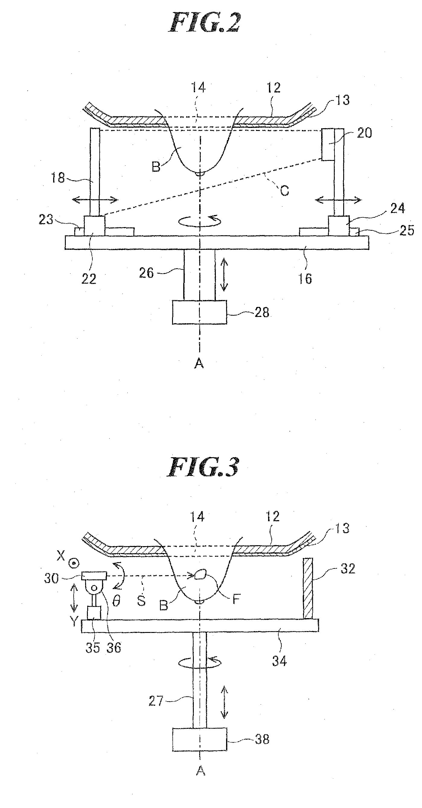
[0027]FIG. 1 is a side view showing an imaging unit and a therapy unit of a radiation imaging and therapy apparatus according to one embodiment of the present invention. As shown in FIG. 1, the radiation imaging and therapy apparatus includes a table 12 having an opening part in which an opening for passing a breast “B” of an examinee (patient) “P” is formed, an X-ray generating unit 20 for applying an imaging X-ray beam (cone beam) “C” toward the breast “B” that has passed through the opening part of the table 12, an X-ray det...


PUM


Abstract

A breast radiation imaging and therapy apparatus for performing radiation imaging of a breast and having a therapy function of applying radiation to an affected part in the breast. The apparatus includes: (i) a table formed with an opening for allowing a breast of an examinee to pass through; (ii) an imaging unit including a first radiation generating unit for applying an imaging radiation beam and a radiation detecting unit for detecting the radiation beam to output detection signals; (iii) a therapy unit including a second radiation generating unit for applying a therapeutic radiation beam, the second radiation generating unit being movable in a tangential direction of a rotational track around a rotational axis and movable in a direction substantially orthogonal to the table; and (iv) at least one rotational driving device for rotating the imaging unit and the therapy unit around the rotational axis.

Description

CROSS-REFERENCE TO RELATED APPLICATION[0001]The present application claims priority from Japanese Patent Application No. 2008-245672 filed on Sep. 25, 2008, the contents of which are incorporated herein by reference in their entirety.BACKGROUND OF THE INVENTION[0002]1. Field of the Invention[0003]The present invention relates to a radiation imaging and therapy apparatus for obtaining a radiation image of a breast and having a therapy function of applying radiation to an affected part in the breast.[0004]2. Description of a Related Art[0005]Recent years, an image-guided radiation therapy (IGRT) apparatus combining a tomographic imaging apparatus and a radiation therapy apparatus has been developed. Thereby, a radiation application method can be planned based on a three-dimensional (3D) image near an affected part, and radiation therapy for applying radiation can be performed according to the plan.[0006]For example, Japanese Patent Application Publication JP-A-9-192245 discloses a rad...

Claims


Application Information

Patent Timeline
IPC IPC(8): A61B6/04A61N5/01G21K5/08
CPCA61B6/04A61B6/0435A61B6/502G21K1/10A61N5/10A61N5/1081A61N5/01
Inventor SENDAI, TOMONARI
Owner FUJIFILM CORP
Why Eureka
Try Eureka