
Method and Device for Thermal Breast Tumor Treatment with 3D Monitoring Function

a breast cancer and 3d monitoring technology, applied in the field of xray machines, can solve the problems of high radiation load caused by high-resolution three-dimensional exposure, inability to control the heating of the tumor in a precise manner, and difficulty in controlling the treatment of the tumor with an instrument of this kind,

Inactive Publication Date: 2010-04-01
AB CT ADVANCED BREAST CT GMBH
View PDF79 Cites 14 Cited by
  • Summary
  • Abstract
  • Description
  • Claims
  • Application Information

AI Technical Summary

Problems solved by technology

However, heating of the tumor cannot be controlled in a precise manner, because the temperature of the tissue is not directly related to the current flow or the energy introduced into the tissue by the electrode.
Controlled therapy of a tumor is, therefore, difficult with an instrument of this kind.
A disadvantage of this method is the high radiation load caused during the high-resolution three-dimensional exposures.

Method used


Image

  • Method and Device for Thermal Breast Tumor Treatment with 3D Monitoring Function
  • Method and Device for Thermal Breast Tumor Treatment with 3D Monitoring Function
  • Method and Device for Thermal Breast Tumor Treatment with 3D Monitoring Function

Examples


Embodiment Construction

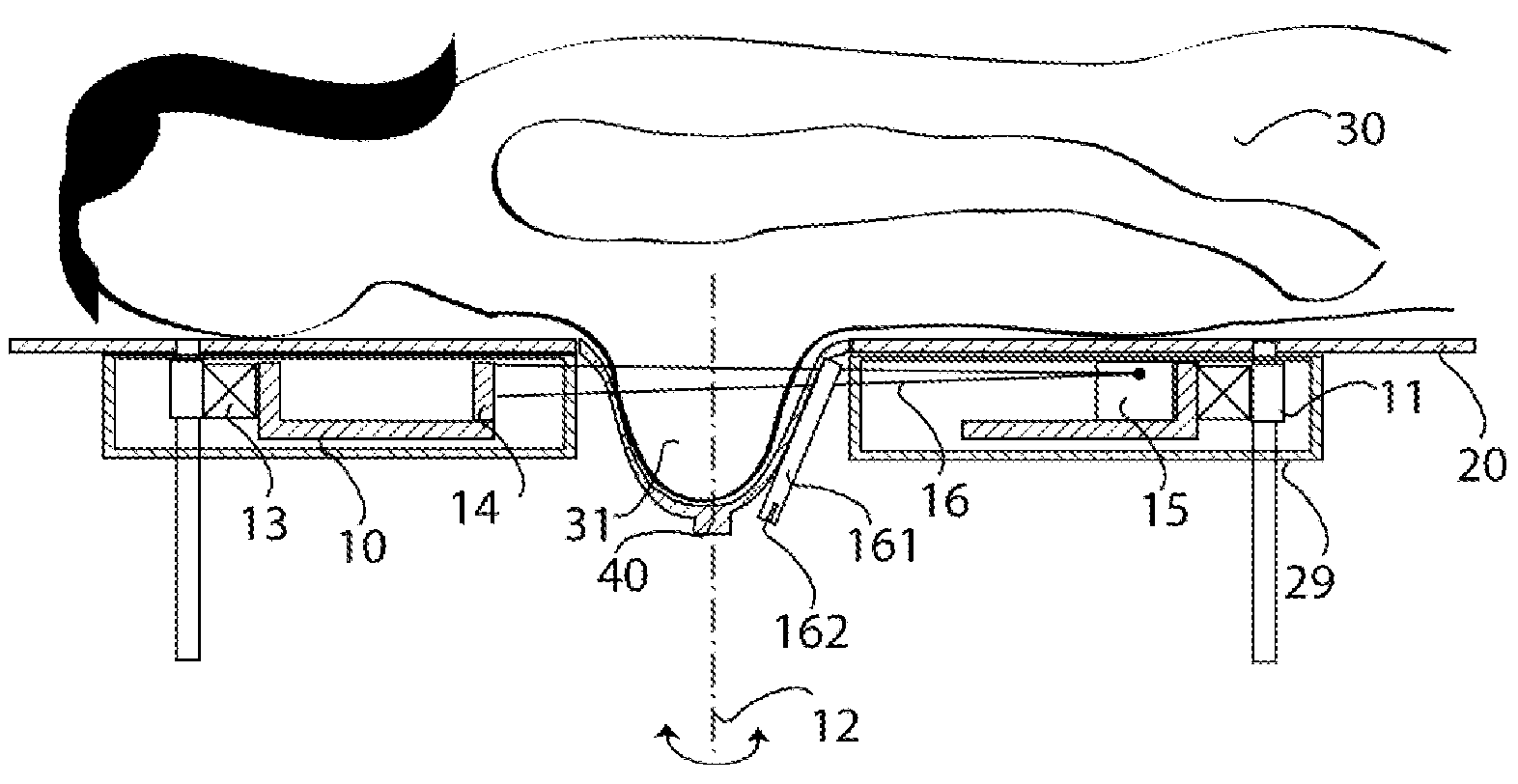
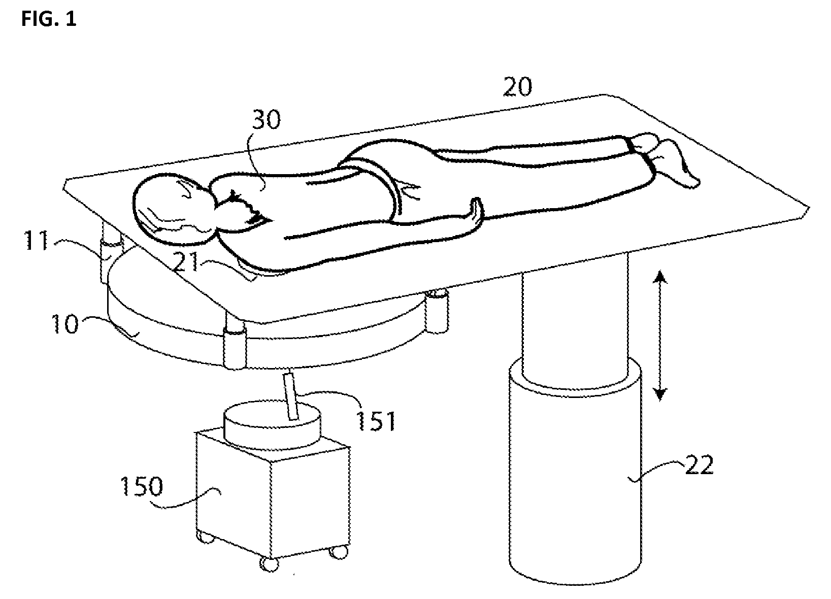
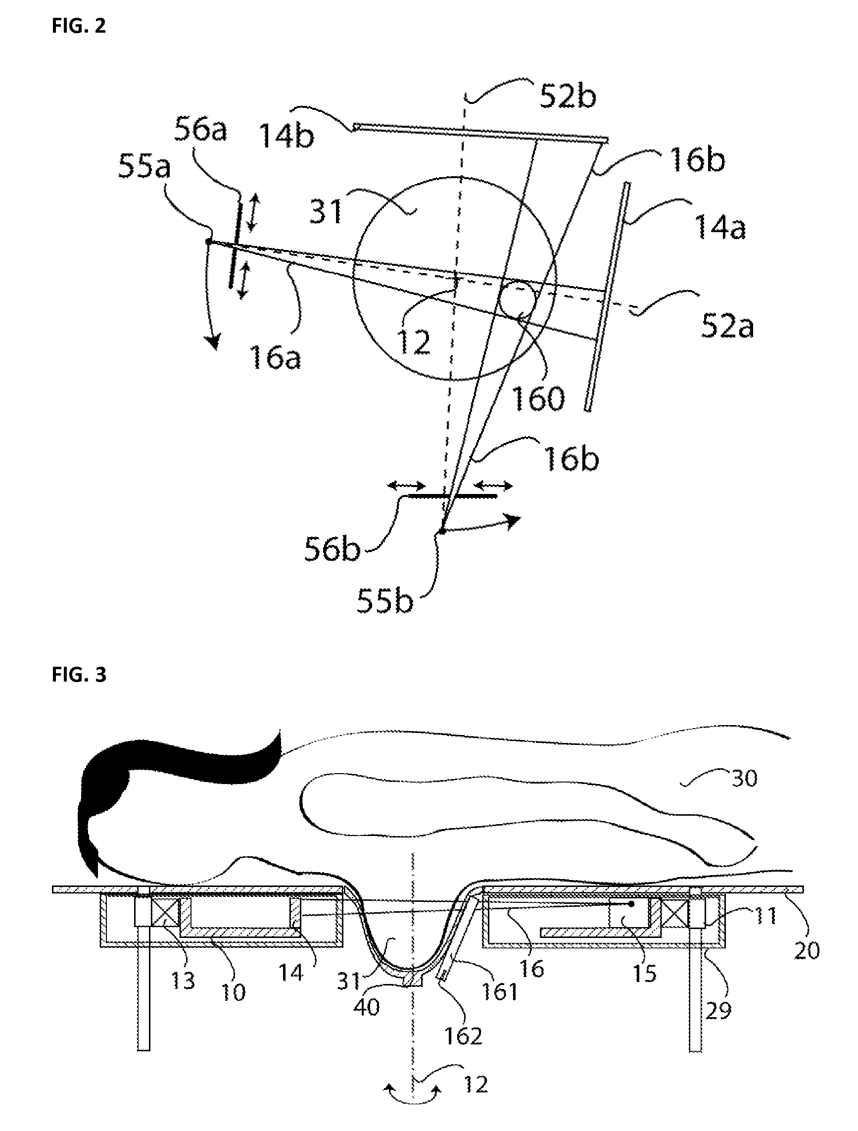
[0018]FIG. 1 illustrates an embodiment of an X-ray machine for examining and / or treating a female breast. A patient 30 rests on a support surface, which in the illustrated embodiment, is a patient's table 20. Although illustrated as having a horizontally disposed support surface 20 for the patient, it is also possible for the support surface disclosed herein to be disposed vertically, or at other angles of inclination.

[0019]The breast to be examined is inserted through a breast cutout portion 21 in the patient's table 20 into an exposure range of a gantry 10. The gantry 10 shown in FIG. 1 is a spiral computer tomograph (CT) gantry having an X-ray tube and an X-ray detector (not shown in FIG. 1), which rotate around the breast to be examined. The breast is imaged with X-rays during rotation of the gantry 10. Simultaneously with the rotation, a displacement of the gantry along a vertical direction is performed via a gantry lift drive 11, so that the breast is scanned with X-rays along...


PUM


Abstract

An X-ray machine for monitoring thermal treatment of human tissue produces a reference exposure of the tissue to be treated prior to commencement of the treatment. Thermal treatment is performed subsequently. Check exposures are made with the X-ray machine during the treatment or also during treatment intervals. The check exposures are performed as partial volume exposures at a lower radiation load than the reference exposures. From a comparison of check exposures with the reference exposures, conclusions can be drawn concerning changes of tissue temperature and also tissue properties.

Description

PRIORITY CLAIM[0001]This application claims priority to pending German Application No. 102008042430.7 filed on Sep. 29, 2008.BACKGROUND OF THE INVENTION[0002]1. Field of the Invention[0003]This invention relates to an X-ray machine and also to a method for monitoring a diathermy treatment. In particular, the invention relates to an X-ray machine for imaging a breast of a female patient (mammography), and also a method for monitoring diathermy treatment of a breast tumor.[0004]2. Description of Related Art[0005]Various X-ray machines are used for performing an examination of a female breast. One type of X-ray machine comprises a rotating gantry, having an X-ray tube and an X-ray detector, located below a patient's table on which a patient to be examined rests. An embodiment of such an X-ray machine is disclosed, for example, in U.S. Pat. No. 4,015,836. The X-ray machine disclosed in U.S. Pat. No. 4,015,836 facilitates diagnosis of diseases of the breast, in particular, of tumors in t...

Claims


Application Information

Patent Timeline
IPC IPC(8): A61B6/04A61N5/10G01D18/00
CPCA61B5/015A61B6/027A61B5/704A61B6/032A61B6/035A61B6/0435A61B6/06A61B6/107A61B6/502A61B6/583A61B18/00A61B2017/00084A61B2019/205A61B2019/5238A61B2019/524G01K11/30G06T7/0012G06T7/0083G06T2207/10081G06T2207/30068G06T2207/30096A61B5/4312A61B6/4275A61B90/17A61B2090/376A61B2090/3762G06T7/74G06T7/12
Inventor KALENDER, WILLISCHILLING, HARRY
Owner AB CT ADVANCED BREAST CT GMBH
Learn More