System and method for 3-d imaging

a technology of system and method, applied in the field of system and method for 3d imaging, can solve the problems of difficult if not impossible, to maintain transthoracic or similar contact and access during a procedure involving the heart below or other similarly situated tissue of the body

Active Publication Date: 2010-04-15
AURIS HEALTH INC
View PDF9 Cites 57 Cited by
  • Summary
  • Abstract
  • Description
  • Claims
  • Application Information

AI Technical Summary

Problems solved by technology

One the challenges with MRI and CT imaging modalities is related to the requisite sampling rate useful for monitoring three dimensional motion of tissues moving with relatively high frequency and high amplitude motion, such as cardiac tissues.
Maintaining transthoracic or similar contact and access during a procedure involving the heart below or other similarly situated tis

Method used


Image

  • System and method for 3-d imaging
  • System and method for 3-d imaging
  • System and method for 3-d imaging

Examples


Embodiment Construction

[0035]In the following detailed description of embodiments of the invention, reference is made to the accompanying drawings in which like references indicate similar elements. The illustrative embodiments described herein are disclosed in sufficient detail to enable those skilled in the art to practice the invention. The following detailed description is therefore not provided, or otherwise to be taken, in a limiting sense, and the scope of the invention is defined only by the appended claims.

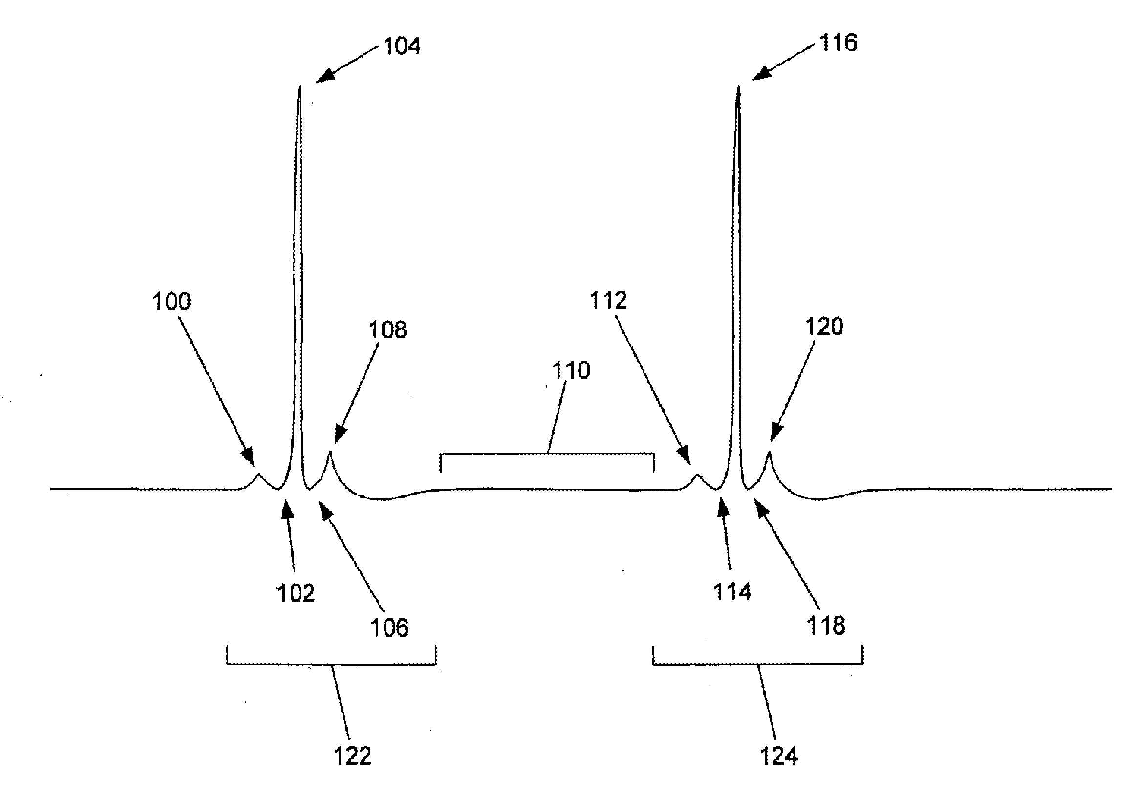
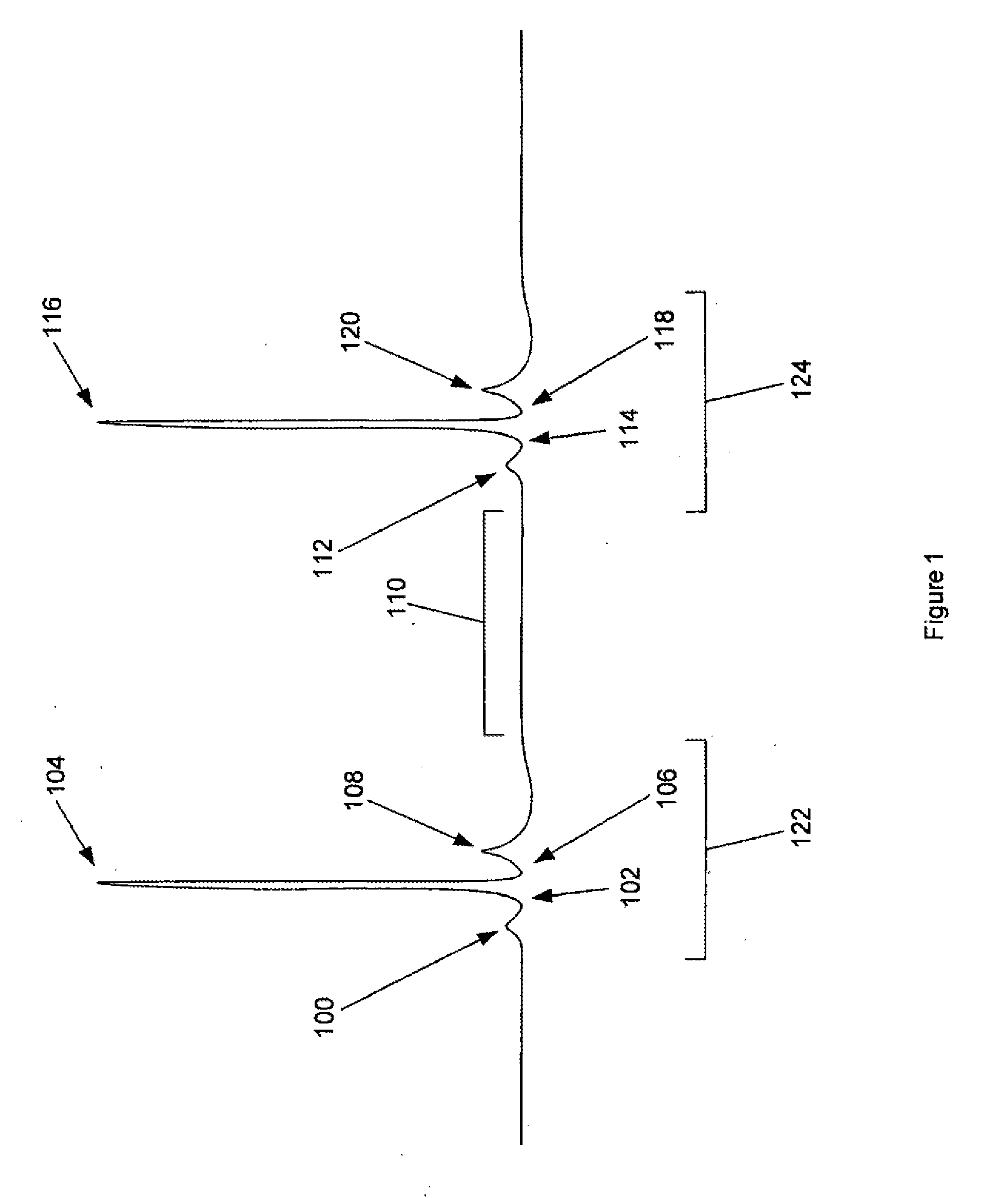
[0036]Referring to FIG. 1, an electrocardiogram (“EKG”) tracing for a human heart is depicted. The two systolic heart cycles (122, 124) depicted are separated in time by a resting period (110) of relatively low heart activity, during the diastolic period of the heart cycle. Each of the active systolic heart cycles (122, 124) in a healthy patient typically comprises a P-wave (100, 112), followed by a Q-wave (102, 114), then a relatively high amplitude R-wave (104, 116), an S-wave (106, 118), and...


PUM


Abstract

A system and method for recording and depicting ultrasound images of a moving object are disclosed. In a preferred embodiment, ultrasound images are acquired as the field of view of an ultrasound probe is advanced across the tissues of interest during a resting period between periods of relatively large-scale heart cycle motion. A series of images acquired during a particular resting period may be represented as a three-dimensional volume image, and the comparison of volume images from adjacent cardiac resting periods enables three-dimensional volume image modulation analysis which may be presented for a user as a moving volume image of the objects of interest within the field of view.

Description

CROSS-REFERENCE TO RELATED APPLICATIONS[0001]This application is a continuation of U.S. patent application Ser. No. 10 / 923,660, filed on Aug. 20, 2004 which claims priority to U.S. Provisional Patent Application Nos. 60 / 496,944 filed Aug. 20, 2003 and 60 / 506,231 filed Sep. 25, 2003 which the contents of which are incorporated herein by reference in their entirety.BACKGROUND OF THE INVENTION[0002]Imaging moving structures such as cardiac tissue walls and nearby medical instrumentation presents a unique set of problems not ideally addressed in three dimensions by conventional devices employing imaging modalities such as magnetic resonance imaging (“MRI”), computed tomography (“CT”), and ultrasound. One the challenges with MRI and CT imaging modalities is related to the requisite sampling rate useful for monitoring three dimensional motion of tissues moving with relatively high frequency and high amplitude motion, such as cardiac tissues. Sophisticated arrays of ultrasound transducers,...

Claims


Application Information

Patent Timeline
IPC IPC(8): A61B8/14A61B5/05A61B8/12G01S7/52G01S15/89
CPCA61B8/12A61B8/4245A61B8/4461A61B8/461A61B8/467G01S15/8993G01S7/52065G01S7/52084G01S7/52087G01S15/8934G01S15/894A61B8/483A61B8/466
Inventor YOUNGE, ROBERT G.WALLACE, DANIEL T.
Owner AURIS HEALTH INC
Why Eureka
Try Eureka