Dopaminergic neuronal survival-promoting factors and uses thereof
- Summary
- Abstract
- Description
- Claims
- Application Information
AI Technical Summary
Benefits of technology
Problems solved by technology
Method used
Image
Examples
example 1
Production and Analysis of VMCL-1 Cells
[0082]The VMCL-1 cell line was made as follows. Rat E14 mesencephalic cells, approximately 2-3% of which are glioblasts, were incubated in medium containing 10% (v / v) fetal bovine serum for 12 hours and subsequently expanded in a serum-free medium, containing basic fibroblast growth factor (bFGF) as a mitogen. After more than 15 DIV, several islets of proliferating, glial-like cells were observed. Following isolation and passaging, the cells (referred to herein as VMCL-1 cells) proliferated rapidly in either a serum-free or serum-containing growth medium. Subsequent immunocytochemical analysis showed that they stained positive for two astrocytic markers, GFAP and vimentin, and negative for markers of oligodendroglial or neuronal lineages, including A2B5, O4, GalC and MAP2. We have deposited the VMCL-1 cell line with the ATCC (Accession No: PTA-2479; deposit date: Sep. 18, 2000).
[0083]Serum-free CM, prepared from the VMCL-1 cells, caused increas...
example 2
Action of VMCL-1 CM on E14 Dopaminergic Neurons in Culture
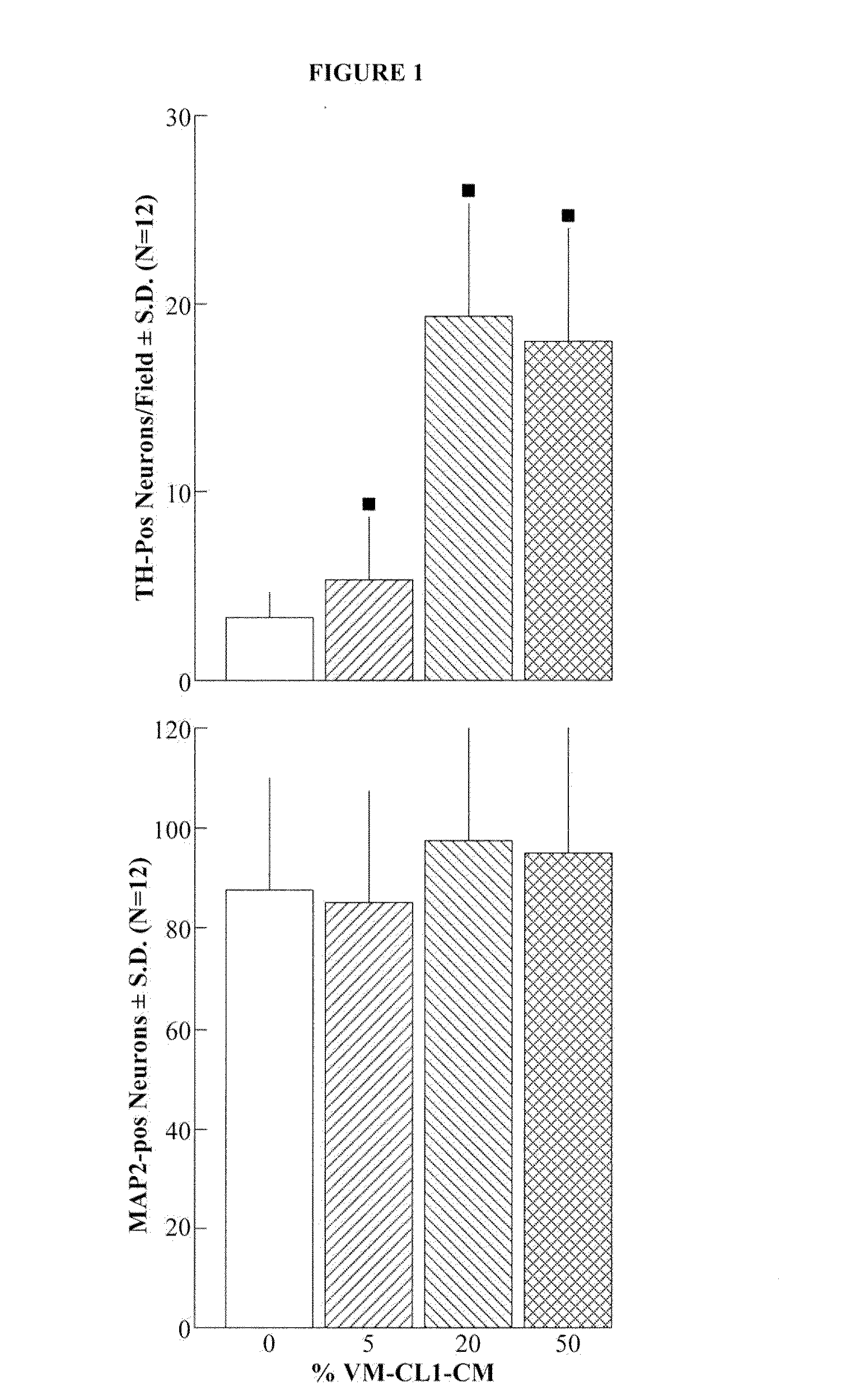
[0085]VMCL-1 CM was tested at 0, 5, 20 and 50% v / v, for its ability to influence survival and development of E14 mesencephalic dopaminergic neurons in culture. The cultures were primed with 10% fetal bovine serum (FBS) for 12 hours, then grown in a serum-free growth medium thereafter, until they were stained and analyzed after 7 DIV. There was a dose-dependent action of the CM on the increased survival of dopaminergic neurons. The CM increased survival by 5-fold. In contrast, there was no significant increase in non-dopaminergic neuronal survival. The profile of the biological action of this putative factor is quite different from that of CM derived from the B49 glioma cell line, the source of GDNF (Lin et al., Science 260: 1130-1132).
example 3
Gene Expression Analysis of VMCL-1 Cells
[0086]To further investigate the similarity between the VMCL-1 cell line and primary cultured astrocytes, we measured the expression of six marker genes characteristic of the mesencephalic region. Analysis of wnt-1, en-1, en-2, pax-2, pax-5, and pax-8 showed that all genes were expressed in both E13 and E14 ventral mesencephalon neural tissue, with the exception of pax-2, which was expressed at E13 but not E14 neural tissue. Both primary astrocytes and VMCL-1 cells expressed wnt-1 at levels comparable with those of E13 and E14 ventral mesencephalic neural tissue. The degree of expression of en-1 was similar in primary astrocytes and VMCL-1 cells, although at a lower level versus expression in E13 and E14 ventral mesencephalic tissue. In contrast, en-2, pax-5 and pax-8 were not expressed in either primary astrocytes or VMCL-1. Pax-2 was expressed in E13 but not E14 ventral mesencephalon, and in primary astrocytes, but not in VMCL-1.
PUM
 Abstract
Description
Claims
Application Information
 - R&D
- Intellectual Property
- Life Sciences
- Materials
- Tech Scout
- Unparalleled Data Quality
- Higher Quality Content
- 60% Fewer Hallucinations
Browse by: Latest US Patents, China's latest patents, Technical Efficacy Thesaurus, Application Domain, Technology Topic, Popular Technical Reports.
© 2025 PatSnap. All rights reserved.Legal|Privacy policy|Modern Slavery Act Transparency Statement|Sitemap|About US| Contact US: help@patsnap.com


