
Systems and Methods for Automated Diagnosis and Decision Support for Breast Imaging

a breast imaging and decision support technology, applied in the field of medical imaging system and decision support system, can solve the problems of not being able to image every patient using multiple imaging modalities, patients may be severely stressed in anticipation of potentially obtaining negative results,

Inactive Publication Date: 2010-05-13
KRISHNAN SRIRAM +3
View PDF3 Cites 110 Cited by
  • Summary
  • Abstract
  • Description
  • Claims
  • Application Information

AI Technical Summary

Benefits of technology

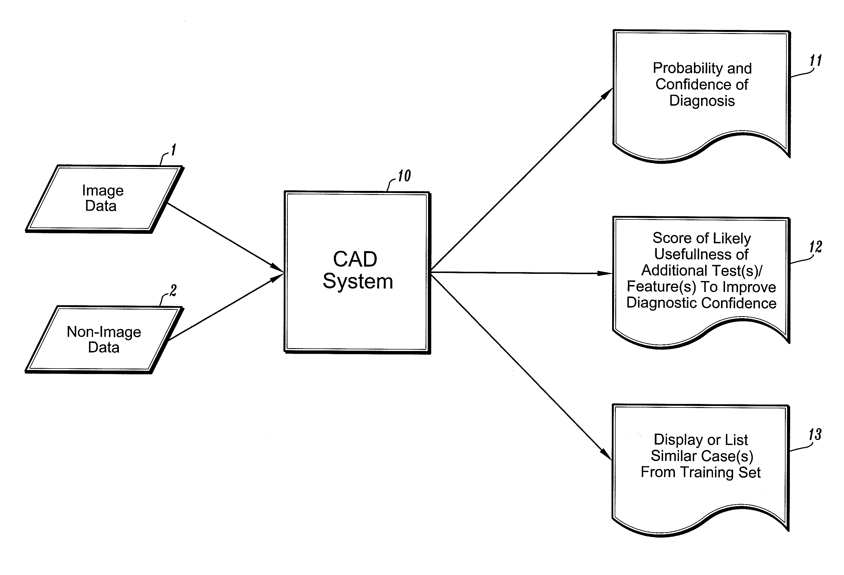
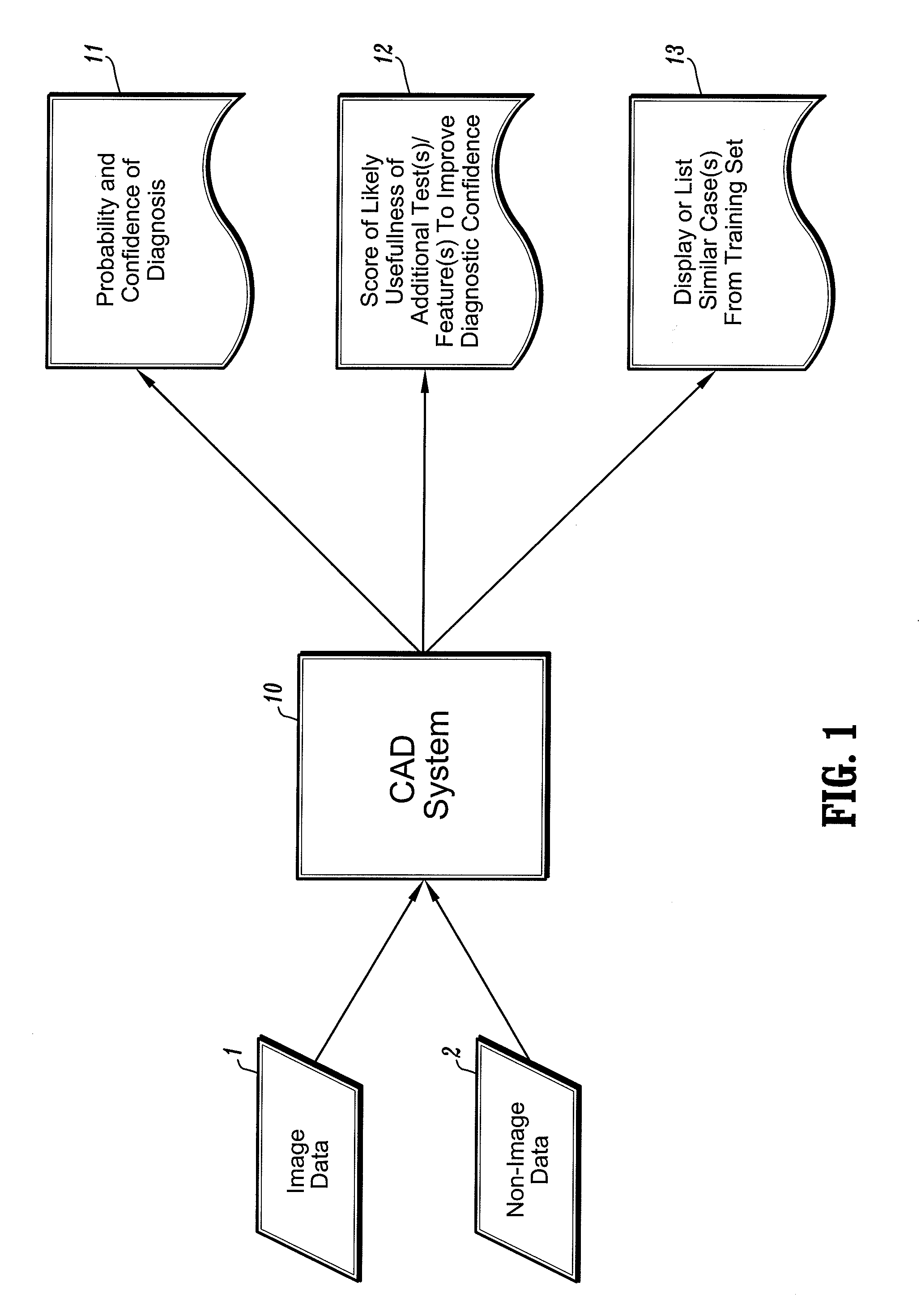
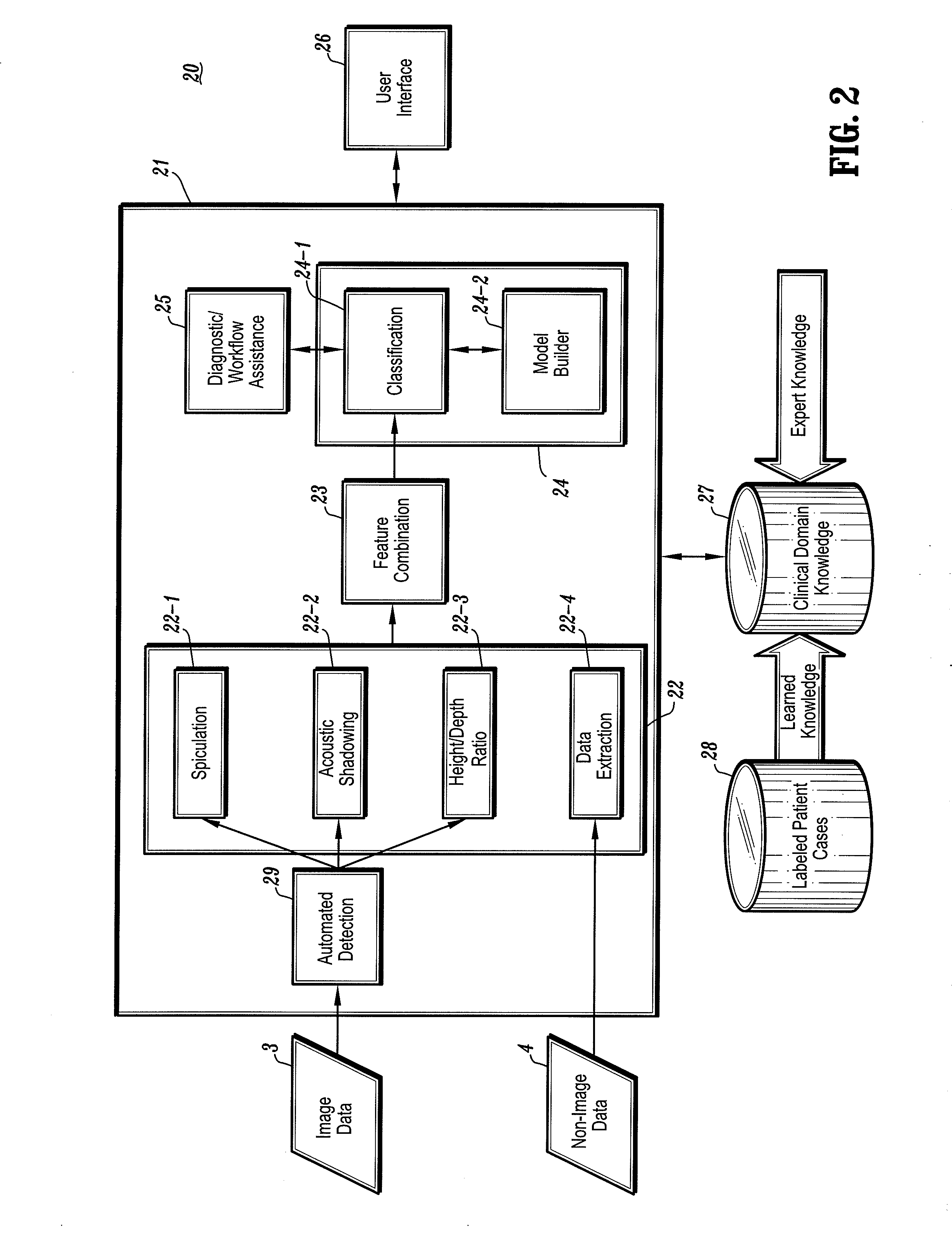
[0008]Exemplary embodiments of the invention generally include systems and methods for providing automated diagnosis and decision support for breast imaging. More specifically, exemplary embodiments of the invention include CAD (computer-aided diagnosis) systems and applications for breast imaging, which implement automated methods for extracting and analyzing relevant features / parameters from a collection of patient information (including image data and / or non-image data) of a subject patient to provide automated assistance to a physician for various aspects of physician workflow in breast care. For example, a CAD system can provide automated diagnosis of breast cancer and other related conditions, assessments with regard to the risk of a subject patient having breast cancer and / or developing breast cancer in the future, and other automated decision support functions to assist a physician in determining efficacious healthcare or diagnostic or therapeutic paths for a subject patient based on a current state of the patient.
[0009]In other exemplary embodiments of the invention, CAD systems and methods for breast imaging implement machine-learning techniques which use training data that is obtained (learned) from a database of previously diagnosed (labeled) patient cases in one or more relevant clinical domains and / or expert interpretations of such data to enable the CAD systems to “learn” to properly and accurately analyze patient data and make proper diagnostic and / or therapeutic assessments and decisions for assisting physician workflow.

Problems solved by technology

For instance, a biopsy procedure causes pain and scarring for the patient, and the long period of time between the time of the biopsy procedure and the time the results are provided to the patient (usually at least a few days), the patient may be become severely stressed in anticipation of potentially obtaining negative results.
Unfortunately, due to cost, it is not possible to image every patient using multiple imaging modalities, and then choose which modality would provide the optimal balance between sensitivity and specificity.

Method used


Image

  • Systems and Methods for Automated Diagnosis and Decision Support for Breast Imaging
  • Systems and Methods for Automated Diagnosis and Decision Support for Breast Imaging
  • Systems and Methods for Automated Diagnosis and Decision Support for Breast Imaging

Examples


Embodiment Construction

[0016]In general, exemplary embodiments of the invention as described below include systems and methods for providing automated diagnosis and decision support for breast imaging. More specifically, exemplary embodiments of the invention as described below with reference to FIGS. 1-4, for example, include CAD (computer-aided diagnosis) systems and applications for breast imaging, which implement automated methods for extracting and analyzing relevant features / parameters from a collection of patient information (including image data and / or non-image data) of a subject patient to provide automated assistance to a physician for various aspects of physician workflow including, for example, automated assistance to a physician for various aspects of physician workflow where decisions must be made respecting healthcare or diagnosis paths and / or therapeutic paths for the patient. Various methods have been developed which attempt to provide decision support for physicians using only informati...


PUM


Abstract

CAD (computer-aided diagnosis) systems and applications for breast imaging are provided, which implement methods to automatically extract and analyze features from a collection of patient information (including image data and / or non-image data) of a subject patient, to provide decision support for various aspects of physician workflow including, for example, automated diagnosis of breast cancer other automated decision support functions that enable decision support for, e.g., screening and staging for breast cancer. The CAD systems implement machine-learning techniques that use a set of training data obtained (learned) from a database of labeled patient cases in one or more relevant clinical domains and / or expert interpretations of such data to enable the CAD systems to “learn” to analyze patient data and make proper diagnostic assessments and decisions for assisting physician workflow.

Description

CROSS-REFERENCE TO RELATED APPLICATION[0001]This application is a Continuation of co-pending U.S. patent application Ser. No. 10 / 877,129, filed Jun. 25, 2004, which claims priority to U.S. Provisional Application Ser. No. 60 / 482,293, filed on Jun. 25, 2003, and U.S. Provisional Application Ser. No. 60 / 541,360, filed on Feb. 3, 2004, all of which are fully incorporated herein by reference.TECHNICAL FIELD OF THE INVENTION[0002]The present invention relates generally to systems and methods for providing automated diagnosis and decision support for medical imaging and, in particular, to CAD (computer-aided diagnosis) systems and applications for breast imaging, which use machine-learning techniques that enable such systems and application to “learn” to analyze parameters extracted from image data and / or non-image patient data of a subject patient for purposes of providing automated decision support functions to assist a physician in various aspects of physician workflow including, but n...

Claims


Application Information

Patent Timeline
Patent Type & Authority Applications(United States)
IPC IPC(8): A61B5/055A61B8/14A61B8/00G06T7/00G16H30/20G16H50/20
CPCG06F19/321G06F19/3406G06F19/345G06T2207/30068G06T2207/10088G06T2207/10116G06T2207/20084G06T7/0012Y10S128/92Y10S128/922Y10S128/924Y10S128/923G16H40/63G16H50/20G16H30/20
Inventor KRISHNAN, SRIRAMRAO, R. BHARATDUNDAR, MURATFUNG, GLENN
Owner KRISHNAN SRIRAM
Learn More