Device for Locating a Female Breast for Diagnostic Imaging and Intervention

a technology for diagnosing imaging and intervention, applied in the field of female breast locating devices for diagnostic imaging and intervention, can solve the problems of inability to precisely reproduce the position and shape of the breast, inability to take invasive diagnostic or therapeutic measures, such as tissue samples, and inability to examine close to the breast wall. , to achieve the effect of affecting the position and representation of tissue structures

Inactive Publication Date: 2010-05-27
MIR MEDICAL IMAGING RES HLDG
View PDF47 Cites 17 Cited by
  • Summary
  • Abstract
  • Description
  • Claims
  • Application Information

AI Technical Summary

Benefits of technology

[0007]The objectives of the devices and systems described herein include fixing or locating a breast of a female patient in a reproducible position during a diagnostic examination and / or an invasive therapy, and releasing the breast as far away as possible from a chest without adversely affecting the position and representation of tissue structures.

Problems solved by technology

However, owing to compression of the breast an examination close to a breast wall is not possible.
With this device an exactly reproducible position and shape of a breast cannot be achieved.
Here too, in the case of hard vessels, an invasive diagnostic or therapeutic measure, such as taking a tissue sample, is not possible.
The size of the bowl must be adapted to that of a breast, which makes handling difficult.

Method used


Image

  • Device for Locating a Female Breast for Diagnostic Imaging and Intervention
  • Device for Locating a Female Breast for Diagnostic Imaging and Intervention
  • Device for Locating a Female Breast for Diagnostic Imaging and Intervention

Examples


Embodiment Construction

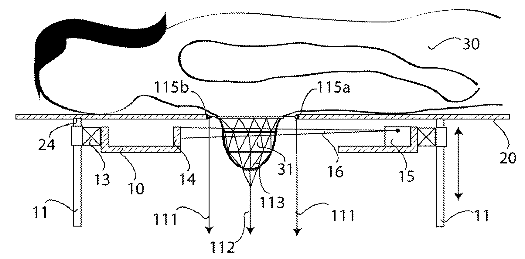
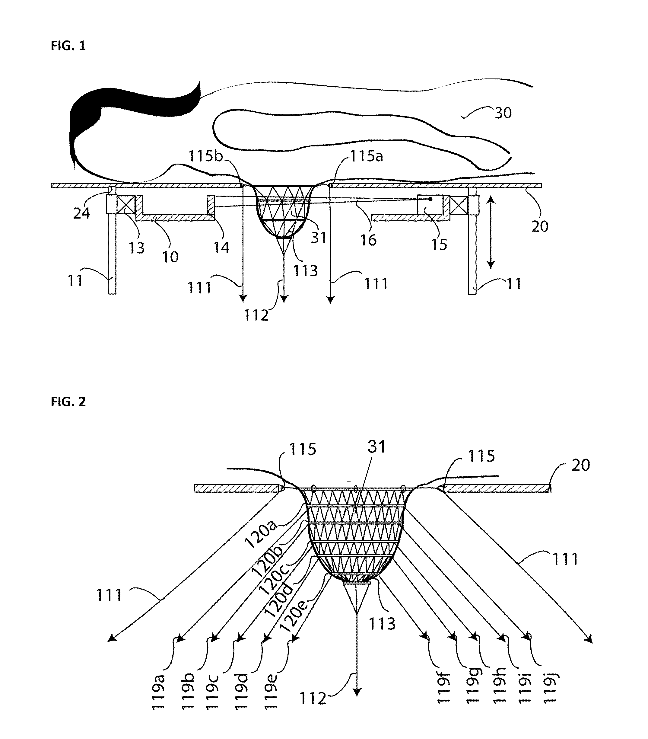
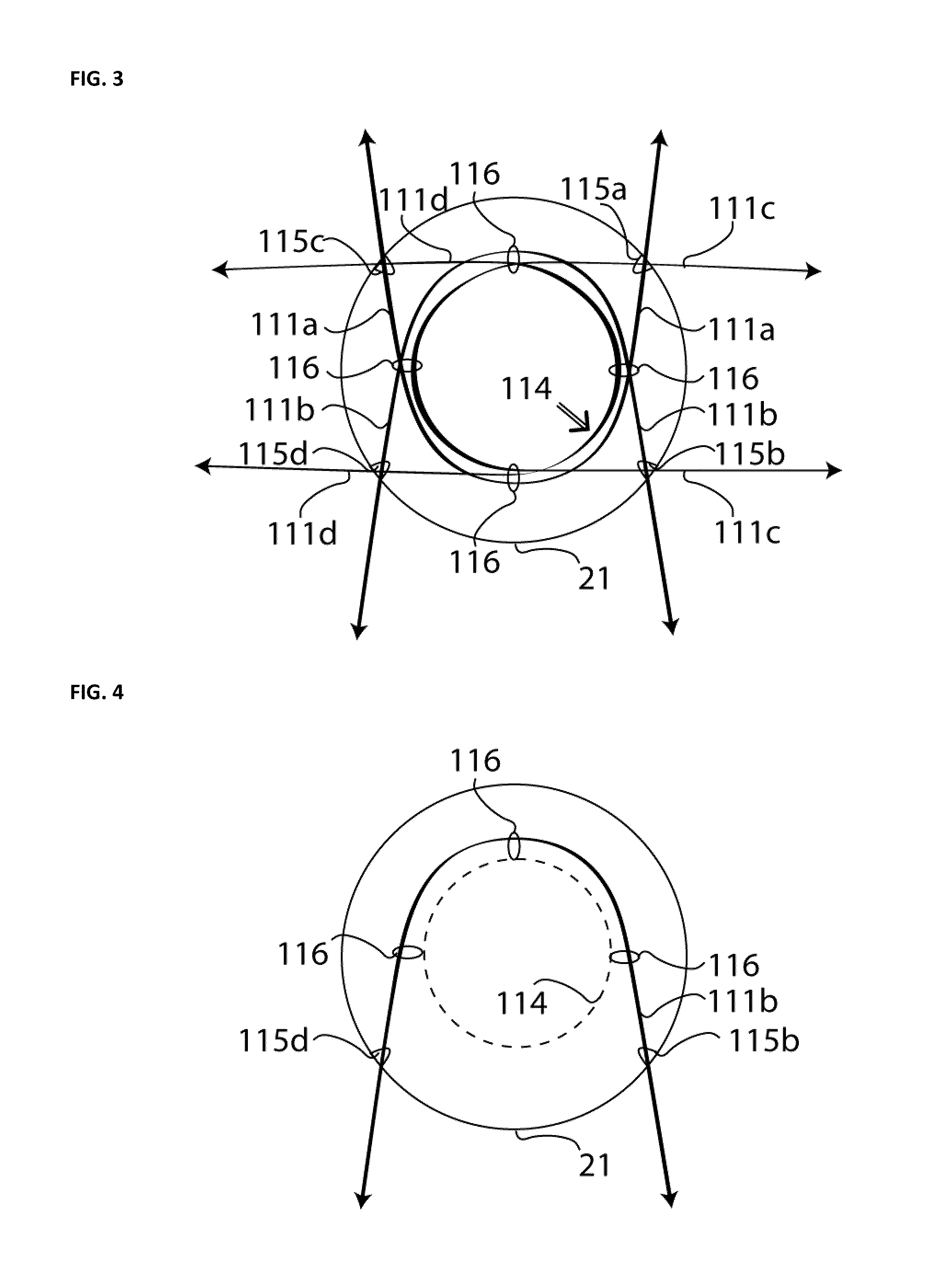
[0018]FIG. 1 illustrates a side view of a cross-section through a device for fixing or locating a breast of a female patient in an X-ray machine. A patient 30 is supported on a patient table 20. A spiral computer tomography gantry 10 is located below the patient. A breast 31 of the patient is suspended through a breast cut-out portion of the patient table into an exposure region of the gantry. Within a gantry housing 24, the gantry 10 has an X-ray tube 15 which generates a fan beam 16. Radiation of this fan beam penetrates the breast 31 and is intercepted by a detector 14. For imaging the entire breast, the gantry can be rotated via a gantry pivot bearing 13. Simultaneously with the rotation, a movement of the gantry in direction towards or away from the patient, here in a vertical direction, is effected by a gantry lift drive 11, so that the breast is scanned along a spiral-shaped track. The breast is fixed or located in a fixing or locating device which comprises at least one fixi...


PUM


Abstract

A device for fixing or locating a breast of a female patient in a medical diagnostic examination instrument comprises an elastic annular-shaped fixing means and also a net fastened thereto which tightly encircle a base of a breast close to a chest wall. A force exerted exerted on the elastic annular-shaped fixing means is applied in a direction away from the patient in order to compress the breast and raise the breast from the chest wall. An X-ray machine including such a device is also provided.

Description

PRIORITY CLAIM[0001]This application claims priority to pending German Application 102008058587.4 filed on Nov. 22, 2008.BACKGROUND OF THE INVENTION[0002]1. Field of the Invention[0003]The invention relates to a device for locating, i.e. fixing, a breast of a female patient in a diagnostic instrument, which is for use in diagnostic imaging and interventional therapy. A device of this kind serves for fixing a breast in a position that is advantageous and reproducible in an imaging diagnostic examination. Moreover, this fixing can be used also for taking a tissue sample (biopsy), or for another kind of diagnosis of a tissue under suspicion, or for a therapy.[0004]2. Description of Related Art[0005]Various instruments such as X-ray machines or also CT scanners are known for examining a female breast. A CT scanner of this kind is disclosed for example in U.S. Patent Application Publication No. 2006 / 0094950. An X-ray facility with a rotating gantry which has an X-ray tube and a detector ...

Claims


Application Information

Patent Timeline
Patent Type & Authority Applications(United States)
IPC IPC(8): A61B6/04H05G1/00
CPCA61B6/0414
Inventor TITA, RALF
Owner MIR MEDICAL IMAGING RES HLDG
Why Eureka
Try Eureka