Anti-igf antibodies
a technology of anti-igf antibodies and antibodies, which is applied in the field of anti-igf antibodies, can solve the problems of increasing the risk of colorectal cancer and adenomas, and the elevated circulating levels of igf-2, and achieve the effect of increasing the total serum igf-1 level
- Summary
- Abstract
- Description
- Claims
- Application Information
AI Technical Summary
Problems solved by technology
Method used
Image
Examples
example 1
[0219]Selection of High Affinity Antibodies that Bind IGF-1
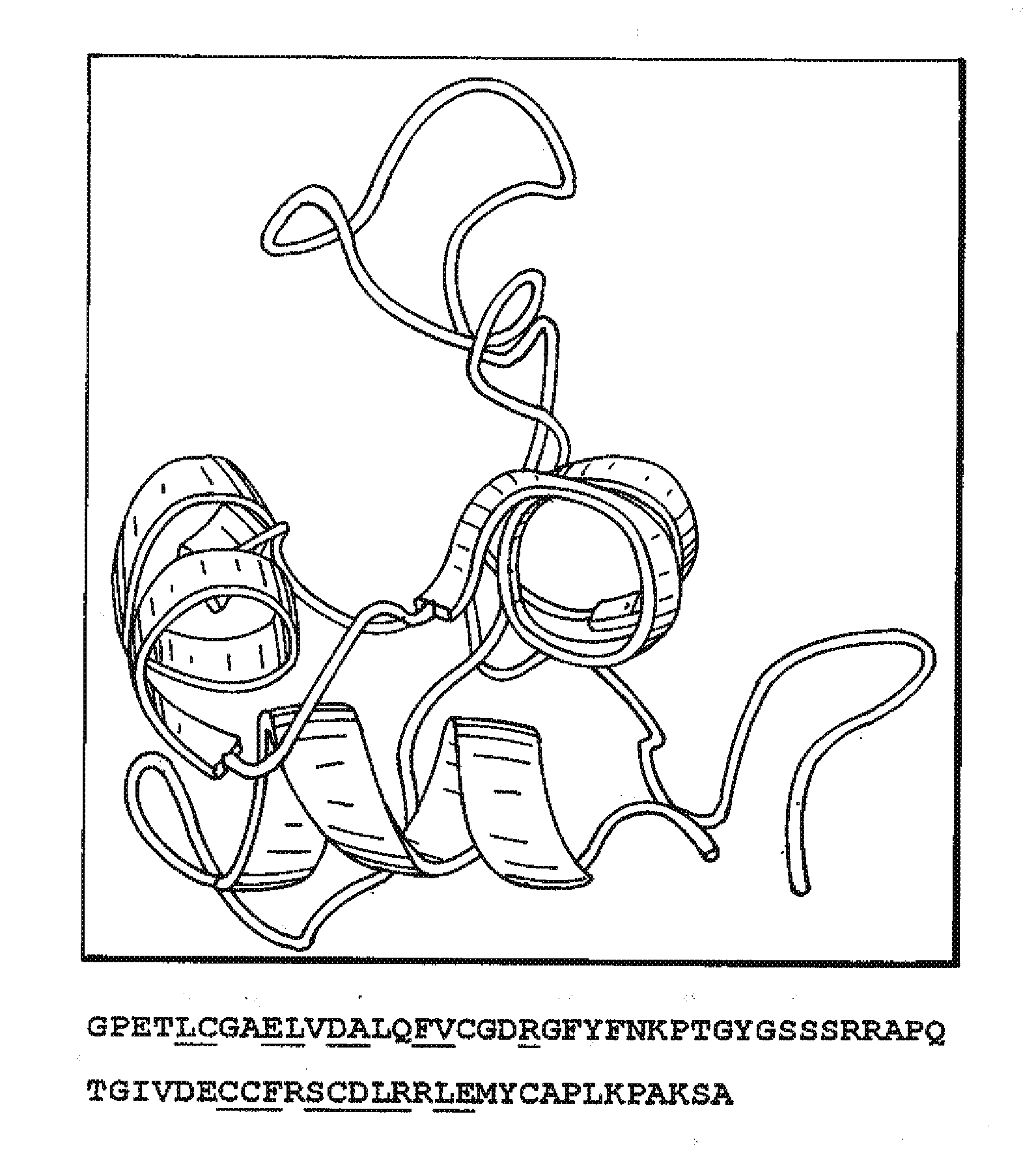
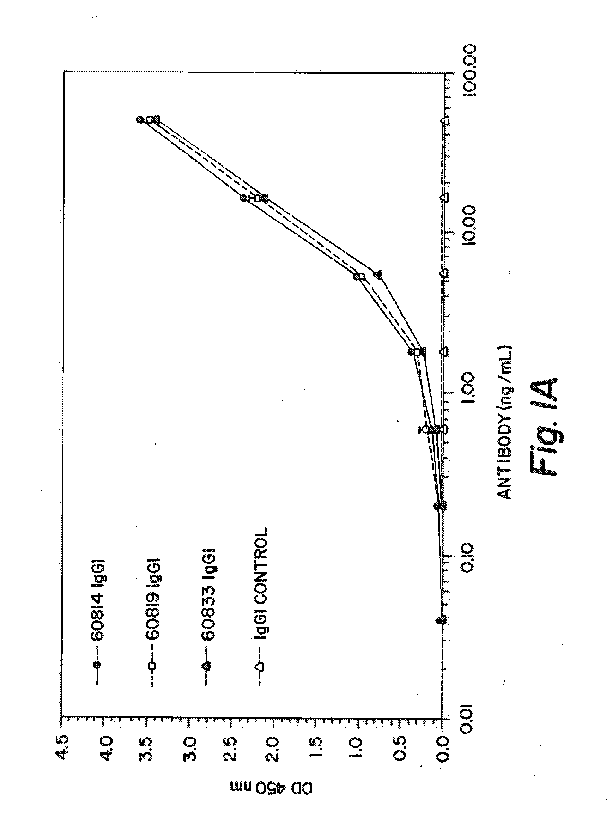
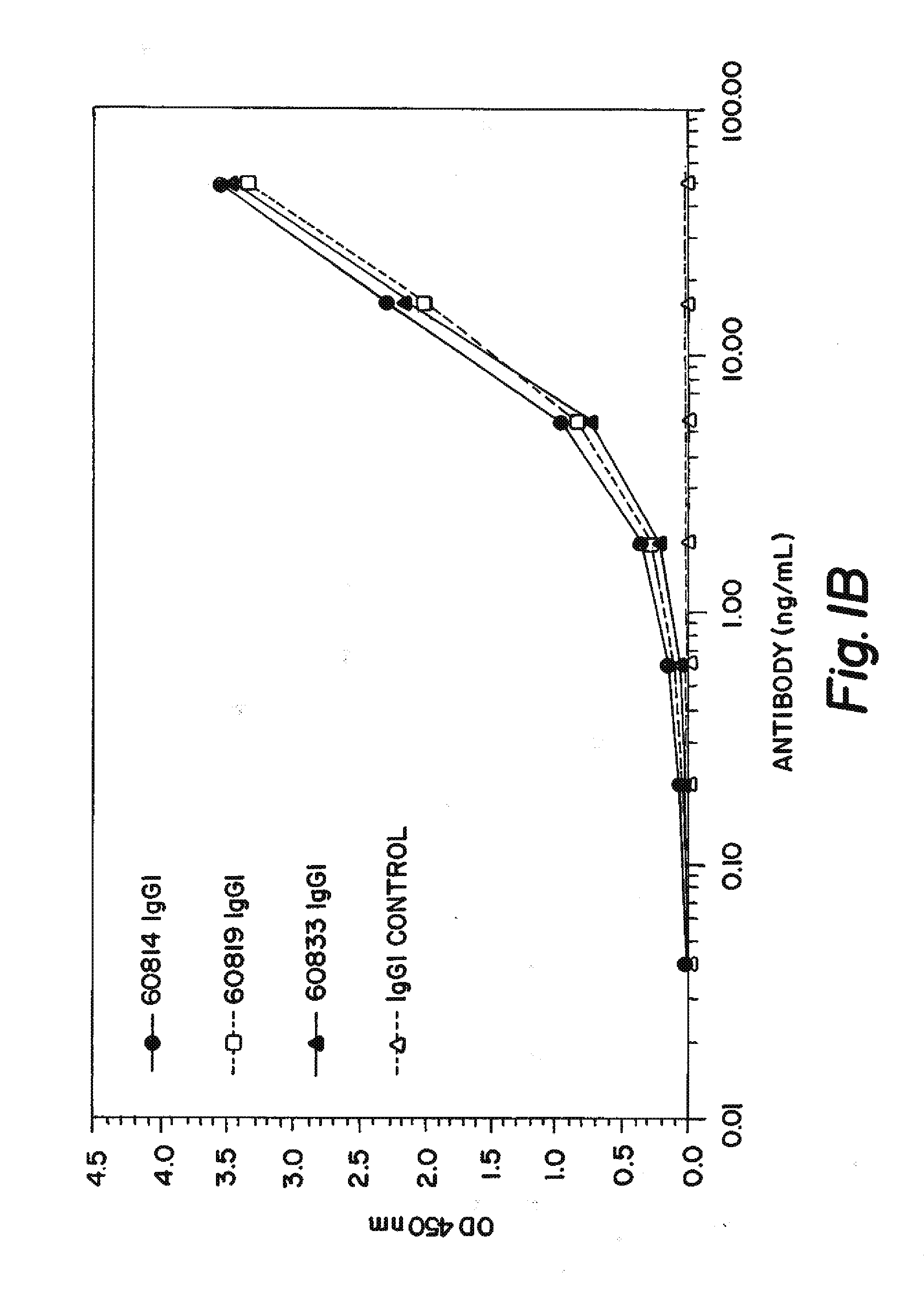
[0220]In order to identify Fab fragments with improved affinity to human IGF-1, several ‘parental’ Fab clones that are identified to bind IGF-1 with low nanomolar affinity are subjected to ‘in vitro affinity maturation’ where the L-CDR3 and H-CDR2 sequences of each clone are separately diversified by substituting the parental sequence with a library of new L-CDR3 and H-CDR2 sequences. The resultant ‘maturation libraries’ are subjected to solution pannings on human IGF-1 and the clones with the best affinity are selected for convertion into IgG1 antibodies and tested further. The three antibodies with the best human IGF-1 affinities are 60814, 60819, and 60833 which had affinities (KD) of 180, 190, and 130 pM respectively (shown in Table 1) as determined by an electrochemiluminescence-based equilibrium titration method.
TABLE 1IGF-1 BINDING SUMMARYAntibodyAffinity (pM)608141806081919060833130
[0221]The antibodies are also teste...
example 2
[0223]Inhibition of IGF Signalling
[0224]The first signalling event which occurs following binding of IGFs to the IGF-1R is the phosphorylation of the IGF-1R. A cell-based ELISA assay is used to measure the inhibition of IGF induced IGF-1R phosphorylation by the antibody 60833. The potency and effectiveness (of up to 15 μg / mL (100 nM)) of 60833 in neutralising recombinant bioactive IGF-1 and IGF-2 induced IGF-1R phosphorylation is determined As shown in Table 3 and example FIG. 2 60833 potently and effectively inhibits IGF-1 (FIG. 2A) and IGF-2 (FIG. 2B) induced signalling. In the same assay the IGF-1R targeted mAb αIR3 is much less potent and effective with respect to IGF-1 induced signalling, and displays a very weak effect on IGF-2 induced signalling.
[0225]A similar cell based IR-A phosphorylation ELISA is used to demonstrate that 60833 can also inhibit IGF-2 signalling via IR-A. As shown in Table 4 and example FIG. 3A, 60833 potently and effectively inhibits IGF-2 induced IR-A ph...
example 3
[0227]Effects on IGF-1 and IGF-2-Induced Cell Proliferation
[0228]The effects of antibodies 60814, 60819, and 60833 on IGF-1 and IGF-2 induced MCF-7 (breast cancer derived) and COLO 205 (colon cancer derived) cell line proliferation is determined Examples of the effects of antibodies 60814 and 60819 are shown in FIGS. 4A-D. All three antibodies show a dose dependent inhibition of IGF-1 (FIGS. 4A and 4C) and IGF-2 (FIGS. 4B and 4D) induced MCF-7 (FIGS. 4A and 4B) and COLO 205 (FIGS. 4C and 4D) cell proliferation. The concentration of each antibody required to inhibit 50% of the IGF-1 or IGF-2 induced proliferation of each cell line is shown in Table 5.
TABLE 5INHIBITION OF IGF-1 AND IGF-2 INDUCED PROLIFERATIONOF THE MCF-7 AND COLO 205 CANCER CELL LINESIC50 (ng / ml)Cell LineStimulation608146081960833MCF7IGF-124.154.038.6MCF7IGF-278.240.881.2COLO-205IGF-1135.0216.9165.1COLO 205IGF-2576.1100.8632.3
PUM
| Property | Measurement | Unit |
|---|---|---|
| molecular weight | aaaaa | aaaaa |
| time | aaaaa | aaaaa |
| concentration | aaaaa | aaaaa |
Abstract
Description
Claims
Application Information
 - R&D
- Intellectual Property
- Life Sciences
- Materials
- Tech Scout
- Unparalleled Data Quality
- Higher Quality Content
- 60% Fewer Hallucinations
Browse by: Latest US Patents, China's latest patents, Technical Efficacy Thesaurus, Application Domain, Technology Topic, Popular Technical Reports.
© 2025 PatSnap. All rights reserved.Legal|Privacy policy|Modern Slavery Act Transparency Statement|Sitemap|About US| Contact US: help@patsnap.com


