
Single fiber endoscopic full-field optical coherence tomography (OCT) imaging probe

a single fiber, full-field optical coherence tomography technology, applied in the field of single fiber full-field optical coherence tomography (oct) imaging probes, can solve the problem of limited biopsy samples

Inactive Publication Date: 2010-06-24
UNIV OF FLORIDA RES FOUNDATION INC
View PDF4 Cites 64 Cited by
  • Summary
  • Abstract
  • Description
  • Claims
  • Application Information

AI Technical Summary

Benefits of technology

[0009]An optical coherence tomographic (OCT) probe-based imaging system for viewing a sample comprises the probe recited in claim 2, and a control module including at least one broadband light source for coupling the broadband light into the single fiber, and signal processing and synchronization electronics for coordinating, detecting and processing opti

Problems solved by technology

Thus they are bulky and are accordingly limited biopsy samples, or for external use.

Method used


Image

  • Single fiber endoscopic full-field optical coherence tomography (OCT) imaging probe
  • Single fiber endoscopic full-field optical coherence tomography (OCT) imaging probe
  • Single fiber endoscopic full-field optical coherence tomography (OCT) imaging probe

Examples


Embodiment Construction

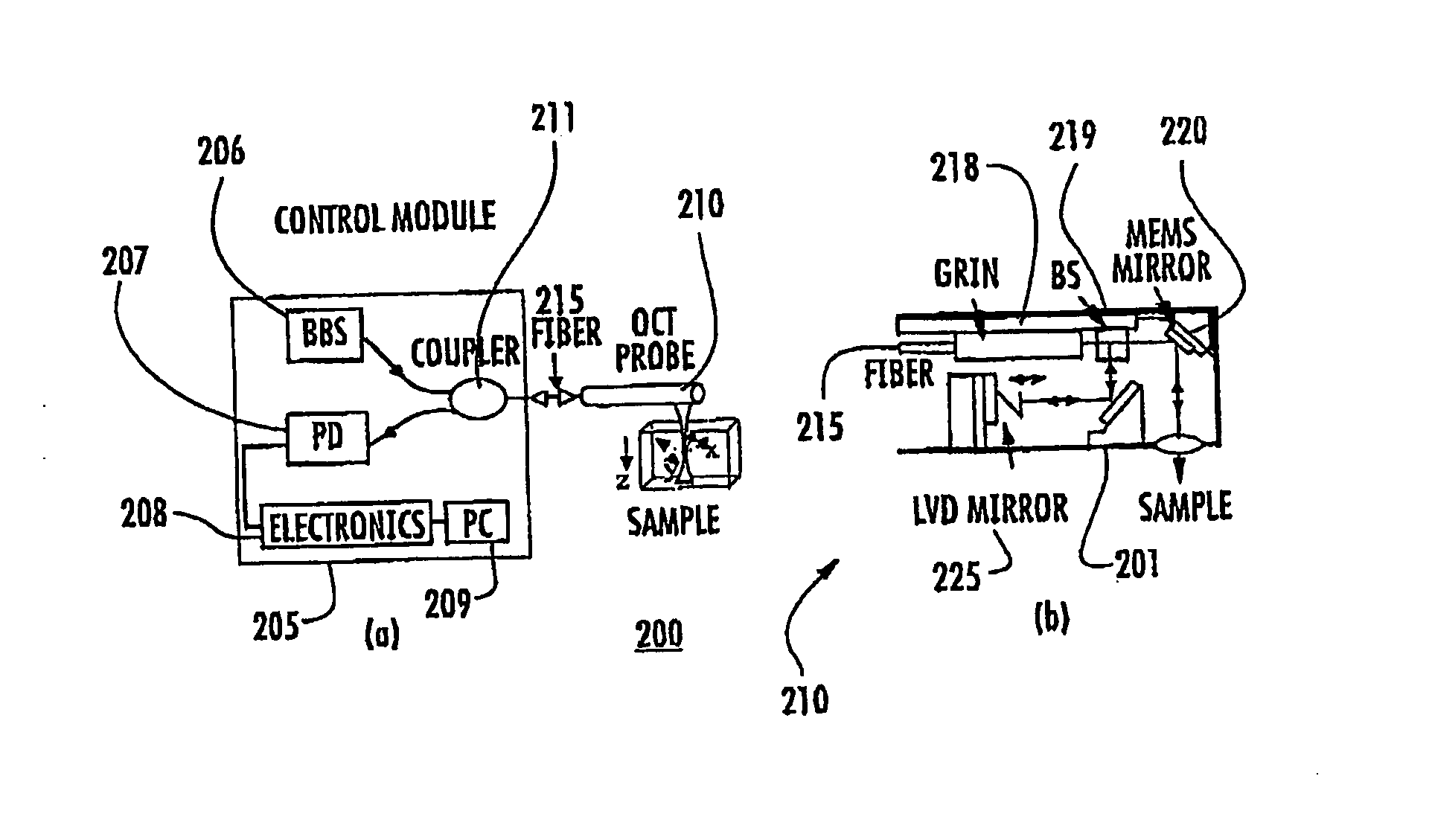
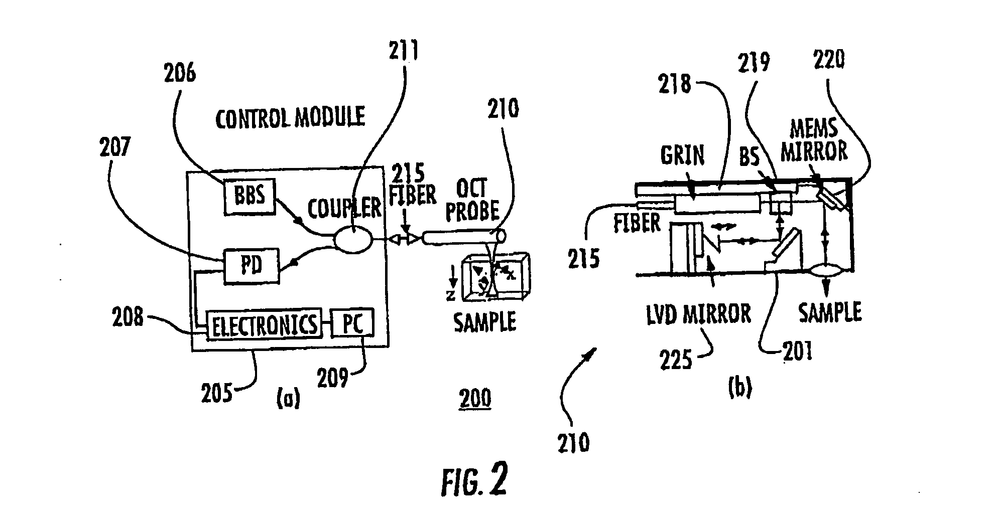
[0019]An optical coherence tomography (OCT) probe for probing a sample comprises a hollow and preferably flexible tube, and a single fiber within the tube for transmitting light received from a broadband light source to a beam splitter in the tube. The beam splitter splits light from the broadband light source into a first and a second optical beam. The first beam is directed to a reference arm and the second beam is directed to a sample arm for probing the sample to be imaged, wherein the reference arm and the sample arm are both embedded in the tube, thus permitting single optical fiber operation. A photodetector is also provided for forming an image of the sample through receipt of the reflected beam from the MEMS reference micromirror and the scattered beam from the sample. Although the photodetector can be disposed external to the probe, in a preferred embodiment the photodetector array is disposed inside the tube.

[0020]The single-fiber full field OCT imaging probe is based on ...


PUM


Abstract

A single fiber full-field optical coherence tomography (OCT) imaging probe (300) includes a hollow tube (301), and a single fiber (305) disposed within the tube for transmitting light received from a broadband light source to a beam splitter (350) in the tube optically coupled to the single fiber (305). The beam splitter (350) splits the light into a first and a second optical beam, wherein the first beam is optically coupled to a reference arm including a MEMS reference micromirror (335) which provides axial scanning and the second beam is optically coupled to a sample arm for probing a sample to be imaged. Both the reference arm and the sample ami are disposed in the tube. A photodetector array (315) is preferably disposed inside the tube (301) optically coupled to the beam splitter (350). The photodetector array (315) receives a reflected beam from the MEMS reference micromirror (335) and a scattered beam from the sample to form an image of the sample.

Description

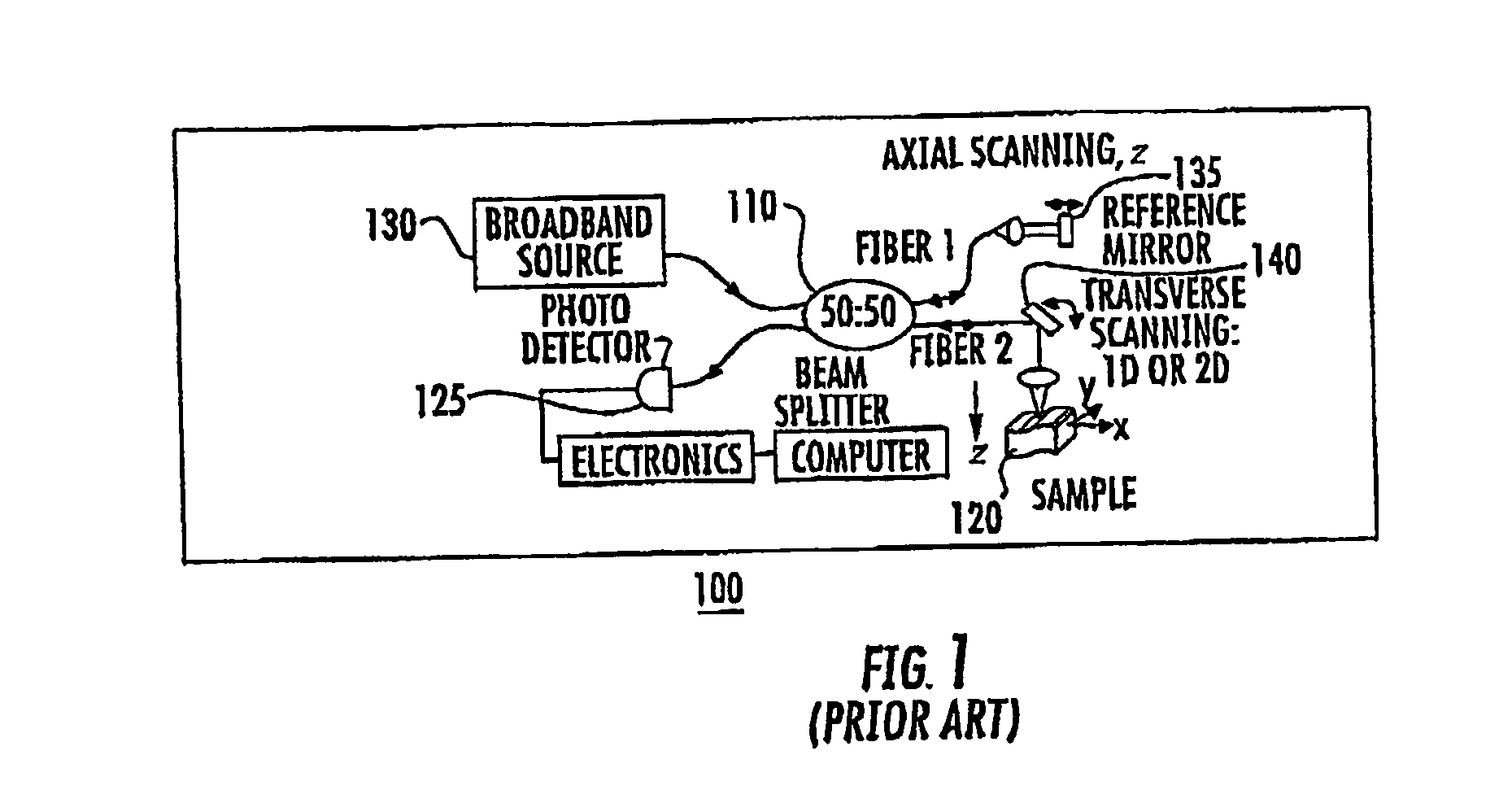
FIELD OF THE INVENTION[0001]The invention relates to single-fiber full field optical coherence tomography (OCT) imaging probes based on embedded MEMS mirrors.BACKGROUND OF THE INVENTION[0002]Optical coherence tomography (OCT), sometimes referred to as “optical biopsy”, can be used to obtain high-resolution (˜10 μm) cross-sectional imaging of scattering biological tissues up to 3 mm deep. OCT is based on low-coherence interferometery and fiber optic technology. The core of an OCT system is a Michelson interferometer, a simplified schematic of which is shown in FIG. 1. For a typical fiberoptic OCT imaging system, such as system 100 shown in FIG. 1, two optical fibers after the beam splitter 110 are required. A first optical fiber (fiber 1) is used for the reference arm of the interferometer, while a second optical fiber (fiber 2) is used for the sample arm of the interferometer which scans the sample 120. The reference arm is external to the probe, while the sample arm including fiber...

Claims


Application Information

Patent Timeline
IPC IPC(8): G01B9/02
CPCA61B5/0066G01B9/02091G01B9/0205A61B5/6852
Inventor XIE, HUIKAI
Owner UNIV OF FLORIDA RES FOUNDATION INC
Learn More