
System and method for computer aided polyp detection

a computer-aided polyp and detection system technology, applied in the field of system and method for computer-aided polyp detection, can solve the problems of inability to follow the advice of most people, time-consuming and laborious colon inspection, and difficult task of automatic detection of colonic polyps

Active Publication Date: 2010-08-26
THE RES FOUND OF STATE UNIV OF NEW YORK
View PDF9 Cites 33 Cited by
  • Summary
  • Abstract
  • Description
  • Claims
  • Application Information

AI Technical Summary

Benefits of technology

[0010]The step of determining the sub-surface density values can take place using volumetric ray casting to integrate sub-surface density values along a projected ray. To easily distinguish different sub-surface density values, a transfer function can be applied to the integrated sub-surface density values to map density values into color values indicative of object density. In this way, the shape of the sub-surface high density regions can readily be determined to aid in the identification of malignancies.

Problems solved by technology

Unfortunately, most people do not follow this advice because of the discomfort and inconvenience of the traditional optical colonoscopy.
However, because of the complicated geometric and topological structure of colon models, inspecting the whole colon can be time consuming and prone to perceptual errors.
However, the automatic detection of colonic polyps is a very challenging task because the polyps can have various sizes and shapes.
Moreover, false positives (FPs) can arise since the colon exhibits numerous folds and residual colonic materials on the colon wall often have characteristics that mimic polyps.
However, these high density areas reside beneath the colon wall and cannot be seen during optical colonoscopy and are not identified in surface rendered virtual colonoscopy.

Method used


Image

  • System and method for computer aided polyp detection
  • System and method for computer aided polyp detection
  • System and method for computer aided polyp detection

Examples


Embodiment Construction

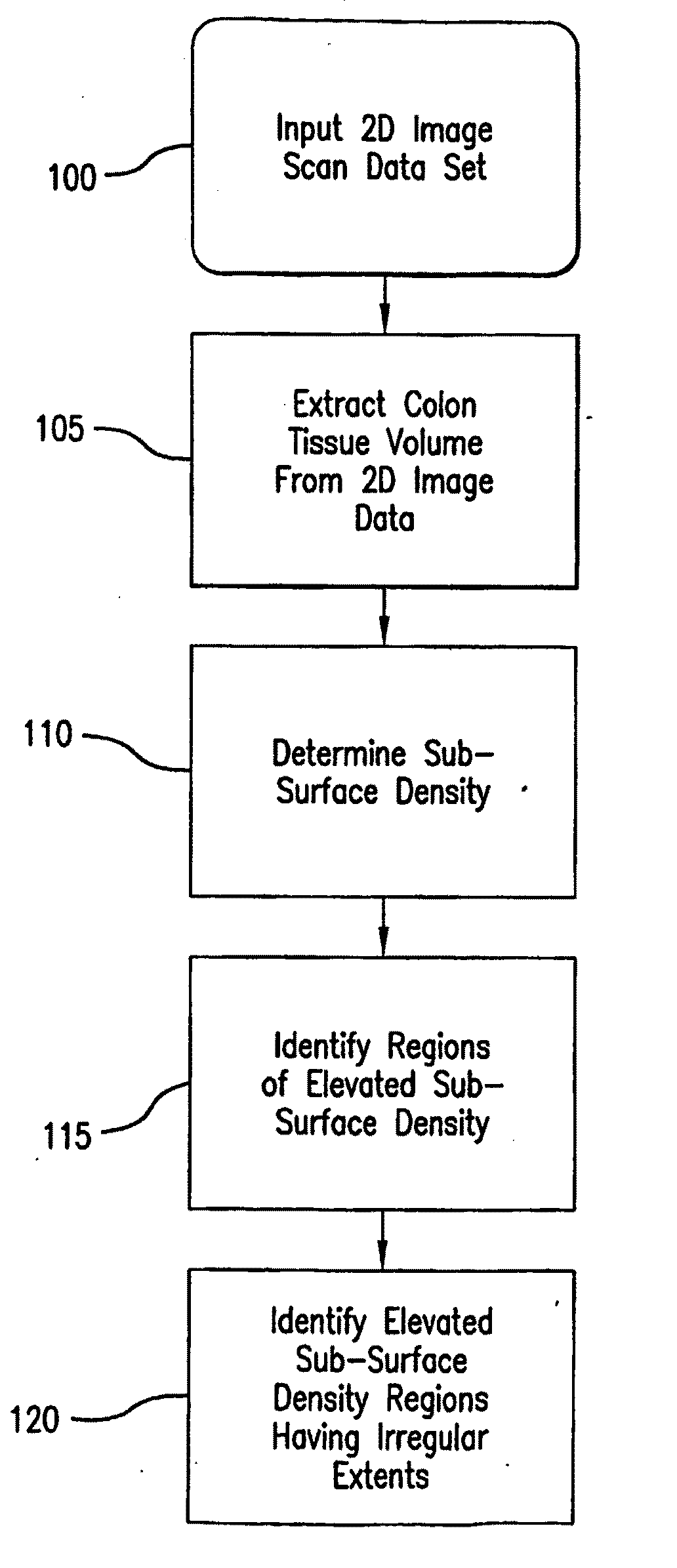
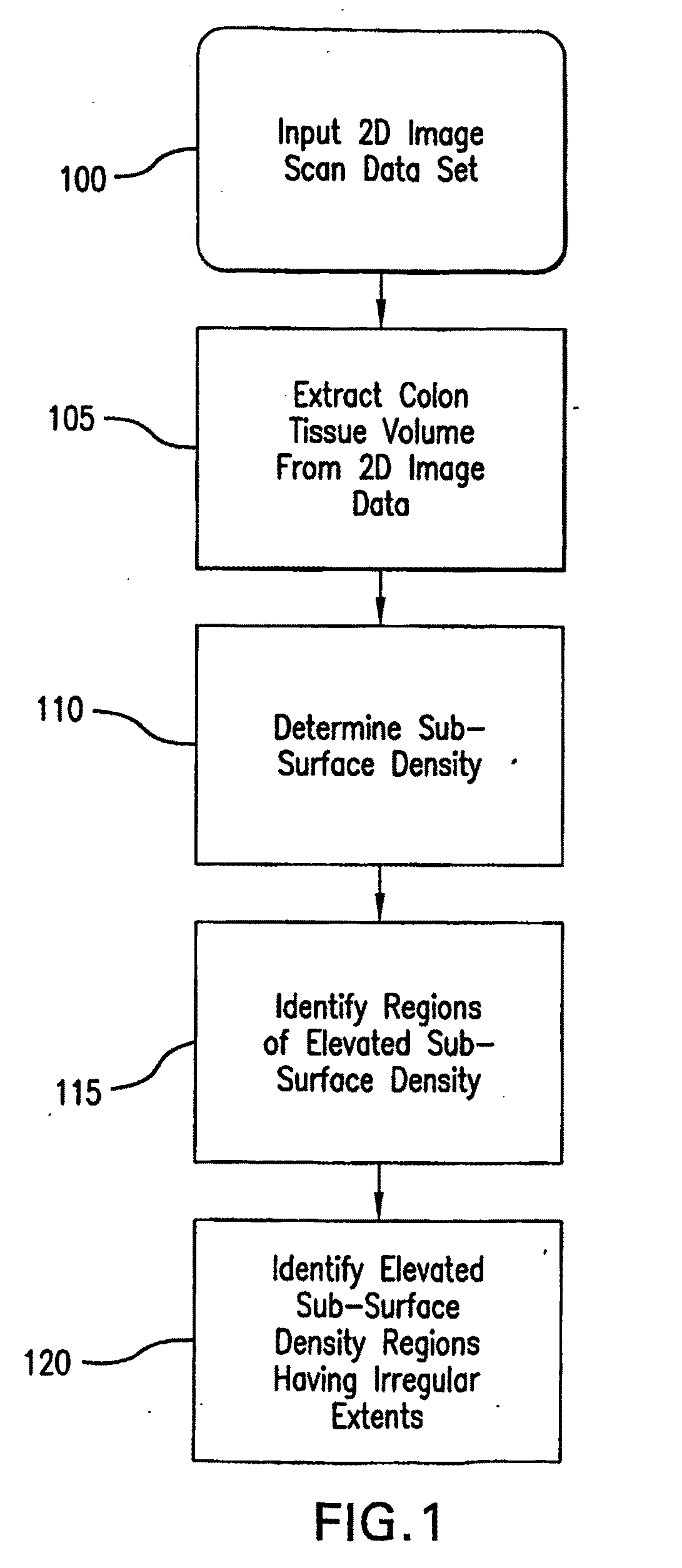
[0030]An overview of a preferred embodiment of the present method for computer aided detection (CAD) of polyps is shown in the simplified flow chart of FIG. 1. The method assumes that appropriate 2D image data has been acquired, such as through the use of a spiral CT scan or other suitable method known in the art of virtual colonoscopy (step 100). From the 2D image data, the colon tissue volume is extracted in step 105, in a manner generally known in the art. After the colon tissue volume has been extracted, the sub-surface density can then be determined (step 110). As is explained in further detail below, the step of determining the sub-surface density is preferably performed on a conformal flattened mapping of the colon surface. The sub-surface density can be determined by integrating voxel values using a ray casting technique which steps through the surface of the colon wall into the depths of the colon tissue and accumulates the voxel values along the ray.

[0031]After the sub-sur...


PUM


Abstract

In the present methods, the automatic detection of polyps is converted into a 2D pattern recognition problem using conformal mapping and direct volume rendering. The colon surface is first segmented and extracted from the CT data set of the abdomen, which is then mapped to a 2D plane using conformal mapping. Ray casting is used to determine sub-surface density values and the flattened image is rendered using a volume rendering technique with a translucent electronic biopsy transfer function. Polyp candidates are detected by a clustering method which identifies regions of elevated sub-surface density. The potential for false positives is reduced by analyzing the volumetric shape and texture features of the polyp candidate regions.

Description

STATEMENT OF PRIORITY AND RELATED APPLICATIONS[0001]This application claims priority to U.S. Provisional Application 60 / 692,903 filed on Jun. 22, 2005, entitled Systems and Methods for Computer Aided Polyp Detection, which is hereby incorporated by reference in its entirety and also claims priority to U.S. Provisional Application 60 / 811,163 filed on Jun. 5, 2006, entitled Improvements in Computer Aided Polyp Detection, which is hereby incorporated by reference in its entirety.STATEMENT OF GOVERNMENT RIGHTS[0002]This work has been supported is part by NSF grant CCR-0306438 and NIH grants CA082402 and CA110186. The United States government may have certain rights to the invention described and claimed herein.BACKGROUND OF THE INVENTION[0003]Colorectal cancer is the second leading cause of cancer-related deaths in the United States. Most colorectal cancers are believed to arise within benign adenomatous polyps that develop slowly over the course of many years. Accepted guidelines recom...

Claims


Application Information

Patent Timeline
Patent Type & Authority Applications(United States)
IPC IPC(8): G06K9/00G06F3/01G06T15/08
CPCG06K9/342G06K2209/053G06T2207/30028G06T7/0012G06T15/08G06T3/0037G06V10/267G06V2201/032G06T3/067G06T2210/41G06T2219/021G06T19/00
Inventor KAUFMAN, ARIE E.HONG, WEIGU, XIANFENGQIU, FENG
Owner THE RES FOUND OF STATE UNIV OF NEW YORK
Learn More