Magnetic resonance method and apparatus for automated analysis of joint inflammation, joint proteoglycan proportion, and joint osteoarthritis

a magnetic resonance and automated analysis technology, applied in the field of magnetic resonance, can solve the problems of inability to provide reproducible, inability to perform reproducible classification or analysis of osteoarthritis, and inability to provide such reproducible analysis or quantification of osteoarthritis in an mr

Inactive Publication Date: 2010-08-26
INSELSPITAL STIFTUNG +1
View PDF1 Cites 6 Cited by
  • Summary
  • Abstract
  • Description
  • Claims
  • Application Information

AI Technical Summary

Benefits of technology

[0014]An object of the present invention is to alleviate the above-mentioned disadvantages associated with such known procedures, in order to provide improved processes and devices for the purpose of generating an analysis of osteoarthritis.

Problems solved by technology

It is not yet possible to perform a reproducible classification or analysis of osteoarthritis that measures the damage of the cartilage and other joint structures (including the synovial fluid).
Techniques are indeed known to determine and to quantify a degree of inflammation of the synovial fluid and a degeneration of the respective cartilage structures, but these techniques are not able to supply reproducible results.
For example, it is not yet possible to provide such reproducible analysis or quantification of osteoarthritis in an MR imaging procedure.
Thus it is almost impossible to reproduce the results since even the same physician in examining his or her results would have difficulties to repeat the MR measurements at the same sections of the joint.
It takes at least 30 minutes until an adequate amount of T1 contrast medium has been absorbed in the region of the joint, which results in a respective waiting period for the patient.
Because of the above-mentioned period of waiting until an adequate amount of T1 contrast medium has been absorbed in the region of the joint, the patient has to be placed twice in a respective magnetic resonance imaging scanner, which is cumbersome, time-consuming and therefore expensive.
What is more, it is extremely difficult for the same physician to perform the two T1 measurements before and after administering the T1 contrast medium, if a manual process is used to measure the same region of the joint.
A further problem involves the feeding of the information obtained through the T1 measurement(s) consistently and reproducibly into a rating system.
In summary the known procedures of analyzing osteoarthritis art are not ideal for providing reproducible results because they are cumbersome and have limited properties.

Method used


Image

  • Magnetic resonance method and apparatus for automated analysis of joint inflammation, joint proteoglycan proportion, and joint osteoarthritis
  • Magnetic resonance method and apparatus for automated analysis of joint inflammation, joint proteoglycan proportion, and joint osteoarthritis
  • Magnetic resonance method and apparatus for automated analysis of joint inflammation, joint proteoglycan proportion, and joint osteoarthritis

Examples


Embodiment Construction

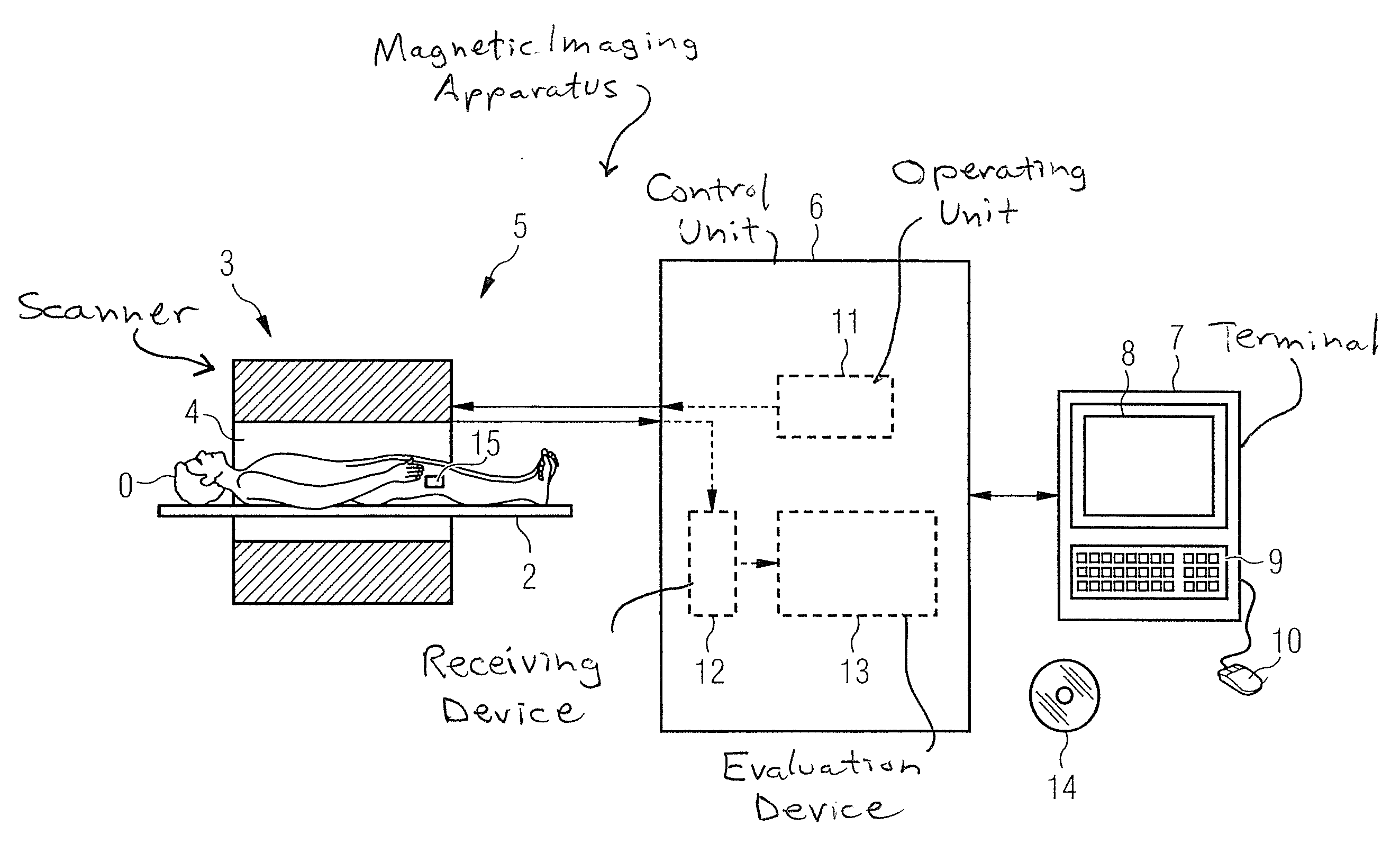
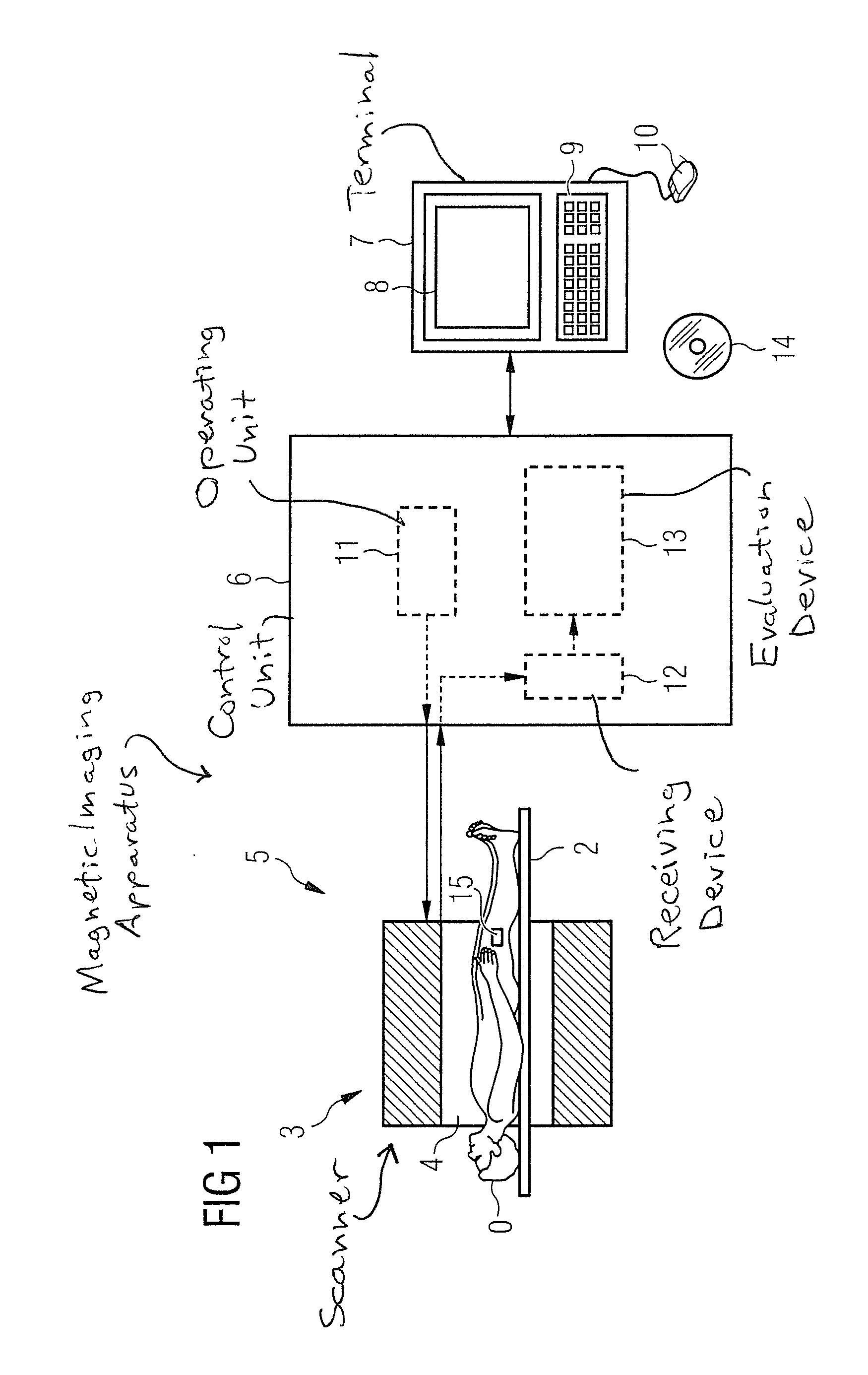
[0052]FIG. 1 shows an inventive magnetic resonance imaging apparatus 5. Basically, the magnetic resonance imaging apparatus 5 includes a scanner 3 that generates, in an examination region 4, the magnetic field required for the MR examination, a table 2, a control unit 6 that controls the scanner 3 and records MR data from the scanner 3, and a terminal 7 connected to the control unit 6.

[0053]The control unit 6, in turn, includes an operating unit (controller or sequencer) 11, a receiving device 12 and an evaluation device 13. During an MR examination, MR data are acquired from the receiving device 12 by means of the scanner 3. In the process, the operating unit 11 controls the scanner 3 to acquire MR data acquired from a volume 15 inside the body of a patient 0 lying on the table 2.

[0054]The evaluation device 13 processes the MR data so as to be presented graphically on the monitor 8 of the terminal 7 with the analyses generated according to the invention being included in the graphi...


PUM


Abstract

In a magnetic resonance method and apparatus for generating an analysis of an inflammation of a joint, for determining a proteoglycan proportion of a joint, and for generating an analysis of osteoarthritis of a joint, for a predetermined region within a body of a patient, a three-dimensional MR image data set is generated with a magnetic resonance imaging scanner. At least, a portion of a joint is located within the region. For each of several different automatically determined layers, a two-dimensional MR image is calculated from the three-dimensional MR image data set. For each layer, the thickness of a predetermined tissue, which is represented in the two-dimensional MR image corresponding to the layer, is determined, by means of which an analysis of a joint inflammation is automatically determined depending on the respective thicknesses. A further layer in the region is also automatically determined and, after injecting a contrast medium, a two-dimensional T1-weighted MR image is generated for this layer, from which a proteoglycan proportion is automatically determined. From the analysis of the joint inflammation and the proteoglycan proportion, it is then possible to perform a reproducible analysis of osteoarthritis of the joint.

Description

BACKGROUND OF THE INVENTION[0001]1. Field of the Invention[0002]The present invention concerns magnetic resonance methods and devices for analyzing a joint inflammation, for determining a proteoglycan proportion in the region of the joint, and / or for analyzing osteoarthritis.[0003]2. Description of the Prior Art[0004]Osteoarthritis (OA) is a process of joint deterioration encompassing of various factors and involves the loss of functional capability of the joint. It is not yet possible to perform a reproducible classification or analysis of osteoarthritis that measures the damage of the cartilage and other joint structures (including the synovial fluid). Techniques are indeed known to determine and to quantify a degree of inflammation of the synovial fluid and a degeneration of the respective cartilage structures, but these techniques are not able to supply reproducible results. For example, it is not yet possible to provide such reproducible analysis or quantification of osteoarthr...

Claims


Application Information

Patent Timeline
Patent Type & Authority Applications(United States)
IPC IPC(8): A61B5/055
CPCA61B5/055A61B5/4514G01R33/5601A61K49/06G01R33/20A61B5/4528
Inventor HUGHES, TIMOTHYMAMISCH, TALLAL CHARLES
Owner INSELSPITAL STIFTUNG
Why Eureka
Try Eureka